
Abstract

Orbital apocrine hidrocystoma is rare and usually found in children. Although superomedial lesions are described, this case illustrates the intricate occurrence at the tendon-trochlea interface, an anatomical niche not reported yet. Meticulous dissection while ensuring complete excision is essential to avoid complications such as iatrogenic Brown's syndrome.
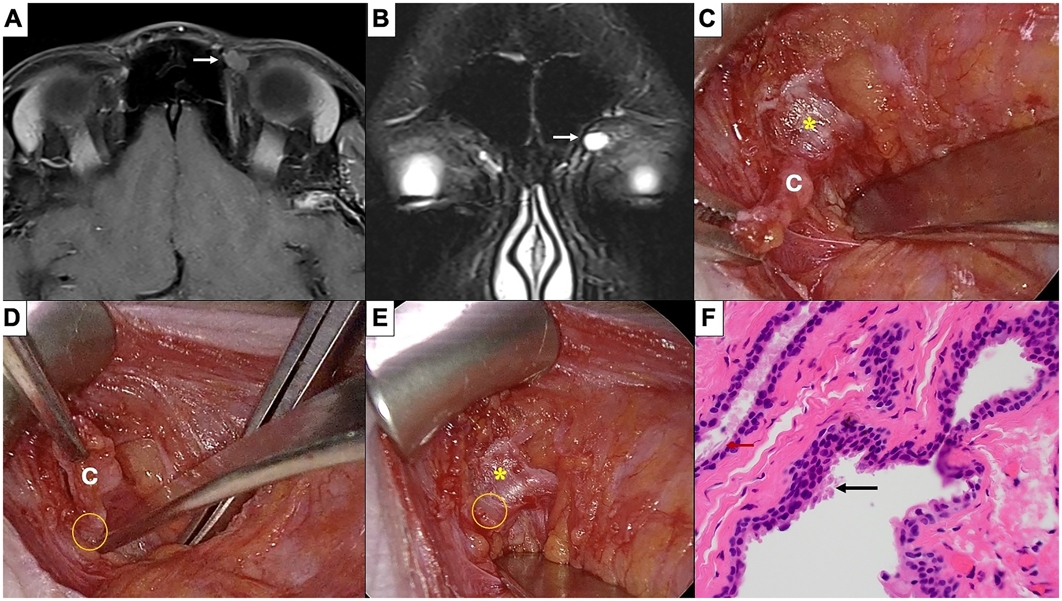
A 33-year-old lady was referred for an incidental left-sided orbital lesion. Visual acuity was 20/20 in both eyes. Examination revealed a well-defined, non-tender, firm mass in the superomedial left orbit with normal ocular motility. Magnetic resonance imaging (MRI), T1-weighted fat-suppressed, axial view, demonstrated a well-circumscribed, non-enhancing, extraconal lesion (arrow), closely related to the superior oblique (SO) tendon (Panel A), appearing hyperintense on T2-weighted sequence (arrow), suggestive of an orbital cyst (Panel B). Excision biopsy was performed through upper eyelid crease incision. Intra-operatively, a thin-walled cyst (labelled C) was found nestled between the distal SO tendon laterally (asterisk) and the trochlear spine medially (circle) (Panel C and D), with clear fluid leakage at the site of adherence. The lesion was completely excised without injury to the tendon-trochlea complex (Panel E). Histopathology (H&E, ×200), revealed a cystic lesion lined by double layer of epithelium with decapitation secretions and apical snouts (arrow), consistent with apocrine hidrocystoma (Panel F).
Footnotes
Consent to participate
Written informed consent was obtained from the patient for publication of this case report and accompanying images.
Funding
The authors received no financial support for the research, authorship, and/or publication of this article.
Declaration of conflicting interests
The authors declared no potential conflicts of interest with respect to the research, authorship, and/or publication of this article.