
Abstract

We read with great interest the article by Dersch et al. on the effect of lateral scaling on measurements in Heidelberg Spectralis spectral domain optical coherence tomography (SD-OCT). 1 In this study, the authors measured the distance between the Bruch's membrane opening at the temporal margin of the optic disc to the center of the fovea by the built-in caliper function. Then, the authors performed ocular magnification according to the axial length (AL) by applying an additional formula to the obtained results for the adjusted lateral scaling. The importance of lateral scaling, especially in retinal imaging, is required to get more accurate measurements, as the authors stated. 2 However, is it essential in Spectralis OCT to adjust the lateral scaling with an additional formula that includes the AL to get an accurate measurement?
It has been previously reported that Spectralis OCT automatically estimates individual lateral scaling using the spherical equivalent (SE) of total refractive error and corneal curvature (CC) with the built-in HEYEX software based on the Gullstrand schematic eye.3,4 With this software, ocular adjustment is performed automatically for accurate lateral scaling without the requirement for additional formula/calculation. In summary, lateral scaling in Spectralis OCT is not constant, and eye-specific scan lengths are produced. A detailed description of the ocular magnification in Spectralis OCT is presented in the study of Delori et al. 4
We believe that Figure 1 in our letter is an illustrative example indicating that lateral scaling is not constant. Retinal imaging was performed by Spectralis OCT separately for three different refractive errors at the same imaging session from a 43-year-old emmetropic volunteer with a CC of 7.64 (informed consent obtained). Following the registration, the CC value was recorded in the HEYEX software, and the first posterior pole imaging was performed at 0.00 diopter (D) SE. Then, consecutive retinal imaging was performed at +3.00 D and −3.00 D SE by using the focus knob. After the retinal imaging, a visible reciprocal vascular bifurcation location was identified on all three near-infrared en-face images, and the distance from this location to the Bruch's membrane opening at the temporal margin of the optic nerve head was measured by caliper for each image individually. As a result of the measurements, a longer distance was detected in the myopic-adjusted image, and a shorter distance was detected in the hyperopic-adjusted image compared to the emmetropic image. It can be seen from the size difference of the scale marker in the lower-left side of the images that dissimilar scaling is performed automatically by the software for all three imaging.
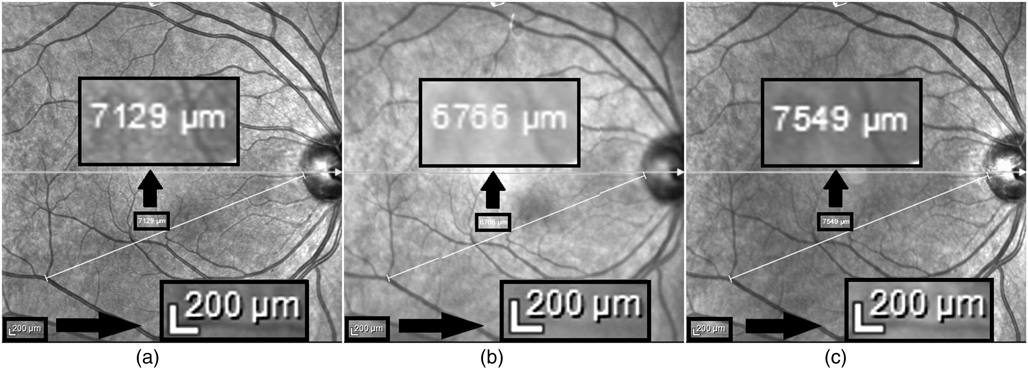
Posterior pole imaging with Heidelberg Spectralis SD-OCT of an emmetropic patient adjusted to different spherical equivalents using the focus knob, and the measurements of the distance from selected vascular bifurcation to Bruch's membrane opening at the temporal margin of the optic nerve head. The distances measured with the built-in caliper function were 7129 µm when refraction was set to 0.00 D (a), 6766 µm when refraction was set to +3.00 D (b), and 7549 µm when refraction was set to −3.00 D (c).
In addition, the authors, citing the study of Ctori et al., stated that the accuracy of lateral measurements could be improved by including CC in Spectralis OCT. 3 However, in the study of Ctori et al., the scan length obtained by recording the CC and SE on Spectralis OCT was compared with the simulated scan length produced by an optical-designed software, and no additional formula was applied to the values obtained from Spectralis OCT.
Footnotes
Statements and declarations
Declaration of conflicting interests
The authors declared no potential conflicts of interest with respect to the research, authorship, and/or publication of this article.
Funding
The authors received no financial support for the research, authorship, and/or publication of this article.
Ethical statement and consent to participate
Informed consent was obtained from the healthy volunteer.