
Abstract
Objectives
This study aimed to evaluate the association of anaemia and thrombocytopenia with the presence and severity of ocular lesions in cats.
Methods
Cats were prospectively recruited into four groups in this cross-sectional study: anaemic (A) (haematocrit level ⩽24%, n = 20), thrombocytopenic (T) (platelet count of ⩽200 × 109/l, n = 10), anaemic and thrombocytopenic (AT) (n = 33) and healthy controls (C) (n = 20). All cats underwent a comprehensive ophthalmic examination, with lesions classified as absent, minor (isolated retinal haemorrhages) or major (diffuse retinal haemorrhages, hyphaema or intravitreal bleeding). Signalment, clinical signs, diagnostic test results and final diagnoses were collected from the medical records.
Results
Ocular lesion prevalence was 20%, 10%, 48% and 0% in the A, T, AT and C groups, respectively. Among affected cats, minor and major lesions occurred in 67% and 33% of cases, respectively. All major lesions were observed in groups T and AT. Anaemia and thrombocytopenia were significantly associated with both the presence (Panaemia = .001, Pthrombocytopenia = .002) and severity (Panaemia <.001, Pthrombocytopenia = .006) of ocular lesions. A haematocrit value of ≤18.5% had a sensitivity of 91% and specificity of 58% for detecting ocular lesions.
Conclusions and relevance
Anaemia and thrombocytopenia were significantly associated with the presence and severity of ocular lesions in cats in this study. Cats with concurrent anaemia and thrombocytopenia exhibited a higher prevalence of ocular lesions than those with either condition alone. This finding warrants further investigation into potential underlying mechanisms. Based on these results, any cat presenting with anaemia or thrombocytopenia should undergo a complete ophthalmic examination.
Introduction
Anaemia is a common clinical issue in cats, with reported prevalences of 3.6% (blood samples from cats that fulfilled criteria for anaemia) 1 and 10% (within the reference population). 2 It is characterised by a decrease in haematocrit (HCT) level below the reference interval (RI) 2 and develops because of haemorrhage, haemolysis or decreased bone marrow red blood cell (RBC) production. 3 Common anaemia aetiologies in cats include immune-mediated haemolytic anaemia (IMHA), 4 chronic kidney disease, 5 gastrointestinal disease, 6 retroviruses 7 and neoplasia. 8 Its clinical importance can vary from being the patient’s primary life-threatening problem to being relatively clinically inconsequential. 9 Ocular manifestations of severe anaemia include pale retinal vasculature, varying degrees of retinal haemorrhage and subtle changes in tapetal reflectivity. 10 Only two studies have investigated the association between anaemia and intraocular haemorrhages in cats, reporting retinal haemorrhages in 20/26 and 14/80 anaemic animals.11,12
Thrombocytopenia is the most commonly encountered platelet (PLT) disorder in feline medicine, 13 with a reported prevalence in the range of 1.2–5.9%.14–17 These estimates are based on retrospective analyses of medical records from referral clinics or veterinary teaching hospitals, with prevalence calculated based on the total caseload during the respective study periods. Thrombocytopenia can result from decreased PLT production, increased PLT removal or increased consumption. A retrospective study of 194 cats with thrombocytopenia reported that the most common aetiologies were infectious, immune-mediated and neoplastic. 16 Thrombocytopenia is associated with ocular and periocular haemorrhages. Although the PLT count threshold at which bleeding occurs varies among individuals, it is typically below 50 ×109/l. 10 Several studies in humans have investigated the association between anaemia, thrombocytopenia and ocular lesions; however, their findings remain inconsistent.18–22
A prospective study in dogs found no association between anaemia and ocular lesion presence or severity but reported a significant relationship between ocular lesions and thrombocytopenia. 23 No studies have directly investigated the association between ocular lesions and thrombocytopenia in cats. Therefore, this study evaluated the association of anaemia and thrombocytopenia with ocular lesion presence and severity in cats.
Materials and methods
Study design and ethical considerations
This cross-sectional study was approved by the VetAgro Sup Committee for Ethics in the Use of Animals of the Veterinary University of Lyon (protocol number 2383). Owners provided written consent for the use of their pets’ clinical data and performance of an ophthalmic examination and blood pressure measurement. The study population comprised cats presented to the Internal Medicine or Emergency and Critical Care services of the Fregis Veterinary Hospital Centre in Gentilly (France) between October 2018 and October 2024.
All animals underwent a comprehensive haematological evaluation, including blood smear analysis. Cats were classified as anaemic and thrombocytopenic if the HCT value was ≤24% and the PLT count was ≤200 × 109/l, respectively. The HCT threshold represents the lower limit of the RI established by the clinic’s ANYDIAG laboratory. Although the laboratory’s lower RI for PLT count is 300 × 109/l, this cut-off point was decreased in this study based on the inclusion criteria of other research investigating thrombocytopenia in cats.14,16 Healthy controls were also included, consisting of animals hospitalised by the Surgery Department for various elective procedures, with blood work results within RIs. Based on these criteria, cats were assigned to groups A (anaemia), T (thrombocytopenia), AT (anaemia and thrombocytopenia) and C (healthy controls). Cats were excluded if clumping was reported on blood smear evaluation or if a manual PLT count had not been performed. The laboratory’s manual PLT count was used to determine the eligibility for inclusion where the manual and automated PLT counts were discrepant.
Clinical examination
Each cat underwent a comprehensive ophthalmic examination, including menace response, pupillary, dazzle and palpebral reflexes, rebound tonometry, slit-lamp biomicroscopy (Kowa SL17) and indirect ophthalmoscopy after pupil dilation with 0.5% topical tropicamide solution. Additional diagnostic testing was performed in some cats on a case-by-case basis, including fluorescein staining. Ocular involvement was classified as minor or major based on its presumed impact on vision or the globe. Minor lesions were characterised by isolated or sparse intraretinal or preretinal haemorrhages. In contrast, major lesions were characterised by diffuse retinal haemorrhages, hyphaema or intravitreal bleeding.
Conscious systolic blood pressure (SBP) measurement was performed in all cooperative cats using Doppler methodology, in accordance with the American College of Veterinary Internal Medicine consensus guidelines. 24 A comprehensive range of diagnostic tests was used to achieve a conclusive diagnosis for each cat, at the discretion of the attending clinician. These included various combinations of blood testing, urinalysis, faecal analysis, diagnostic imaging, cytology and histopathology. The signalment, clinical signs at presentation and final diagnoses were obtained from the medical records. Furthermore, the attending clinicians determined the final diagnoses, while the ophthalmologist remained unaware of the systemic diagnosis during the ophthalmic examination.
Statistical analysis
All statistical analyses were performed using SPSS Statistics for Windows version 29 (IBM Corp). A χ2 test was employed to assess the association between anaemia and ocular lesions by comparing the prevalence of ocular lesions in cats with anaemia (groups A and AT combined) and those without anaemia (groups T and C combined). Similarly, a χ2 test was used to evaluate the association between thrombocytopenia and ocular lesions by comparing the prevalence of ocular lesions in cats with thrombocytopenia (groups AT and T combined) and those without thrombocytopenia (groups A and C combined). Furthermore, a χ2 test was used to assess the association between the prevalence of ocular lesions across all four groups, as well as between ocular lesion prevalence and cytopenia severity. HCT value and PLT count were categorised into moderate (HCT 16–24%, PLT 51–200 × 109/l) and severe (HCT 0–15%, PLT 0–50 × 109/l). The Kruskal
Results
Epidemiology
A total of 83 cats were included, comprising 47 males (37 neutered, 10 entire) and 36 females (31 neutered, five entire). Among these, 46 (55%) were European Shorthairs, with the remainder comprising 13 other breeds. Table 1 summarises the median and interquartile range values for age, HCT and PLT count across the four groups. Animals in group AT were the youngest across all groups. The median HCT was lower in group AT than in group A, while the median PLT count was lower in group T than in group AT. SBP measurements were conducted in 14/21 cats with ocular lesions, of which only one was hypertensive (190 mmHg, RI 120–160) and belonged to group T. Of the 43 cats with thrombocytopenia, only one (2%) had dermatorrhagia (subcutaneous ecchymoses and petechiae), and this cat was in group AT.
Age, haematocrit (HCT) value and platelet (PLT) count across the study groups

Data are n (%) or median (interquartile range)
A = anaemic group; AT = anaemic and thrombocytopenic group; C = control group; T = thrombocytopenic group
Ophthalmic findings
Table 2 summarises the signalment and clinical characteristics of the 21 cats with anaemia and/or thrombocytopenia and ocular lesions. Ocular lesions were observed in 4/20 (20%), 1/10 (10%), 16/33 (48%) and 0/20 (0%) cats in groups A, T, AT and C, respectively. In group A, four cats exhibited minor ocular lesions, characterised by small, isolated or scattered intraretinal or preretinal haemorrhages (Figure 1). Major and minor ocular lesions were observed in six (18%) and 10 (30%) cats within group AT, respectively. The major lesions in this group were characterised by extensive retinal haemorrhages, hyphaema (Figure 2) or intravitreal bleeding. One cat in group T with ocular pathology had major lesions, comprising a haemorrhage in the iris stroma and concurrent hyphaema, which prohibited a complete fundic examination. Anaemia and thrombocytopenia were significantly associated with both the presence (Panaemia = .001, Pthrombocytopenia = .002) and severity (Panaemia <.001, Pthrombocytopenia = .006) of ocular lesions. A significant association was also found between ocular lesion prevalence across all four groups (P <.001). The Kruskal–Wallis test revealed significantly lower median HCT values and PLT counts in the major ocular lesion group (Table 3). When assessing the relationship between cytopenia severity and ocular lesion presence, the χ² test revealed a significant association for anaemia but not for thrombocytopenia (Panaemia = .002, Pthrombocytopenia = .834) (Figures 3 and 4). The area under the ROC curve (AUC) for HCT was 0.793 (95% confidence interval [CI] 0.691–0.894) (Figure 5). Specifically, the Youden index provided an optimal HCT cut-off value of 18.5%, resulting in sensitivity and specificity values of 91% and 58%, respectively. The AUC was markedly different from 0.5, indicating a significant ability of HCT to discriminate between cats with and without ocular lesions.
Clinical data of cats with anaemia and/or thrombocytopenia and ocular lesions
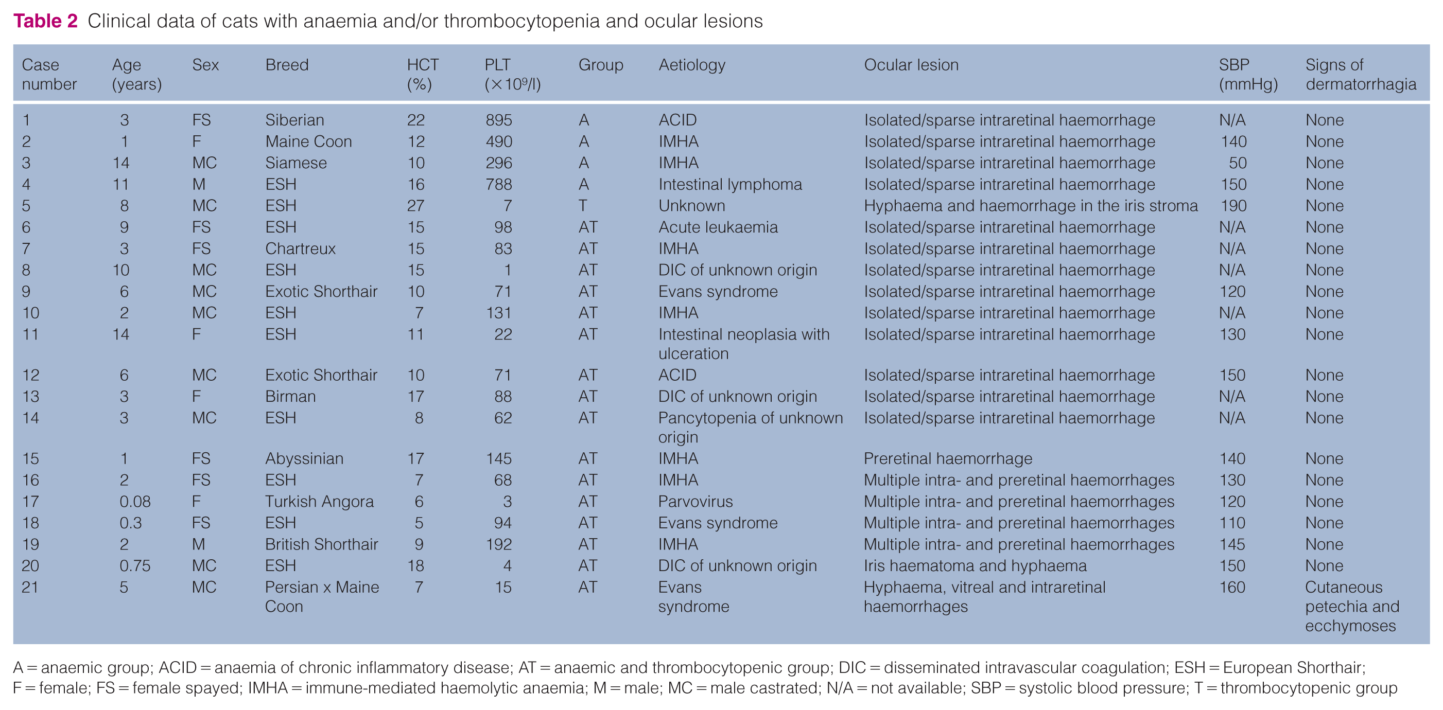
A = anaemic group; ACID = anaemia of chronic inflammatory disease; AT = anaemic and thrombocytopenic group; DIC = disseminated intravascular coagulation; ESH = European Shorthair; F = female; FS = female spayed; IMHA = immune-mediated haemolytic anaemia; M = male; MC = male castrated; N/A = not available; SBP = systolic blood pressure; T = thrombocytopenic group

(a) Multiple intraretinal haemorrhages (red arrows) in the right fundus of a 9-year-old female spayed European Shorthair cat with a haematocrit (HCT) value of 15% and a platelet (PLT) count of 98 × 109/l with acute lymphoblastic leukaemia (case 6). (b) Preretinal (keelboat-shaped) haemorrhage (red arrow) in the left fundus of a 2-year-old female spayed European Shorthair cat with an HCT value of 7% and a PLT count of 68 × 109/l with immune-mediated haemolytic anaemia (case 16). This cat was bilaterally affected. OD = oculus dexter; OS = oculus sinister

Hyphaema in the right eye of a 9-month-old male castrated European Shorthair cat with a haematocrit value of 18% and a platelet count of 4 × 10⁹/l with disseminated intravascular coagulation (case 20)
Comparison of haematocrit (HCT) values and platelet (PLT) counts across the ocular lesion severity groups using the Kruskal–Wallis test

Data are median (interquartile range)
Df = degrees of freedom

Box plot of haematocrit (HCT) values (%) for cats with and without ocular lesions
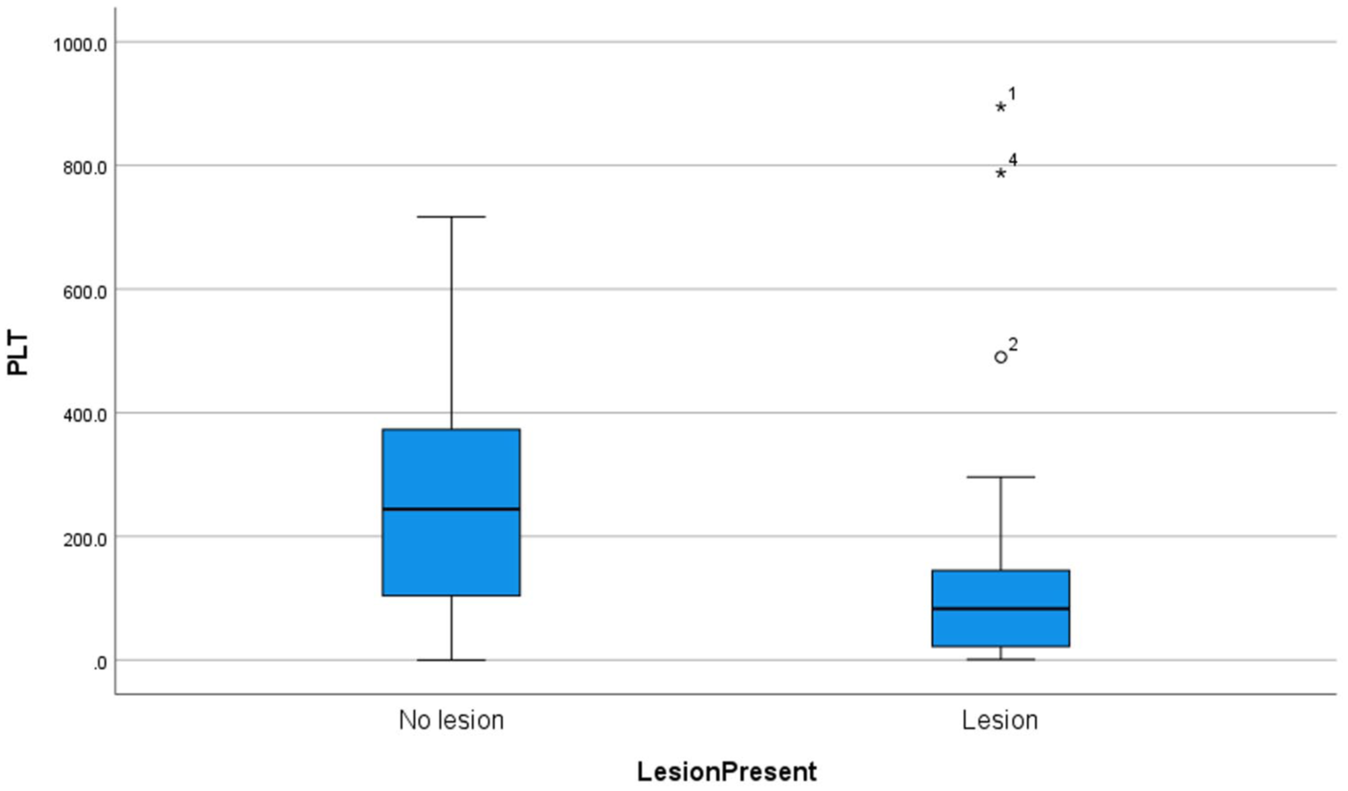
Box plot of platelet (PLT) counts (× 109/l) for cats with and without ocular lesions

Receiver operating characteristic (ROC) curve for haematocrit (HCT) in discriminating between cats with and without ocular lesions. The area under the ROC curve was 0.793 (95% confidence interval 0.691–0.894), demonstrating a significant discriminatory ability. A Youden index analysis identified an optimal HCT cut-off of 18.5%, resulting in a sensitivity of 91% and specificity of 58%
Aetiologies
The most common causes of anaemia were anaemia of chronic inflammatory disease, haemolysis and bone marrow suppression, while those of thrombocytopenia were immune-mediated destruction and bone marrow suppression. Among the 22 cats with immune-mediated disease (IMHA, n = 13; immune-mediated thrombocytopenia [IMTP], n = 4; Evans syndrome, n = 5), 10 (45%) had ocular lesions. Immune-mediated disease was diagnosed in 10/21 (48%) cats with ocular lesions and in 12/62 (19%) cats without ocular pathology.
Discussion
This study describes the association of anaemia and thrombocytopenia with ocular lesion presence and severity in cats. Although the pathway through which thrombocytopenia results in ocular haemorrhage is evident, given the essential function of PLTs in primary haemostasis, the role of anaemia in the development of ocular lesions is not fully understood.19,23 Anaemia-associated vascular hypoxia is hypothesised to induce endothelial fragility, leading to increased vascular permeability and subsequent retinal haemorrhages.19,25–27 The overall ocular lesion prevalence in this study (21/83, 25%) aligns closely with findings documented in a canine report and in human research.19,23,28 The ocular lesion prevalence in group A (20%) observed in the current study aligns with findings from previous studies in cats 12 and humans,19,29 whereas an older feline study reported a higher prevalence of 77%. 11 In contrast, other studies in dogs and humans reported a lower prevalence of 12% 23 and 10%, respectively. 28
The current study found a significant relationship between anaemia and the presence of ocular lesions. However, this and the following significant results should be interpreted with caution since no a priori sample size calculation was performed, and statistically significant findings in studies with potentially low power warrant careful consideration. 30 An HCT of 18.5% could predict the presence of ocular lesions with a sensitivity and specificity of 91% and 58%, respectively. The AUC was 0.793, indicating good model performance across all possible thresholds. 31 A recent study in human patients with anaemia reported a sensitivity and specificity of 80% and 52%, respectively, for an HCT of 30.5%. 32 Although previous studies have examined the correlation between anaemia and retinal haemorrhages in cats, they do not report any association with thrombocytopenia.11,12 Furthermore, several studies in humans have identified a correlation between anaemia and ocular lesions.18,19,21,28,32,33 However, the present finding of a positive correlation between anaemia and ocular lesion prevalence in cats differs from the result of a similar study in dogs, where ocular lesion prevalence and severity were not significantly associated with anaemia. 23
The current study’s ocular lesion prevalence in group T was 10%. Two human studies reported prevalence rates of 0% and 12%.19,28 To our knowledge, no similar data in cats have been published in the veterinary literature. The present study found a significant association between thrombocytopenia and ocular lesion presence. In human studies, the association between thrombocytopenia and retinal lesions remains contentious. Some investigations have demonstrated a significant relationship between severe thrombocytopenia and the presence of retinal lesions,19,21 while others reported no such correlation.18,28,32,34 Despite severe thrombocytopenia (PLT count <50 × 109/l) being sufficient to cause spontaneous bleeding, three studies suggest that isolated thrombocytopenia rarely induces significant retinal haemorrhage in individuals, even in the presence of cutaneous haemorrhages.20,33,35 In contrast to the current study, a previous canine study found a higher ocular lesion prevalence (42%) in animals with isolated thrombocytopenia. 23 One potential explanation for this discrepancy might be that feline platelets are more easily stimulated by aggregation inducers, including collagen or thrombin, than canine platelets,17,36 and less bleeding is usually observed in cats with thrombocytopenia than in dogs.14,36–38 In this study population, only 2% (1/43) of cats with thrombocytopenia showed signs of dermatorrhagia, while 6.3% of dogs with thrombocytopenia presented this clinical sign in one study. 39 Thrombocytopenia severity is likely a more critical factor influencing bleeding tendencies. Without stratifying for PLT count severity, direct comparisons of bleeding prevalence between species or studies should be cautiously interpreted.
Ocular lesion prevalence in group AT (48%) aligns with findings in both canine21,23 and human literature.19,28 More ocular lesions were identified in group AT than in groups A and T. Carraro et al 19 reported a similar observation, indicating that ocular lesion prevalence in humans was highest in group AT. Furthermore, Rubenstein et al 28 reported that only 2/187 human patients with anaemia without thrombocytopenia had retinal haemorrhages; however, 44% of 39 patients presented with retinal haemorrhages when anaemia and thrombocytopenia were combined. A similar study in dogs reported no significant difference in lesion prevalence between groups T and AT. 23
Human studies suggest that anaemia aetiology influences ocular lesion prevalence.40,41 In the current study, an immune-mediated disorder was more frequently identified in animals with ocular lesions (48%) than in those without (19%). This finding suggests a potential link that warrants further investigation in a future prospective study focusing on the association of immune-mediated anaemia and/or thrombocytopenia with the presence of ocular lesions. Besides thrombocytopenia, PLT dysfunction has been described in cats with IMTP, which might increase the bleeding tendency. 42 These results should be interpreted with caution, as IMHA, IMTP and Evans syndrome are highly likely to be associated with significantly lower HCT values and PLT counts.4,38 The Kruskal–Wallis test in the current study revealed that the median HCT values and PLT counts were significantly lower in the major lesion group, suggesting that immune-mediated conditions have a pronounced impact on lesion severity and prevalence due to these markedly reduced values. Korman et al 43 reported a significant association between anaemia severity and underlying aetiology. Nevertheless, investigating the potential relationships between cytopenia severity, aetiology and ocular lesion presence was beyond the scope of this study. A previous study in cats found no correlation between the underlying cause of anaemia and the presence of ocular lesions. 12
Minor lesions were overrepresented in this study, consistent with findings in humans, cats and dogs.12,18,19,23,28,32 These were characterised in this cat population by single or sparse, small intraretinal and/or preretinal haemorrhages, which aligns with observations in dogs, cats and humans.19,21,23 Ocular lesion severity is not well described in previous human studies, although the impact on vision is usually minimal,19,32 which aligns with our results. Most retinal haemorrhages secondary to haematological abnormalities are described as ‘flame-shaped’ in human literature.19,21 In contrast to the absence of retinal haemorrhages in dogs with isolated anaemia reported by Shelah-Goraly et al, 23 intraretinal haemorrhages were observed in four cats in group A in this study. Major lesions were only identified in groups AT (n = 6) and T (n = 1). The major lesions identified in this study encompassed several types of intraocular haemorrhages, including hyphaema, intravitreal haemorrhages and diffuse retinal haemorrhages; however, no retinal detachments were observed. A previous study in cats with anaemia identified similar major lesions but also reported a bullous retinal detachment in one case. 12 The concomitant presence of systemic hypertension in the cat precludes definitive attribution of the lesion solely to anaemia. 12 Although retinal detachments were also absent in human reports, hyphaema was not documented, and some human cases in the literature presented with intraretinal inflammatory exudates.19,21,28 This contrasts with findings in dogs, 23 where several retinal detachments were observed.
A potential explanation for this difference could be the overrepresentation of canine ehrlichiosis in the canine population, a condition known to induce thrombocytopenia and vasculitis. However, this study did not identify any cases of ehrlichiosis, and disease due to Ehrlichia in cats remains poorly understood and appears to be rare compared with dogs. 44
We found a significant association of anaemia and thrombocytopenia with ocular lesion severity, consistent with results from two human studies,19,21 but contrasting with findings in dogs. 23 In addition, this study revealed a significant association between anaemia severity and ocular lesion occurrence, which aligns with a previous study in cats. 12
The present study has some limitations. First, since isolated thrombocytopenia is relatively uncommon in cats, the sample size of group T was relatively small. Although IMTP is a well-known cause of isolated thrombocytopenia in dogs,39,45–47 it is considered rare in cats, with most publications consisting of small case series.14,36,48–53 Feline thrombocytopenia is usually associated with concurrent conditions, including anaemia, which is frequently linked to viral infections or neoplasia, 14 and IMHA in cats typically occurs alongside mild thrombocytopenia. 54 In this study, the HCT values were primarily obtained from automated analysers, and a systematic comparison with packed cell volume (PCV) was not performed in all cases. Although both HCT and PCV are measures of RBC volume, discrepancies can occasionally arise because of differences in measurement techniques or sample handling.
Blood pressure measurement was not conducted in a significant proportion of the cats with ocular lesions (7/21, 33%). Although their clinical presentation suggested a low likelihood of systemic hypertension, this cannot be excluded as a potential cause or contributing factor for their ocular lesions. Only one cat with ocular lesions had documented systemic hypertension, making it challenging to confirm whether this or its thrombocytopenia was responsible. Finally, causality cannot be fully confirmed because of the cross-sectional nature of the study. The ocular lesions may have predated the development of anaemia or thrombocytopenia, suggesting that these findings represent an association rather than a definitive causal relationship. However, since the ocular lesions identified appeared to be recent in the fundus and anterior chamber, they are likely associated with the identified cytopenias.
Conclusions
Anaemia and thrombocytopenia were significantly associated with ocular lesion presence and severity in cats. Cats with concurrent anaemia and thrombocytopenia demonstrated a higher ocular lesion prevalence than those with either condition alone. This observation warrants further investigation to explore potential underlying mechanisms. Based on these findings, any cat presenting with anaemia and/or thrombocytopenia should undergo a comprehensive ophthalmic examination.
Footnotes
Acknowledgements
We wish to thank IVC Evidensia Research Fund for providing the funding for this study.
Conflict of interest
The authors declared no potential conflicts of interest with respect to the research, authorship, and/or publication of this article.
Funding
This study received funding from the IVC Evidensia Research Fund.
Ethical approval
The work described in this manuscript involved the use of non-experimental (owned or unowned) animals. Established internationally recognised high standards (‘best practice’) of veterinary clinical care for the individual patient were always followed and/or this work involved the use of cadavers. Ethical approval from a committee was therefore not specifically required for publication in JFMS. Although not required, where ethical approval was still obtained, it is stated in the manuscript.
Informed consent
Informed consent (verbal or written) was obtained from the owner or legal custodian of all animal(s) described in this work (experimental or non-experimental animals, including cadavers, tissues and samples) for all procedure(s) undertaken (prospective or retrospective studies). No animals or people are identifiable within this publication, and therefore additional informed consent for publication was not required.