
Abstract
Objectives
The aims of this study were to evaluate the prevalence and to describe the clinical and diagnostic imaging features of the different types of feline intervertebral disc herniation (IVDH).
Methods
Medical records and imaging studies were retrospectively reviewed for cats diagnosed with IVDH between January 2008 and October 2020. Information obtained from the clinical records included signalment, clinical presentation, the presence of spinal hyperaesthesia and neurolocalisation. Diagnostic imaging findings, including type (ie, intervertebral disc extrusion [IVDE], intervertebral disc protrusion [IVDP] or acute non-compressive nucleus pulposus extrusion (ANNPE), site and number of IVDHs, were recorded. The association between breed, age, sex, duration and severity of neurological signs, the presence of spinal pain and MRI features was evaluated.
Results
Forty-three cats were included. A total of 50 IVDHs were identified: 39 cats were diagnosed with a single IVDH and four with multiple IVDHs. The most common type of IVDH was ANNPE (n = 22), followed by IVDP (n = 19) and IVDE (n = 9). Neuroanatomical localisation included L4–S3 (n = 19/43), T3–L3 (n = 18/43) and C1–C5 (n = 6/43). Cats with a single IVDH were statistically significantly associated with a diagnosis of ANNPE (P = 0.023) compared with cats with multiple IVDHs affected by IVDP (P = 0.004). Males were more commonly affected by IVDE (P = 0.020) and females by ANNPE (P = 0.020). Cats with IVDP had a longer duration of clinical signs (P <0.001) than cats with ANNPE and demonstrated milder neurological deficits (P = 0.005). IVDEs were statistically significantly associated with spinal hyperaesthesia (P = 0.013), while ANNPEs were not (P = 0.014).
Conclusions and relevance
ANNPE, IVDP and IVDE are each associated with distinctive clinical scenarios. Thoracolumbar and mid-to-caudal lumbar regions are the most affected, followed by the cranial cervical spine segment.
Introduction
Several post-mortem studies have reported intervertebral disc herniation (IVDH) as a common finding in asymptomatic older cats.1–4 In a cadaveric investigation of spinal pathology in 1958, intervertebral disc protrusions (IVDPs) were identified in about 25% of randomly selected cats, none with neurological signs. 1 However, the relevance of these post-mortem studies in clinical neurology is questionable as IVDH in symptomatic cats has been estimated to represent only 4–5% of feline spinal disease.5,6
The first report of symptomatic feline IVDH dates back to 1971. 7 Since then, several clinical cases have been published, the vast majority as case reports8–29 or case series numbering 10 or fewer cats,30–34 mirroring the low prevalence of this condition in the feline population. In the past 5 years, only three larger retrospective studies have been published, two describing IVDHs limited to the thoracolumbar region of the spine in a population of 31 35 and 35 cats,35,36 and the other focusing on a single type of IVDH (namely, acute non-compressive nucleus pulposus extrusion [ANNPE]). 37 Furthermore, in 2015, an essential contribution to the knowledge of feline IVDH was published. This work described signalment, clinical and diagnostic imaging features, and treatment in 46 cases and reviewed 40 previously published cases, making use of descriptive statistics. 38 Although available information about this condition in cats has increased over the years, the published evidence on IVDH in the feline population remains limited.
Therefore, the overall goal of the present study was to determine the clinical characteristics of all the types of IVDH in a large feline population referred to a single institution in a 13-year period. More specifically, the aims of the study were to (1) analyse the prevalence of clinically significant IVDHs in both the general hospital population and among cats with neurological disease; (2) report and analyse signalment, historical data, clinical and diagnostic imaging findings of all the different types of IVDH; and (3) identify statistically significant differences between the subgroups of IVDHs. We hypothesised that IVDH would be considered an uncommon condition in cats. Additionally, we supposed that cats could suffer from different types of IVDH and that each one would be associated with a distinctive clinical presentation.
Materials and methods
The medical records of cats diagnosed with IVDH referred to the Neurology Unit of the Anicura Portoni Rossi Veterinary Hospital between January 2008 and October 2020 were retrospectively reviewed. To be eligible for inclusion, cats needed to have had a complete medical evaluation, including a physical and neurological examination performed by either a board-certified neurologist (FB, MB, SC) or a neurology resident (LB), a spinal cord neurolocalisation and MRI exploring the selected segments of the spine conclusive of a diagnosis of IVDH, and be available for re-evaluation. For each cat, the following information was recorded: signalment (breed, age and sex); onset of clinical signs; type and severity of neurological deficits; presence of hyperaesthesia on spinal palpation; and diagnostic imaging findings (type, site and number of IVDHs). IVDHs included in the MRI field of view (FOV) but outside the segments of the neurolocalisation were counted. Any traumatic event preceding the onset of clinical signs was recorded. The rate of onset of clinical signs was arbitrarily categorised as peracute (<24 h), acute (24 h–5 days) or chronic (>5 days). A neurological grade had been assigned in accordance with a previously reported 6-point scoring system in cats, ranging from normal neurological examination (grade 0) to most severe degree of neurological dysfunction (plegia with absent nociception – grade 5). 38 For all included cats, MRI of the spine was performed using either low- or high-field units (0.22 T MrV [Paramed] and 1.5 T Vantage Elan [Canon Medical Systems], respectively) under general anaesthesia and included at least T2- and T1-weighted images on the sagittal and transverse planes. In selected cases, to obtain a confident diagnosis, additional sequences were obtained at the discretion of the radiologist. The location of the affected intervertebral disc space (IVDS) was recorded and each IVDH was further characterised as intervertebral disc extrusion (IVDE), IVDP, ANNPE or intradural/intramedullary intervertebral disc extrusion (IIVDE) based on criteria discussed in the current literature. 39 Cats were further divided into two groups based on the number of IVDHs diagnosed (ie, single or multiple).
Statistical analyses were performed with SAS 9.4 (SAS Institute). Count data were analysed as percentages. Comparisons among types of IVDH, neurolocalisation and the number of IVDHs were performed using a k-proportions test. Post-hoc pairwise comparisons among levels were calculated with the Marascuilo approach. A multivariate logistic regression analysis was carried out to assess the effects of sex, onset, neurological score, pain score and number of IVDHs (single vs multiple) on the type of IVDH. Odds ratios and 95% confidence intervals were calculated. Values of P <0.05 were considered to be statistically significant.
Results
Signalment
Forty-three cats fulfilled the inclusion criteria. Thirty-six (83.7%) were domestic shorthair (DSH) cats and seven (16.3%) were purebreds (Siamese = 3, Exotic Shorthair = 2, Persian = 1, Maine Coon = 1) (Table 1).
Signalment, clinical findings, neuroanatomical localisation and diagnosis in 43 cats affected by intervertebral disc herniation

DSH = domestic shorthair; FN = female neutered; ANNPE = acute non-compressive nucleus pulposus extrusion; MN = male neutered; IVDP = intervertebral disc protrusion; IVDE = intervertebral disc extrusion; ME = male entire; FE = female entire
A total of 18,618 cats were referred to our institution in the same period, resulting in a 0.23% prevalence of IVDH in the general population of cats. Furthermore, 1524 cats were referred to the hospital’s neurology unit, resulting in a 2.8% prevalence of IVDH among neurological cases. Among the neurological cases, 213 cats were presented with a spinal neurolocalisation, and 43/213 (20%) were diagnosed with IVDH. Compared with the general hospital population and neurological cases, among the purebred cats, the prevalence of IVDH for Exotic Shorthairs was 4.17% and 18.2%, respectively.
There were 24 (55.8%) males and 19 (44.2%) females (all but two neutered) with a mean age of 8.7 years (range 4 months–15 years). The mean ages of DSH and purebred cats were 8.4 years and 10.6 years, respectively.
Clinical presentation
Details about the onset of clinical signs were available in 41 cats (95.3%). The clinical presentation was peracute in 25 cats (61.0%), chronic in 12 (29.3%) and acute in the remaining four (9.7%). The severity of neurological signs varied between grade 1 (n = 7), grade 2 (n = 22), grade 3 (n = 7), grade 4 (n = 4) and grade 5 (n = 3). There was no significant influence of age, sex, onset of clinical signs, presence of spinal pain or type of IVDH on the neurological score, except for grade 1, which was significantly associated with IVDP (P = 0.005; Table 2).
Results of the univariate analysis for age, sex, onset, neurological score and pain relative to the type of intervertebral disc herniation

Data are n (%). Different letters along rows indicate statistically significant different proportions
P <0.05
IVDE = intervertebral disc extrusion; IVDP = intervertebral disc protrusion; ANNPE = acute non-compressive nucleus pulposus extrusion
Neuroanatomical localisation
The most common neuroanatomical localisations were L4–S3 (n = 19) and T3–L3 (n = 18), followed by C1–C5 (n = 6). No cats had the lesion localised at C6–T2. Hyperaesthesia on spinal palpation could be elicited in half the cats (Table 1). There was no significant influence of age or sex on the onset of clinical signs, neurological score or presence of pain.
Types of IVDH
Of 43 cats with IVDH, 22 (51.2%) were diagnosed with ANNPE, 12 (27.9%) with IVDP and nine (20.9%) with IVDE (Figure 1). No cats were diagnosed with IIVDE.

Examples of different types of intervertebral disc (IVD) herniation. (a) Sagittal and (b) transverse T2-weighted MRI images of case 7, which was diagnosed with acute non-compressive nucleus pulposus extrusion. A focal intraparenchymal hyperintensity is present immediately dorsal to the L4–L5 IVD (long arrow). The L4–L5 IVD nucleus pulposus has a markedly reduced volume and signal intensity. (c) Sagittal and (d) transverse T2-weighted MRI images of case 22, which was diagnosed with IVD protrusion. A midline hypointense disc material confined to the L1–L2 intervertebral disc space is present, causing mild spinal cord compression (arrowhead). Note the degenerative changes of multiple IVDs. (e) Sagittal and (f) transverse T2-weighted MRI images of case 42 diagnosed with IVD extrusion. A large volume of lateralised and markedly compressive hypointense disc material is located at the level of the L5–L6 IVD, extending over the adjacent vertebral bodies (short arrow)
A total of 50 IVDHs were identified: 39 cats were diagnosed with a single IVDH (single lesion) and four cats with two or more IVDHs (multiple lesions). Two of these four cats presented an IVDH outside the segments of the neurolocalisation (Table 3).
Distribution of intervertebral disc herniation among the 43 study cats

IVDS = intervertebral disc space; IVDP = intervertebral disc protrusion; IVDE = intervertebral disc extrusion; ANNPE = acute non-compressive nucleus pulposus extrusion
Cats with a single IVDH were significantly associated with both a diagnosis of ANNPE (P = 0.023) and a peracute onset of clinical signs (P = 0.014). In contrast, cats with multiple IVDHs were statistically more affected by IVDP (P = 0.004; Table 4).
Results of the univariate analysis for age, sex, onset, neurological score, neurolocalisation, type of intervertebral disc herniation (IVDH) and pain relative to number of IVDHs (ie, single or multiple)
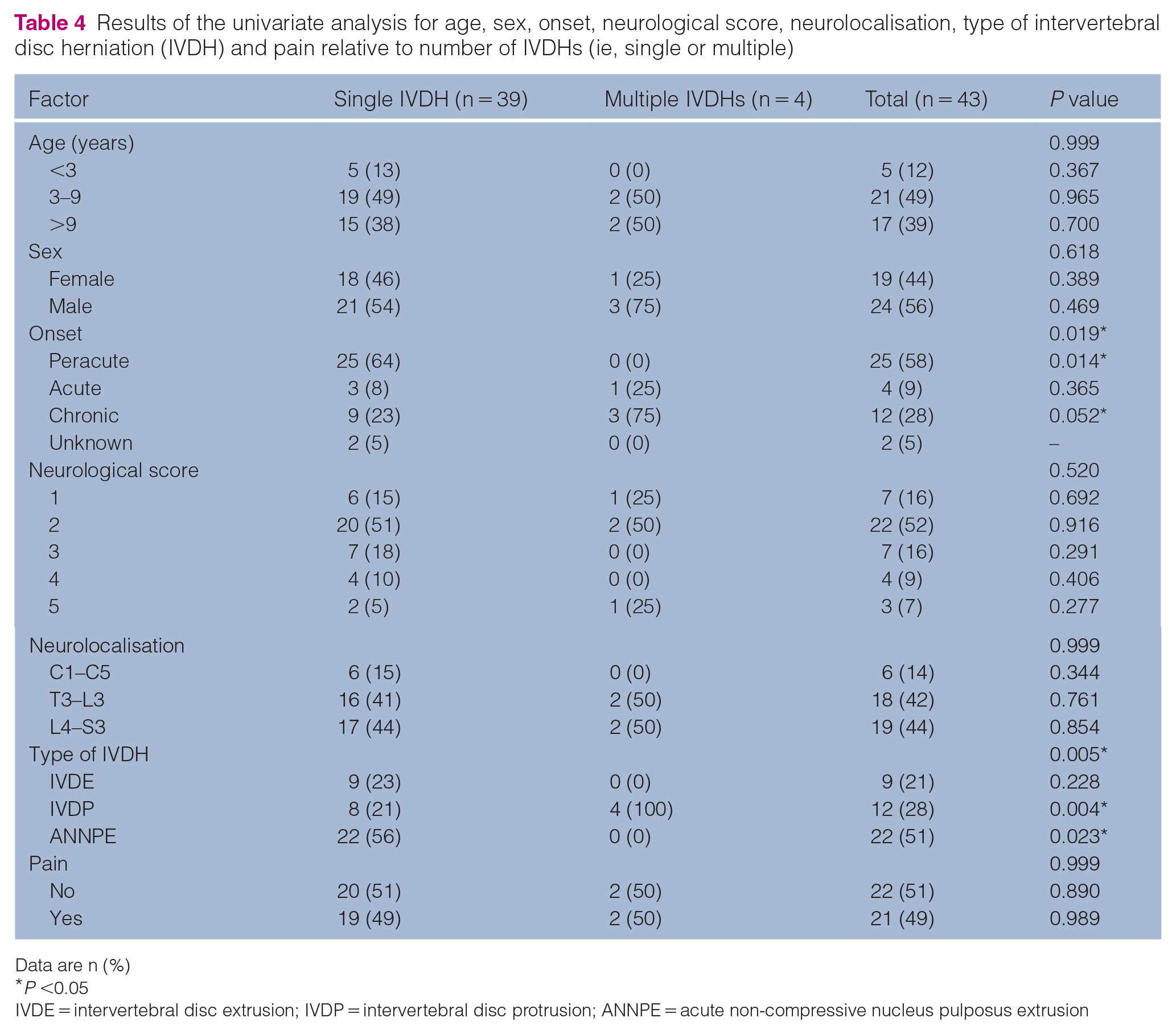
Data are n (%)
P <0.05
IVDE = intervertebral disc extrusion; IVDP = intervertebral disc protrusion; ANNPE = acute non-compressive nucleus pulposus extrusion
No significant differences between DSH and purebred cats for age, neuroanatomical localisation or type of IVDH were found.
The type of IVDH was significantly associated with the type of onset: ANNPE was more commonly associated with a peracute onset (P <0.001) compared with the chronic clinical presentation of IVDP (P <0.001; Table 2). Moreover, there was a significant association between type of IVDH and sex: ANNPE was more common in females (P = 0.020) than IVDE, which mainly affected males (P = 0.020; Table 2).
Of the 22 cats with a diagnosis of ANNPE, two had an unknown history, two had sustained a witnessed traumatic injury (falling from a height) and two had signs consistent with external trauma (pelvic fractures or skin lesions). The onset of the clinical signs was preceded by vigorous exercise, such as jumping or playing, in nine cats.
Spinal pain
Spinal hyperaesthesia was significantly associated with both neurolocalisation and type of IVDH. Cats with spinal hyperaesthesia more frequently had an L4–S3 neurolocalisation (P <0.001; Table 5) and a diagnosis of IVDE (P = 0.013; Table 2). However, the absence of spinal pain was associated with a C1–C5 neurolocalisation (P <0.001; Table 5) and a diagnosis of ANNPE (P = 0.014; Table 2).
Results of the univariate analysis for age, sex, onset, neurological score, type of intervertebral disc herniation (IVDH) and pain relative to neuroanatomical localisation

Data are n (%). Different letters along rows indicate statistically significant different proportions
P <0.05
IVDE = intervertebral disc extrusion; IVDP = intervertebral disc protrusion; ANNPE = acute non-compressive nucleus pulposus extrusion
Multivariate analysis
Due to our group’s distribution and small sample size, factors included in the multivariate logistic regression model were limited to sex and spinal pain (Table 6).
Results of the multivariate logistic regression of the predictors (sex and spinal pain) to detect the type of intervertebral disc herniation (final diagnosis)

Acute non-compressive nucleus pulposus extrusion was the reference level
P <0.05
OR = odds ratio; CI = confidence interval; IVDP = intervertebral disc protrusion; IVDE = intervertebral disc extrusion
Both variables retained a significant association with the type of IVDH. Specifically, male cats were predisposed to IVDE, being >18 times more likely to be affected by IVDE than ANNPE. Furthermore, cats with spinal hyperaesthesia had 6- and 12-fold higher odds of a diagnosis of IVDP or IVDE, respectively, than ANNPE.
Discussion
This retrospective study described the clinical and diagnostic imaging features of different types of IVDH in a large population of cats. In the veterinary literature, relatively few data are available on feline IVDH. Only 185 cats clinically affected by IVDH have been reported in the past 50 years, most of them described from 2011 to date.24–29,33–38 Moreover, several studies have separately investigated various features of each different type of IVDH, mainly as individual case reports or case series including low numbers of cats. Thus, a comprehensive overview regarding the overall clinical and MRI characteristics of this disease is lacking.
The prevalence of IVDH in the overall population of cats referred to our institution was 0.23%. The first report on the frequency of IVDH in cats dates back over 30 years and estimated an incidence of 0.02%. 40 Since then, few studies have investigated the prevalence of IVDH in cats, reporting a rate of 0.26% or lower.30,34,35 The prevalence of feline IVDH at our institution agrees with these latter studies and reflects the low predisposition of cats to IVDHs. By contrast, in previous post-mortem studies,1–4 IVDHs have been more frequently detected in cats, with IVDP being the more common type. 1 This difference could be related to the underlying pathophysiological mechanism of IVDP, which is mainly associated with slowly progressive spinal cord compression and consequent milder clinical signs, such as pain or discomfort, that are probably more difficult to assess by owners.
IVDH was diagnosed in 2.8% of all neurological cases and in 20% of cats with a spinal neurolocalisation presented at our institution, according with a previous study evaluating, through a clinical reasoning process, the main differential diagnoses in cats with spinal disease. 41 This finding is different from previous data suggesting that IVDH should represent only 4% of feline spinal disease. 5 This disagreement might be explained by the different study design and inclusion criteria, as the previous study included only cats with a histologically confirmed diagnosis.
Many breeds of cats have been reported to be clinically affected by IVDH, although non-purebred cats constitute roughly two-thirds of the 185 published feline cases.8,10,11,13,15–28,30–38 However, a recent study identified Persian and British Shorthair cats as being predisposed to thoracolumbar IVDH when compared with the general feline population of a referral hospital. 35 In agreement with previous studies,8,10,11,13,15–28,30–38 the DSH was the most common breed affected by IVDH in our study, reflecting the greater proportion of this breed among the overall feline population. However, when taking exclusively purebred cats into account, Exotic Shorthairs seem to be over-represented, although the low number of cases limits the strength of this observation. The Exotic Shorthair cat belongs to the Persian family group, which includes the British Shorthair and the Persian. 42 Members of the different breeds of this family share genetic and general health features, 42 and a possible relationship between the typical brachycephalic morphology and IVDH should be explored in further, larger studies.
The mean age of the cats in our study was 8.7 years, which is in agreement with the mean age of all the published feline IVDH cases thus far (8.3 years).7–38 This occurrence in mature cats has been reported previously,30,35 and is consistent with several post-mortem studies on feline intervertebral disc degeneration and herniation that found an increased occurrence of this condition in older animals.1–4 When considering each type of IVDH separately, previously reported cats affected by IVDP and IVDE had a similar mean age (8.6 years),7–14,16,18–23,25–27,29–36,38 whereas the mean age of cats with ANNPE was slightly lower (6.5 years).15,24,37,38 Within our population, the mean age of cats was 10.1 years for IVDP, 7.8 years for IVDE and 8.4 years for ANNPE. Our findings are in accordance with reported distributions for age for middle-aged and older cats affected by IVDE and IVDP, but a predisposition of younger cats to ANNPE was not found. Additionally, a different age predisposition to IVDH was found in a study comparing non-purebred and purebred cats, with the latter being affected younger. 35 In our study, a statistically significant difference for age between the two groups was not found. One explanation could be the very low number of purebred cats vs DSH cats, which represented 84% of our population. However, it is also possible that statistically significant findings in small populations are no longer significant in larger populations.
In cats, a sex predisposition to IVDH has not been reported, although males seem to be over-represen-ted.7,10,12–19,21,23,25–27,29–38 Among published feline IVDE cases, a male predisposition could be suspected owing to a male-to-female ratio of 1.8.29,34,35,38 In our study, a strong association between sex and type of IVDH was found, with males being significantly more prone to being affected by IVDE and females by ANNPE. However, a female predisposition to ANNPE has not previously been found and only 8/23 published cases of ANNPE were females.24,37,38 Similarly, in dogs, a strong sex predilection to IVDH has not been reported, although males and neutered females seemed to be more affected.43–46 Several explanations have been proposed, such as a within-breed weight factor 43 and the neuter status itself, which is expected to lead to weight gain and reduce the protective role of the sex hormones for intervertebral discs, 47 which is considered a risk factor for IVDH in dachshunds. 47
In our study, body weight, which is probably greater in male and neutered female cats, could be considered a potential risk factor for developing IVDH, but large epidemiological studies are needed to evaluate and confirm this finding.
In cats, four types of IVDH have been reported: IVDE8,9,11–13,18,19,29–32,34–36,38 and IVDP7,10,14,16,20–23,25–27,33,35,36,38 are the most common types, followed by a lower number of ANNPE15,24,37,38 and only two case reports of IIVDE.17,28 In our study, IIVDE was not reported and, surprisingly, ANNPE was the most frequent. These different proportions could be attributable to the design of the study itself, which, for the first time, simultaneously evaluated the distribution of all IVDH types in a feline population.
The hypothesis of a relationship between the type of IVDH and clinical presentation was confirmed by our findings, which agreed with a previous study. 35 Cats with IVDP had a longer duration of milder clinical signs than cats with ANNPE. These findings were expected and reflect the different underlying pathophysiology: IVDP is typically associated with gradual spinal cord compression without contusion, 48 while ANNPE is characterised by a sudden focal contusive injury. 49 In addition, ANNPEs were reported in up to 75% of cats following an external trauma. 37 In contrast, in our study, only 4/22 cats with ANNPE had been involved in a traumatic injury or had signs consistent with an external trauma, while 9/22 cats presented clinical signs during vigorous exercise. According to our findings, strenuous activity should be considered a predisposing factor to ANNPE in cats, as it should be for dogs, 49 and should be investigated while taking the cat’s history in the case of peracute clinical signs of myelopathy to help develop a logical list of differential diagnoses, including ANNPE, even in the absence of a history of trauma.
The majority of cats in our study were diagnosed with presumptive ANNPE based on specific clinical and MRI findings, although MRI differentiation between this IVDH type and ischaemic myelopathy can sometimes be challenging. As specific MRI criteria have been described to discriminate between these conditions, 49 a more confident suspected diagnosis of ANNPE can be reached, as happened in our cases, likely mirroring the real occurrence of this disease in cats. However, as a definitive diagnosis requires histopathological examination, the lack of post-mortem evaluation could have contributed to our results.
Spinal pain was present in half of the cats in this study and was a clinical sign significantly associated with the type of IVDH: cats affected by IVDE were more likely to display signs of pain at presentation than cats with ANNPE. Spinal hyperaesthesia has been variably reported in previous studies of IVDH in cats, most including both IVDP and IVDE.12,14,16,25,30,32,33,35,36,38 However, reported feline clinical cases of ANNPE have usually been non-painful at presentation.15,24,37,38 Spinal pain may thus be considered a useful clinical sign in a cat with a sudden spinal cord disorder and, according to the clinical presentation, represents important clinical information supporting the suspicion of IVDE.
The most frequently affected intervertebral disc (IVD) in our population was L7–S1, followed by those at the thoracolumbar junction (from T11 to L2 IVDs) and L4–L5. The lumbosacral junction accounts for a large part of the overall mobility in the lumbar spine 50 and represents the most common site affected by IVDH in 39/185 previously reported cats.16,32,33,36,38 Moreover, the feline thoracolumbar and mid-to-caudal lumbar spine region have been identified as being frequently affected by IVDH.8,9,11–14,17,18,22,24,28–31,34–38 Our findings are consistent with the higher predisposition of these feline spine locations to IVDH, probably related to the peculiar vertebral column configuration and range of motion. 51 Interestingly, none of our feline cases was affected by IVDHs in the cervicothoracic spine. Likewise, there was no report of C6–T2 IVD involvement among all published feline cases of IVDH. 38 The cervicothoracic spine’s low susceptibility to IVDH could be related to the range of motion and stance configuration of the feline spine. The feline cervical column is highly mobile, especially in dorsiflexion, which is required for the vertical orientation of the head. 51 The other flexible spine region, from T4 to T11, enables cats to undergo extreme torsion. However, the particular sling-like arrangement of trunk muscles that insert near the T2–T3 joint could help to stabilise the spine, particularly against torsion movements. 51 It has been hypothesised that this T2–T3 stability could contribute to the local incidence of IVDH, particularly after trauma. 52 Although it is difficult to explain these findings, it is possible that the specific organisation and mobility of the vertebral column of the cat, combined with the proximity to a more stable area at T2–T3, could protect cervicothoracic IVDs from degenerative changes.
Feline IVDHs have been reported mainly as single lesions, with only 7.5% of cats having more than one IVD involved.9,21,31,35,36,38 In the present study, 91% of cats were affected by a single IVDH; the remaining 4/43 cats had multiple lesions and were all diagnosed with IVDP. This result is in accordance with previous studies in dogs in which the presence of multiple compressive lesions was associated with a diagnosis of IVDP.53,54
The limitations of this study include the inherent nature of a retrospective study. The relatively small sample size, owing to the infrequent occurrence of IVDH in cats, precluded the detection of multivariate statistical significance of some factors. Another limitation was the different types of MRI scanner used, as the strength of the magnetic field influences the spatial resolution, resulting in suboptimal image quality during the scanning of the spine in small patients. Finally, only two cats (cats 20 and 22) presented a concurrent IVDH outside the spinal cord segments of the neurolocalisation but included in the MRI FOV. Both these cats were diagnosed with one or more IVDPs in the clinically affected segments and the additional IVDH was also an IVDP. It is therefore possible that our cats with IVDP could have presented more IVDPs along the spine, as chronic conditions such as IVDP may be asymptomatic. Consequently, the real prevalence of IVDP might have been underestimated.
Conclusions
IVDH should be considered an uncommon disease in cats. However, this study adds significant information to the limited available data about this feline disorder, highlighting that ANNPE could occur more frequently than previously reported, that the cervicothoracic spine is a region not affected by IVDH and that each type of IVDH is associated with distinctive clinical scenarios. Certain clinical features, such as spinal hyperaesthesia and the onset of clinical signs, can help discriminate between these conditions. Moreover, involvement of a single IVD is more frequently associated with ANNPE compared with multiple IVDs affected by IVDP. The thoracolumbar and mid-to-caudal lumbar regions are the most affected, followed by the cranial cervical spine segment.
Footnotes
Conflict of interest
The authors declared no potential conflicts of interest with respect to the research, authorship, and/or publication of this article.
Funding
The authors received no financial support for the research, authorship, and/or publication of this article.
Ethical approval
The work described in this manuscript involved the use of non-experimental (owned or unowned) animals. Established internationally recognised high standards (‘best practice’) of veterinary clinical care for the individual patient were always followed and/or this work involved the use of cadavers. Ethical approval from a committee was therefore not specifically required for publication in JFMS. Although not required, where ethical approval was still obtained, it is stated in the manuscript.
Informed consent
Informed consent (either verbal or written) was obtained from the owner or legal custodian of all animal(s) described in this work (either experimental or non-experimental animals, including cadavers) for all procedure(s) undertaken (prospective or retrospective studies). No animals or people are identifiable within this publication, and therefore additional informed consent for publication was not required.