
Abstract
Practical relevance:
Any congenital or developmental abnormality of any part of the male or female reproductive tract is a ‘disorder of sexual development’ (DSD). The tricolored male cat phenotype, cryptorchidism, gonadal hypoplasia and incidental abnormalities such as cystic remnants or embryonic ducts are well-known feline DSDs.
Clinical challenges:
Full characterization of DSDs requires sex chromosome determination and identification of genes related to development of the gonads, internal tubular genitalia and external genitalia. Fortunately, affected cats are seen sporadically and the clinical effects are usually minimal.
Classification:
The classification nomenclature has changed. In place of intersex, hermaphrodite, pseudohermaphrodite and sex reversal, the newer standard classification, based on sex chromosomes, designates sex chromosome DSD when there is an abnormality in the sex chromosomes, and XX (female) and XY (male) DSDs where there is not. Identification of the gonadal type (testes, ovaries, ovotestes or gonadal dysgenesis) and documentation of the internal and external genital components completes the classification.
Evidence base:
The original basis of the DSD classification was a consensus reached in humans. It was quickly accepted in veterinary pathology, courtesy of its logic and ease of application, and it has subsequently begun to appear in peer-reviewed papers and clinical reviews. This article reviewing the various disorders in cats is based on application of the classification and draws on the feline peer-reviewed literature encompassing chromosome analysis and definition of reproductive abnormalities, syndromes and diseases.
Phasing in of current terminology
A disorder of sexual development (DSD) in human medicine is any abnormality, anomaly, ambiguity or lesion that occurs because of a failure of development of the female or male reproductive systems. This terminology replaces the terms intersex, hermaphrodite, pseudo hermaphrodite and sex reversal,1-4 and is increasingly being used in veterinary medicine and science.
The original purpose of the DSD terminology in humans was to ensure consistency and to avoid the stigma of genital ambiguity and gender assignment in babies born with disorders of the external reproductive system. The classification allowed for all abnormalities of the reproductive tract to be categorized and defined in full. In this review, the broader approach is used to include all disorders, not just external sexual phenotypic ambiguity. Thus DSDs vary from major anomalies that affect fertility or clinical wellbeing to incidental and minor changes to structures.
‘Intersex’ conditions were always defined by sex chromosome type and arrangement, gonadal type and phenotype; this continues. In clinical veterinary medicine and science, access to sex chromosome analysis and gene identification is limited to research settings, so syndromes are often defined by gonadal and phenotypic sex only. The stigma and gender issues of humans are not a factor in cats.

Terms such as sex reversal, male feminization and persistent Müllerian duct syndrome are now only used in veterinary medicine and science when referring to historically defined clinical syndromes and will eventually be phased out.
The classification of DSDs into sex chromosomal, gonadal and phenotypic categories is now specific, accurate and more descriptive. It avoids the variation in the definitions of what constitutes intersex, hermaphroditism and pseudohermaphroditism. The classification begins with identifying the sex chromosomes and their make-up, such as XX, XY, X, or XXY. There are many different genes and gene products involved in normal sexual development in both males and females. SRY is the gene that is the sex-determining region of the Y chromosome which results in the development of a male, and R-spondin1 (RSPO1) is an early gene in the development of a female. The convention in the scientific literature and adopted in this review is to use italics and uppercase for the gene with a human homologue; upper case and no italics is used for the protein product. SRY is the protein product of SRY and RSP01 is the product of RSPO1. Many publications now include the presence or absence of SRY in the classification, and eventually RSPO1 could be a routine contributor to the classification.
The identification of individual disorders (summary box 1) does not usually allow the exact classification to be determined (summary box 2). A single individual disorder may occur with multiple classifications; for example, testicular hypoplasia is seen variously in cases of sex chromosome, XX and XY DSDs.
As yet, no veterinary diagnostic laboratory provides chromosomal analysis or gene and/or protein identification for sexual development. When the various genes and protein products are identified and molecular tests become available, the classification system will expand to incorporate the genomics of sexual development.
There are many reviews, textbook chapters and conference proceedings devoted to feline DSDs.5-9 This article reviews the current state of knowledge and outlines a diagnostic approach when disorders are identified clinically either based on external genital development or changes observed at ovariohysterec-tomy or castration (see summary box 1).
Clinical signs and lesions of disorders of sexual development (DSDs)
The pathogenesis of any disease has three major components: 1) cause, 2) mechanisms and 3) lesions and/or clinical findings. A clinician identifies clinical findings and/or lesions of DSDs by evaluating the external genitalia during a physical examination, and the gonads and other components of the internal genitalia at neutering. To fully characterize a DSD, chromosomal and genomic information is required. The lack of access to such tests greatly restricts this part and thus this review will not include all of the differentiation genes that are known in cats. Gonadal and phenotypic components, including internal tubular genitalia, can be defined, however. This requires careful histopathological evaluation (and sometimes the expertise of a dedicated reproductive pathologist).

Fortunately, cats affected with DSDs are seen sporadically and the clinical effects are usually minimal (compared, say, with psychological effects in humans). Curiosity is often the major reason for attempting to fully define a disorder. The following discussion attempts to work backwards from the lesion or clinical findings to try to identify the underlying DSD. As with many diseases, there is a disconnect between what is seen clinically and the eventual classification or specific cause. Where there are direct correlations between the lesion or observation and the mechanisms and causes, these will be highlighted. A summary of the normal embryonic structures, their origin and the adult counterparts is provided in Table 1 for reference.
Normal embryonic structures, their origin, adult counterparts, and controlling gene and hormone
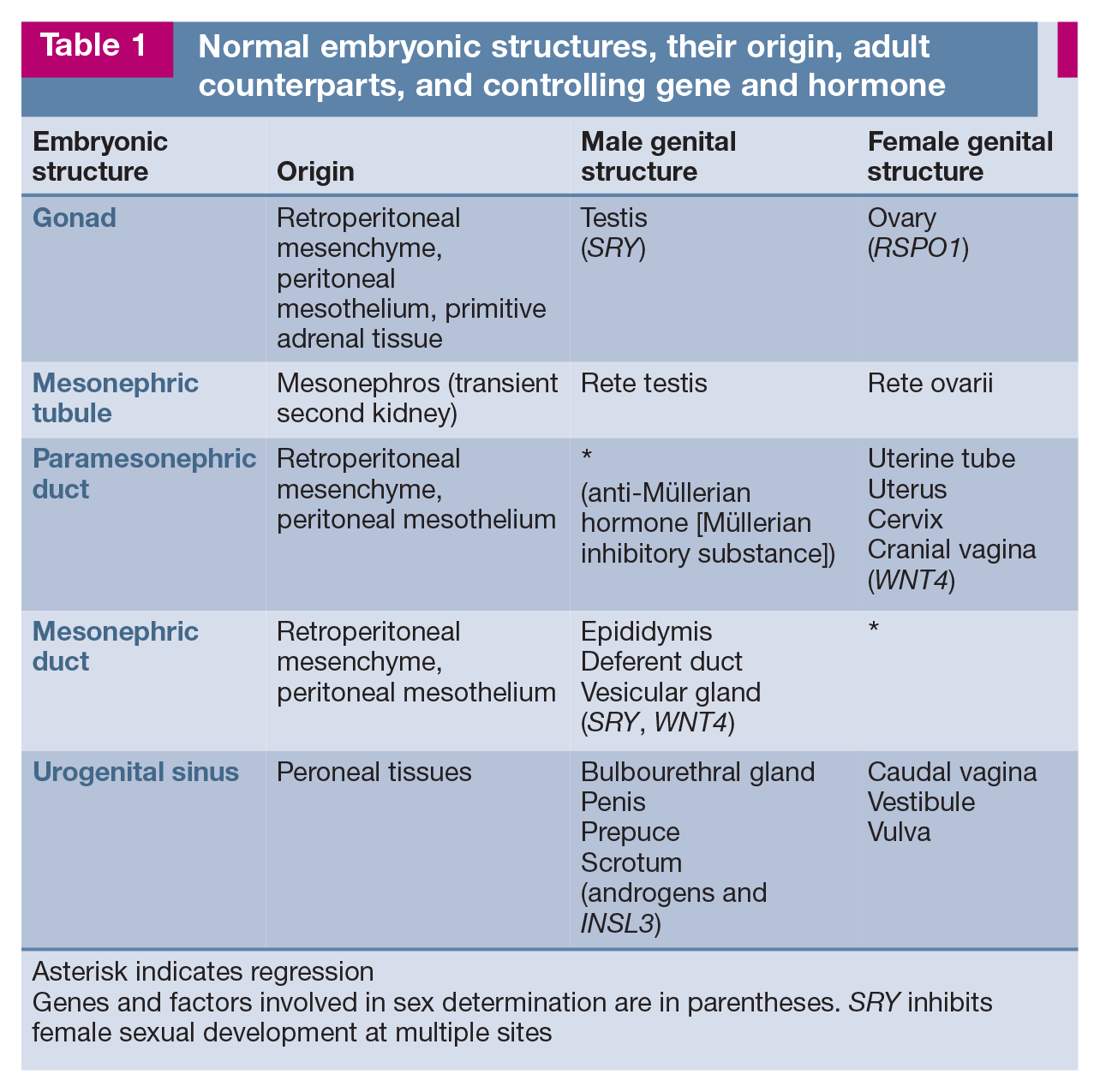
Asterisk indicates regression
Genes and factors involved in sex determination are in parentheses. SRY inhibits female sexual development at multiple sites
Tricolored male cat
Any male cat with three colors in the haircoat must have at least two X chromosomes; thus almost all have a sex chromosome DSD. The majority will have XXY trisomy, or will be a mosaic or chimera (see later discussion of ‘Sex chromosome disorders of sexual development’ for definitions). Some have other poly-poidy. Their gonads can be testes or ovotestes and are usually hypoplastic. The external genitalia can be male, ambiguous with male and female components, and/or hypospadias may be present. Cryptorchidism is also common. These cats are rarely fertile.
Hypospadias
Cats with hypospadias have an opening of the urethra on the ventral surface of the penis. They typically also have a bifid scrotum - a divided scrotal sac, with the penis and its ventral opening between the scrota (Figure 1). Most cats with hypospadias have a sex chromosome DSD.

Cat with hypospadias and ambiguous genitalia.
A cat with ambiguous external genitalia including penis and vulva could have a sex chromosome DSD or, less commonly, an XX (female) chromosome or XY (male) chromosome DSD.
Female external genitalia but internal testes
Most cats with a female phenotype including female genitalia, but with internal testes or ovotestes, are classified as having an XY testicular DSD. There is no testosterone production and no androgen receptor.
Anorchia, monorchia, cryptorchidism, testicular aplasia and hypoplasia
Anorchia, monorchia and cryptorchidism are terms that mean no testes, one testis or hidden testes, respectively. Cryptorchidism is the most common reproductive disease in the male cat; it is an XY chromosomal DSD. The terms anorchia and monorchia are often used incorrectly in clinical veterinary practice, and may convey a misleading message to clients. The hidden testis (or testes) or their remains are almost always found outside of the scrotum and either in the inguinal canal or within the abdomen. A specific diagnosis to indicate the cause, made after careful his-tological examination, is agenesis (testes did not begin development), aplasia (did not grow), extreme hypoplasia (too small to indentify at surgery) or post-necrotic atrophy (testis became necrotic and scarred). Affected cats commonly have an XY testicular DSD.
Failure to find one or both testes in the scrotum presents a clinical challenge. If the results of hormone testing or the presence of penile spines indicate functional testicular tissue, the first condition to exclude is cryptorchidism. Surgery to remove the retained structure will hopefully reveal a testis and epididymis (Figure 2); this can usually be confirmed by histological examination. The main possibilities if a testis is absent from the scrotum are:
✜ Previous surgical removal
✜ Previous testicular necrosis
✜ Testicular agenesis, aplasia or extreme hypoplasia
Should there be testicular hypoplasia or aplasia, and an epididymis present in a cat that is not tricolored, this probably represents an XY testicular DSD.

In cats with abdominal cryptorchidism, the retained testis is often found near the bladder. 6 Courtesy of Susan Little
Ovarian hypoplasia or aplasia
Female cats lacking an ovary or ovaries, or with ovaries that are smaller than normal, are a very rare finding and not fully described in the literature. Most are classified as having XX gonadal dysgenesis or an XX ovarian DSD; gonadal dysgenesis is where the gonad is too primitive to determine its type.
Ovarian/periovarian cysts or cysts near the uterus
There are a variety of retained embryonic ducts or tubules, and these may expand and develop into cysts within or near the ovary or uterus. Cysts are derived from either the parameso-nephric (future female) or mesonephric (future male) ducts. These form from an embryonic duct that is an invagination of the celom, or the mesonephric (male) tubules that derive from the mesonephros. Such cysts are often an incidental finding and this is an XX ovarian DSD.
Segmental absence of the uterus
Phenotypically female cats with a partial absence of the uterus, diagnosed as segmental aplasia, are classified as having an XX ovarian DSD (Figure 3).

Reproductive tract of a cat with segmental aplasia of a uterine horn. Although one uterine horn may be hypoplastic or missing, the ipsilateral ovary is almost always present. 6 In this case, the left uterine horn (L) is replaced by a solid cord of smooth muscle and vascularised loose connective tissue. Arrows denote corpora lutea within the ovaries. One site of placentation is prominent (P). Reproduced, with permission, from Brookshire et al (2017) 10
Classification of disorders of sexual development (DSDs)
The classification of DSDs begins with the sex chromosome composition. A normal cat has 38 chromosomes per cell; these 38 represent 19 matching pairs, 19 from each parent. A cell with 38 normal chromosomes is ‘diploid’. Those cats with an abnormal composition of sex chromosomes have a sex chromosome DSD. Gonadal type is not part of the sex chromosome DSD classification.
Those animals with a congenital abnormality of the reproductive system and with a normal XX (female) or XY (male) chromosome complement have an XX DSD or XY DSD, respectively. Testing for SRY is most commonly reported in the literature; so, if available, the SRY status is included. Accordingly, there are XX SRY-positive, XX SRY-negative, XY SRY-positive and XY SRY-negative DSDs.
The next part of the classification concerns gonadal type. The main possibilities are testes, ovaries, ovotestes and gonadal dysgenesis. This determination is established histologically and accurate classification may require the input of a reproductive pathologist (see summary box 2).
As alluded to earlier, the sex chromosome composition and/or gonadal type cannot always be predicted by the clinical signs or lesions. Similar clinical findings can occur with a variety of chromosome and gonadal types. Thus the information in summary boxes 1 and 2 may not always align with the actual lesions or changes present.
Sex chromosome disorders of sexual development
There are many sex chromosomal disorders identified in cats (see summary box 1) and the most studied and well known is the tortoise-shell/tricolor/calico male cat with DSD. Because the locus responsible for orange (or red) coat color (O/o locus) is located on the X chromosome, normal male cats (XY SRY-positive) can be either orange or not orange. A male cat with a tricolored coat almost certainly has two X chromosomes and an SRY; most are XXY.
There are many case reports of cats with sex chromosome abnormalities. The seminal paper - focusing on male tortoiseshell and calico cats - was published by Centerwall and Benirschke almost 50 years ago. 5 Cats with abnormal numbers or arrangements of sex chromosomes are variously aneuploid, poly-ploid, mosaics or chimeras.
✜
✜
✜
✜
XX disorders of sexual development
Cats with an XX DSD can have ovaries, testes, ovotestes or gonadal dysgenesis. These disorders are categorized as XX ovarian, XX testicular, XX ovotesticular or XX gonadal dysgenesis DSDs, respectively. It is easy to miss the ovarian component of an ovotestis and reproductive pathologists make a concerted effort to identify ovarian tissue, especially if a DSD is suspected. Among XX DSDs, the most common are XX ovarian DSDs, and the lesions are aplasia, hypoplasia, segmental aplasia, embryonic duct and tubule remnants and duplications.
XX ovarian DSD and female phenotype
Cats with a normal diploid chromosomal complement and normal ovaries can have a wide variety of congenital abnormalities of their reproductive system. Some examples are described below.
✜
✜
✜
✜
✜
XX ovarian DSD and male phenotype
Cats with an XX sex chromosome complement, ovaries and female tubular genitalia, but a male phenotype with a penis, prepuce and scrotum, are exceedingly rare. 29 They are only identified when laparotomy for removal of a cryptorchid testis results in the discovery of ovaries and female internal genitalia. The cause is deficiency of one of the enzymes responsible for cortisol formation, which leads to excessive testosterone production and congenital adrenal hyperplasia.
XX testicular DSD
For a cat to have an XX sex chromosome type and a testis and/or male tubular genitalia and evidence of male phenotype there must be an SRY gene or another gene that produces a product similar to SRY. There are two reports of phenotypic male cats, each with a penis and scrotum but with XX sex chromosomes. One was tricolored and XX SRY-negative (Figure 4), 30 and the other was XX and SRY-positive. 12

(a) Tortoiseshell cat documented as having an XX testicular (SRY-negative) DSD - the only such case reported to date. The cat had hypoplastic testes and a normal penis (b). 30
XX ovotesticular DSD
XX ovotesticular DSD has not, to date, been reported in cats.
XX gonadal dysgenesis DSD
Dysgenesis refers to altered development of the gonads, such that the structures are not identifiable as testis or ovary, yet there are gonadal components present (eg, sex cords, interstitial endocrine cells or stroma). Some of these structures are visible at surgery; others are not, but are microscopic. (Agenesis is when there is no development of any part of the gonad - this requires careful histological evaluation to confirm, and is so rare it is not given a separate category here.)
Cats with an XX chromosomal complement and no visible gonads on surgical exploration are exceedingly rare. Often reproductive pathologists are called upon to identify this abnormality, particularly when gonadal tissue appears either to be missing or minimal at ovariohysterectomy. Cats with these abnormalities are phenotypically normal females but have an immature uterus and vagina. They fail to cycle.
XY disorders of sexual development
Cats with an XY DSD can have testes, ovaries, ovotestes or gonadal dysgenesis. These disorders are categorized as XY testicular, XY ovarian, XY ovotesticular or XY gonadal dys-genesis DSDs, respectively. Just as for XX DSDs, it is easy to miss the ovarian component of an ovotestis and reproductive pathol-ogists make a concerted effort to identify ovarian tissue, especially if a DSD is suspected. Among XY DSDs, the most common lesions are aplasia, hypoplasia, segmental aplasia, embryonic duct and tubule remnants and duplications.
XY testicular DSD
Normal tubular genitalia and normal scrotum
✜
✜
✜
✜
✜
Hypoplasia is best diagnosed clinically by identifying that the testis has not increased to its normal size at puberty. However, testicular size is seldom monitored in cats and so the presence of a small testis could indicate either hypoplasia (a potentially genetic or heritable condition - at least in other species) or atrophy (an acquired condition that results from degeneration of the seminiferous tubules).
Hypoplasia is variable in its degree.
All retained testes are hypoplastic because internal temperatures are higher than in the scrotum. There is variation in the degree of hypoplasia in cryptorchid testes; at its extreme, there may be just a microscopic amount of testicular tissue present.
✜
✜
✜
Normal tubular genitalia and external genitalia lacking scrotal testes
✜
Normally, the testes are within the scrotum at or soon after birth. Before puberty, the retained testis is identical to the descended testis in every way except for location. With the onset of puberty, there is no spermatogenesis in cryptorchid testes, so the histological appearance is identical to that of a prepubertal testis. In a postpubertal cat, cryptorchid testes are hypoplastic.
Key Points
✜ Any congenital or developmental abnormality of any part of the male or female reproductive tract is a disorder of sexual development (DSD).
✜ Full characterization requires sex chromosome determination, identification of genes related to development of the gonads, and evaluation of both the internal tubular genitalia and external genitalia.
✜ The standard classification based on sex chromosomes is sex chromosome DSD when there is an abnormality in the sex chromosomes, and XX DSD and XY DSD when there is not.
✜ Identification of the gonadal type (testes, ovaries, ovotestes or gonadal dysgenesis) and listing of the internal and external genital components completes the classification.
✜ Cryptorchidism and incidental abnormalities such as cystic remnants of embryonic ducts are common DSDs.
Normal tubular genitalia and external genitalia, and an additional testis
✜
Male or female tubular genitalia and hypospadias or ambiguous external genitalia
Male cats with hypospadias and/or a vulva and vagina are reported. (Previous terminology for those with a female phenotype and external genitalia was male feminization syndrome.) Many have a sex chromosome DSD. Some have normal male sex chromosomes 37 and, where examined, are also SRY-positive.38-42 They may have a failure of expression of the androgen receptor. 39 The testes are usually cryptorchid or, if scrotal, are hypoplastic. 43
XY ovotesticular DSD
There is one report of a phenotypically male cat with penis and scrotum, and ovotestes in the location of the ovaries. This cat had both male and female tubular genitalia. It was 38,XY, SRY-positive. 44
XY ovarian DSD
This type of DSD is not well described in cats in the literature.
XY gonadal dysgenesis DSD
This type of DSD is not well described in cats in the literature.
Footnotes
Conflict of interest
The author declared no potential conflicts of interest with respect to the research, authorship and/or publication of this article.
Funding
The author received no financial support for the research, authorship and/or publication of this article.
Ethical approval
This work did not involve the use of animals and therefore ethical approval was not specifically required for publication in JFMS.
Informed consent
This work did not involve the use of animals (including cadavers) and therefore informed consent was not required. No animals or people are identifiable within this publication, and therefore additional informed consent for publication was not required.