
Abstract
Objectives
The purpose of this study was to investigate the prevalence of cataracts in the Russian Blue breed of cats in Sweden, and to describe the clinical appearance of this presumed inherited form of cataract.
Methods
A total of 66 Russian Blue cats were examined in Sweden, between March and October 2014, using standard examination techniques. The examined cats were between 3 months and 14 years of age. Pedigrees were collected from all examined cats for genetic studies.
Results
Mild-to-severe forms of mainly bilateral cataracts were observed in 22/66 examined Russian Blue cats of both sexes. Two affected cats were <1 year of age. The most frequently observed appearance of a cataract was a small triangular, Y-shaped or circular opacity at the border of the posterior nucleus and the anterior part of the posterior cortex, which caused no observable visual impairment. More extended forms were observed in 6/22 cats, with involvement of both the nucleus and either the entire cortex or parts of the posterior and/or anterior cortex. Visual impairment or blindness was observed in the latter six cases. Pedigree analyses indicated a simple autosomal recessive mode of inheritance for the defect, although a dominant mode with incomplete penetrance could not be excluded.
Conclusions and relevance
This study indicates that the Russian Blue breed of cat is affected by hereditary cataracts. The high prevalence in young cats and the characteristic location of the most frequently observed defect in the study suggest an early onset type of cataract. The breeders should be aware of this defect and have their cats examined by a veterinary ophthalmologist before breeding of an individual Russian Blue cat is considered.
Introduction
The word cataract comes from the Greek word kataraktes (waterfall), and refers to a distinct opacity of the lens and/or its capsule. Classification of cataracts can be made in various ways, most often according to anatomical localisation of the opacity in the lens, appearance, age of onset, stage of progression and aetiology.1–4 Inherited cataracts are often classified according to age of onset, area of the lens first affected, clinical appearance and mode of inheritance. A characteristic localisation and appearance of the cataract in its early stage, characteristic age of onset and course of progression, bilateral nature, and lack of other ocular disorders that might cause cataracts, support a hereditary basis for the defect. Hereditary cataracts are common in dogs. Heritability has, however, only been conclusively established in a minority of canine breeds.3,4 Cataracts in cats are comparatively uncommon. More often, cataracts in cats are secondary to uveitis, trauma, neoplasia, or nutritional and metabolic disorders.3,5–7 The prevalence of cataracts in cats increases with age, as in dogs, but cataracts are observed at a later age, presumably representing different lifespans of the two species. 8
There are only a few reports of cataracts in cats. Presumed hereditary forms of cataract in cats have all so far been classified as congenital. Congenital cataracts have been reported in the Persian, Birman, British Shorthair, British Shorthair (blue and cream colour variants) and Himalayan cat breeds.5–6,9–11 A familial congenital form of cataract was recently reported in Bengal cats. 12 The defect has also been seen in association with feline Chédiak-Higashi syndrome. 13 Cataracts secondary to inherited progressive retinal atrophy have, however, not been documented in cats.
The Russian Blue is a small breed of cat with approximately 3000 in Scandinavia, according to the local breed clubs (Ryssfolket and The Russian Blue Club, Sweden). The present study was initiated in 2014, in collaboration with breeders mainly in Sweden. The purpose was to investigate the prevalence of cataracts in the breed in Sweden and to describe the clinical appearance of this presumed inherited form of cataract in the Russian Blue breed. A preliminary report of this study was presented at the European College of Veterinary Ophthalmologists meeting in Helsinki 2015. 14
Materials and methods
Sixty-six Russian Blue cats of both sexes were examined; 37 were female and 29 were male. They were between 3 months and 14 years of age. The cats were invited by the breed clubs and encouraged to come to one of two different examinations, one in Stockholm and one in Gothenburg, Sweden.
All cats were examined by one of the authors, a board-certified veterinary ophthalmologist, between March and October 2014. Vision, pupillary light reflexes, dazzle and menace reflexes and responses were evaluated before pupillary dilatation using 0.5% tropicamide (Mydriacyl; SA Alcon-Couvreur NV). Eyes were examined with indirect ophthalmoscopy (Omega 500 or EN 50; Heine Technology) and slit lamp biomicroscopy (Kowa SL-5 or SL-15; Kowa). Some of the eyes were photographed (Smartscope Vet2 PRO [Optomed] and Pentax Q camera attached to a Kowa SL 15). The results of examination of the anterior and posterior segments of the eyes were recorded for all the cats using the Swedish kennel club’s official form for eye examination and the owners’ consent for publication of results was obtained. All examined cats had microchip implants for identification. The identification number was verified and registered on the form. Certified pedigrees were collected for all examined cats.
Seven cats with cataracts were re-examined for signs of progression of the lenticular opacities after another 1.5–3 years.
Results
Of the 66 cats examined in the present study, a total of 22 (33%) were affected with cataracts (Table 1). The form of cataract most frequently observed in the breed (n = 16) was a small triangular, Y-shaped or circular opacity with or without a shallow but more widespread circular disc-like opacity at the border of the posterior nucleus and the anterior part of the posterior cortex (Figures 1 and 2). More extended forms (n = 6) were less common and could involve the entire nucleus and either the entire cortex or parts of the posterior and/or anterior cortex (Figure 3). The cataracts were bilateral in all of the affected cats (n = 22/22).
Distribution of age, sex and type of cataract in 66 Russian Blue cats

Anterior suture line cataract
Retinopathy
Vitreous prolapse
Anterior cortical cataract
M = male; F = female

A Russian Blue cat with a cataract in the posterior nucleus

Appearance of the most frequently observed form of cataract with a centrally located triangular opacity (arrow) and, in this case, a surrounding disc-like opacity. The four white dots are artefacts from the camera

Slit-lamp photography of a posterior nuclear and partial posterior and anterior cortical cataract
The median age at examination of the affected cats was 2.5 years (range 0.5–11) (Table 1). For unaffected cats the median age at examination was 4 years (range 0.25–14). Both sexes were affected and in this study nine affected cats were male and 13 were female. Other abnormal ophthalmic findings noted in the studied group of cats (both in cats affected and unaffected by cataracts) were bilateral retinopathy (n = 2), unilateral anterior suture line cataract (n = 1), unilateral anterior cortical cataract (n = 1) and bilateral vitreous prolapse (n = 1).
Pedigree analyses showed that breeding two unaffected cats produced affected offspring in two different matings (Figure 4). Furthermore, breeding two affected cats with each other produced affected offspring. Individuals of both sexes were affected by the cataracts. These findings support an autosomal recessive mode of inheritance, although it does not rule out an autosomal dominant mode of inheritance with incomplete penetrance.
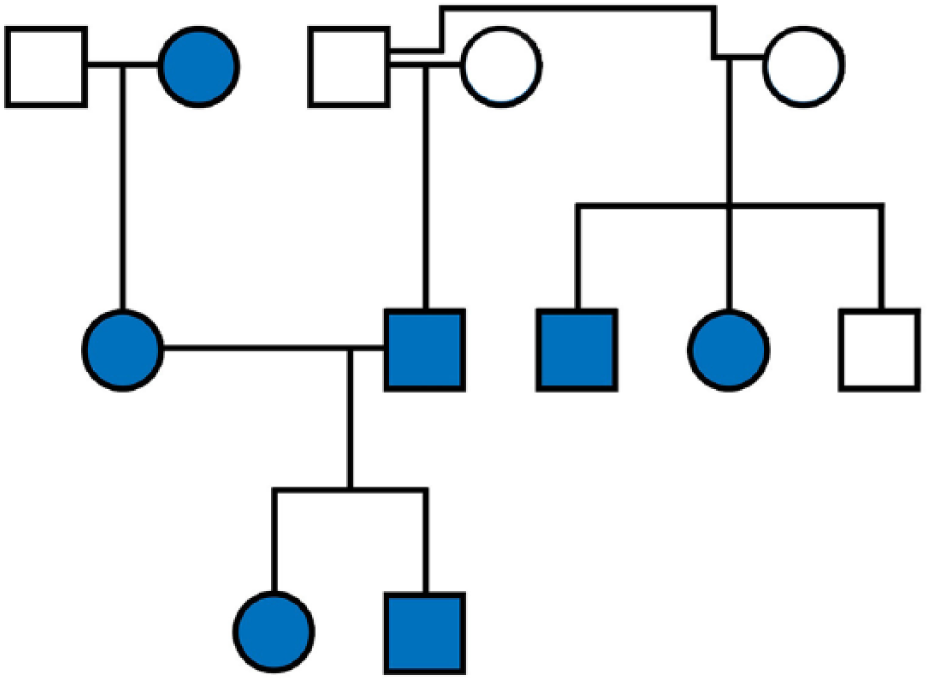
Pedigree of Russian Blue cats with (blue) and without (white) cataracts. Square symbols indicate males and circles indicate females
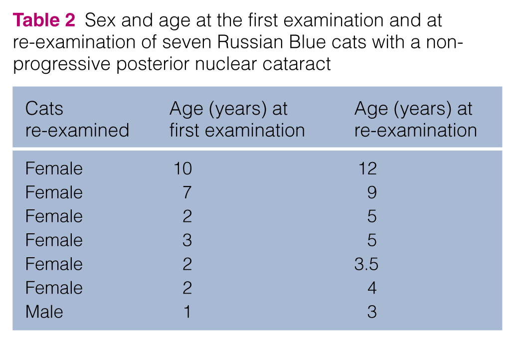
Seven cats (one male, six females) between 1 and 10 years of age at first examination, all of them with the posterior nuclear form of cataract, were re-examined after 1.5–3 years. None showed signs of progression of the cataract (Table 2). Another case with a more extensive cataract had also not progressed at the 3 year follow-up (Figure 3).
Sex and age at the first examination and at re-examination of seven Russian Blue cats with a non-progressive posterior nuclear cataract
Discussion
Twenty-two of the 66 cats examined were affected by cataracts, which amounts to a prevalence of 33% in the examined group of cats. The most prevalent form of cataract seen in the majority of the cases (n = 16/22), was a small triangular, Y-shaped or circular opacity located in the posterior nucleus at the border between the nucleus and the posterior cortex. The area is traditionally described as the adult part of the nucleus. 15 Whether a cataract with this localisation should be termed a posterior nuclear cataract or a posterior cortical cataract remains to be determined. 15 At birth the lens consists almost entirely of lens nucleus with minimal lens cortical material. 16 The localisation of the cataract and the fact that the cataracts were seen in young cats, two below the age of 1 year, suggest that the defect develops early in life and could be congenital. The definition of a congenital condition is that it is present at birth, when the eyelids are open, or is observed during the first 6–8 weeks of the cat’s life. 17
The most prevalent form of cataract in the examined group of cats did not seem to be progressive. Seven cats with this form were re-examined after 1.5–3 years and showed no signs of progression. Six of the 22 cats with cataracts had more extensive forms in which the entire nucleus and either the entire cortex or parts of the posterior and/or anterior cortex were involved. These extended forms impaired vision and one of the cats had gone through bilateral cataract surgery at 8 months of age (Evidensia Animal Hospital Strömsholm, 2013). The relationship between these more extended forms of cataracts and the most frequently observed form is uncertain at the present time and needs further investigation.
The pedigree analyses did not include an extensive number of littermates and other relatives, as comparatively few cats participated in the study. However, the findings supported an autosomal recessive mode of inheritance for the defect. Further studies of the hereditary characteristics of the disease are needed because of the low number of cats in the present study and the fact that the pedigree does not exclude an autosomal dominant mode of inheritance with incomplete penetrance.
Investigations are currently ongoing to elucidate the genetic basis through whole-genome sequence analysis. Thus, work is underway to identify a causative genetic variant. Given this information, we hope that a genetic test can be developed to facilitate selective breeding.
Conclusions
Our data support the fact that the Russian Blue cat is affected by hereditary cataracts with a high prevalence in the breed. The most frequently observed localisation of the cataract is in the central parts of the posterior nucleus, at the border between the nucleus and the posterior cortex. The high prevalence in young cats and characteristic localisation of the cataract in the posterior nucleus suggest an early onset form of cataract. As the Russian Blue cat is a small breed in Scandinavia and some forms of the presently described cataracts have been proven to impair vision, the breeders should be aware of this defect in the breed and have their cats examined by a veterinary ophthalmologist before the breeding of an individual Russian Blue cat is considered.
Footnotes
Acknowledgements
We thank Kaija Merta, the Russian Blue breed clubs and the owners of the Russian Blue cats that participated in the study. Further, we are grateful to Howard Wilson for help with the graphics. Finally, we thank Dr William Murphy for continued research efforts in regard to hereditary cataracts in the Russian Blue cat.
Author note
A preliminary report of this study was presented at the European College of Veterinary Ophthalmologists meeting in Helsinki 2015.
Conflict of interest
The authors declared no potential conflicts of interest with respect to the research, authorship, and/or publication of this article.
Funding
The authors received no financial support for the research, authorship, and/or publication of this article.