




Abstract
Objectives:
This paper, the last in a series of three on ‘feline leprosy’, provides a detailed description of disease referable to the previously unnamed species, Candidatus ‘Mycobacterium lepraefelis’, a close relative of the human pathogens Mycobacterium leprae and Mycobacterium lepromatosis.
Methods:
Cases were sourced retrospectively and prospectively for this observational study, describing clinical, geographical and molecular microbiological data for cats definitively diagnosed with Candidatus ‘M lepraefelis’ infection.
Results:
A total of 145 cases of feline leprosy were scrutinised; 114 ‘new’ cases were sourced from the Victorian Infectious Diseases Reference Laboratory (VIDRL) records, veterinary pathology laboratories or veterinarians, and 31 cases were derived from six published studies. Thirty-eight cats were definitively diagnosed with Candidatus ‘M lepraefelis’ infection. Typically, cats tended to be middle-aged or older when first infected, with a male predilection. Affected cats typically had widespread cutaneous lesions, in some cases after initially localised disease. Advanced cases were often systemically unwell. All cats had outdoor access. The histological picture was lepromatous in the majority of patients, although two cases had tuberculoid disease. In one case that underwent necropsy, lesions were evident in the liver, spleen and lungs. Treatment was varied, although most cats received a combination of oral clarithromycin and rifampicin. Prognosis for recovery was variable, but typically poor.
Conclusions and relevance:
Candidatus ‘M lepraefelis’ typically causes high bacterial index (lepromatous) feline leprosy that in some cases progresses to systemic mycobacteriosis. The disease has a variable clinical course and prognosis. Many cases either died or were euthanased due to the infection. Multilocus sequence analysis reveals a heterogeneous picture and further analysis of draft genome sequencing may give clues to the taxonomy and epidemiology of this organism. Prospective treatment trials and/or additional drug susceptibility testing in specialised systems would further inform treatment recommendations.
Comparative aspects:
This paper finishes with a discussion of comparative aspects of infection caused by the three feline leproid disease agents that have been the subject of this series: Candidatus ’Mycobacterium tarwinense’, Mycobacterium lepraemurium and Candidatus ‘M lepraefelis’.
Introduction
The mycobacterium that is the subject of this paper was first identified by Hughes and colleagues in 1997, in skin biopsy material obtained from two 9-year-old male New Zealand cats. 1 Sequence analysis of the V2 and V3 regions of the 16S rRNA gene revealed a novel species that most closely resembled Mycobacterium malmoense in the GenBank database. Shortly after that study, a case report of a cat with fastidious mycobacteriosis presenting with multiple cutaneous nodules and suspected liver involvement was published by investigators at the University of Sydney, Australia. 2 Genetic analysis of variable regions of the 16S rRNA gene revealed that it was most closely related to Mycobacterium haemophilum. It was not recognised at the time that this organism was the same as the one reported by Hughes et al.
These two groups subsequently collaborated to further characterise the disease in Australian cats. 3 From a small cohort of nine cases, it was observed that cats with this infection were typically older (>9 years of age) and tended to experience a more protracted, and potentially aggressive, clinical disease than those diagnosed with Mycobacterium lepraemurium. Infected cats showed a propensity to develop widespread, numerous, nonulcerated cutaneous nodules after initially presenting with localised disease. Histological examination of biopsy material from these cases demonstrated lep-romatous pathological change, often with enormous numbers of mycobacteria in macrophages and giant cells within the skin lesions. The authors speculated as to whether these aged cats potentially had decreased immunological competence, particularly given that some cases presented with concurrent (or subsequently developed) conditions such as chronic kidney disease.

This paper, the last in a series of three,4,5 concerns feline patients with skin lesions proven to be caused by Candidatus ‘Mycobacterium lepraefelis’ using molecular methods. This represents the largest collection of such cases reported to date, and greatly extends the previous observations, both in terms of case numbers and duration of follow-up.
Materials and methods
Case recruitment, demographics, data retrieval, cytological and histological assessment, molecular microbiology methods, genetic analysis, mycobacterial culture and statistical analysis were essentially the same as described in the first paper in this series, and interested readers are referred to that publication. 4 Different primers were used to partially sequence the sodA gene of Candidatus ’M lepraefelis’: ‘ECsodF’ 5′-CTTGCCAAACTTGACGAGGC-3′ (forward) and ‘ECsodR’ 5′-ATTGAGCGCGGAATTTGTCG-3′ (reverse).
Results
Clinical data
A total of 145 cases of feline leprosy were analysed; 114 cases were sourced from the Victorian Infectious Diseases Reference Laboratory (VIDRL) records, veterinary pathology laboratories or veterinarians, with an additional 31 cases derived from six published studies.1,3,6–9 Of these, 38 had Candidatus ‘M lepraefelis’ infection, comprising 30 ‘new’ cases and eight cases recorded previously (Table 1).
Detailed case data for 38 cats infected with Candidatus ‘Mycobacterium lepraefelis’
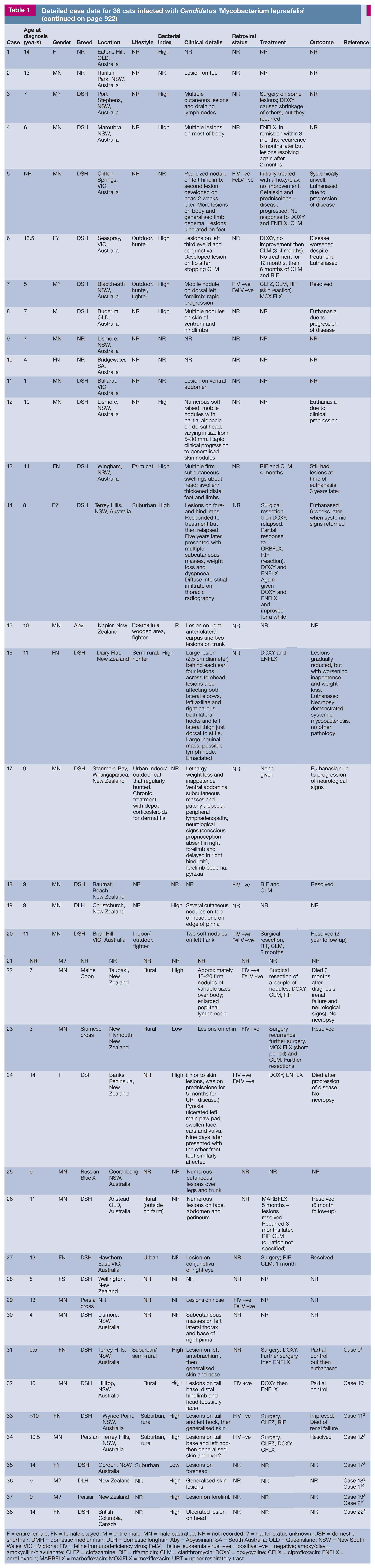
F = entire female; FN = female spayed; M = entire male; MN = male castrated; NR = not recorded; ? = neuter status unknown; DSH = domestic shorthair; DMH = domestic mediumhair; DLH = domestic longhair; Aby = Abyssinian; SA = South Australia; QLD = Queensland; NSW = New South Wales; VIC = Victoria; FIV = feline immunodeficiency virus; FeLV = feline leukaemia virus; +ve = positive; −ve = negative; amoxy/clav = amoxycillin/clavulanate; CLFZ = clofazamine; RIF = rifampicin; CLM = clarithromycin; DOXY = doxycycline; CFLX = ciprofloxacin; ENFLX = enrofloxacin; MARBFLX = marbofloxacin; MOXIFLX = moxifloxacin; URT = upper respiratory tract
The median age at diagnosis was 9.5 years (range 1–14). Only five cats were 6 years of age or less. Where gender was recorded, 24/37 (65%) Candidatus ‘M lepraefelis’ infections were in male cats, which is significantly more than would be expected based on the age/gender pyramid for Australian cats. 11 Where breed was recorded, 24/33 cats were domestic shorthair, 2/33 were domestic longhair, 3/33 were Persian or Persian cross, and there was one each of Maine Coon, Abyssinian, Siamese cross and Russian Blue cross; the crossbreed to pedigree ratio was similar to that of the Australian feline population. 11 Information in the dataset was generally too sparse to assess whether there was association between signalment and clinical presentation or outcome.
Geographical location data were recorded for 36 cats, 24 from Australia, 11 from New Zealand and one from Canada. In the Australasian region, Candidatus ‘M lepraefelis’ was documented to have a geographical range that typically extended along the east coast of Australia, with one case as far west as Adelaide, South Australia, and it was present on both islands of New Zealand (Figure 1). The infection was not documented in Tasmania, despite a similar longitude, latitude and daily maximal temperature to other endemic areas in this region. Again, lack of data prevented analysis for associations between location of domicile and signalment, clinical presentation or outcome.

Geographical distribution of feline infections caused by Candidatus ‘M lepraefelis’ for which accurate location data were available
Where lifestyle was recorded, all cats had unsupervised outdoor access and many were known hunters and/or fighters. Feline immunodeficiency virus (FIV) status was only recorded for 11 cats, with three (27%) being seropositive. As far as could be ascertained from the case records, none of the FIV-positive cases had received prior FIV vaccination. This FIV prevalence is higher than published rates of endemicity in pets cats from both eastern Australia (8%) 12 and New Zealand (10%); 13 however, numbers were too small to make statistical inferences and the observation may represent an epiphenomenon related to the outdoor lifestyle. None of the six cats tested for feline leukaemia virus (FeLV) were seropositive.
Where clinical features and clinical course were well documented, 12/31 (39%) cats had a few lesions, with the remainder having multiple regional or generalised nodular skin disease (the latter occurring in 13/31 [42%] cases). There was no difference in age within this cohort between cats with one to two lesions (median 11 years, range 1–14) vs those with three or more nodules (median 9.5 years, range 6–14) (P = 0.9681). No anatomical region had a predilection for involvement (Figures 2a–c). In 11 cases, lesions were anatomically localised (Figure 2d). Ulceration of skin lesions was rarely reported (two cases). Diffuse regional subcutaneous oedema was noted in four cases, particularly in those with widespread lesions and systemic signs of illness (eg, emaciation, pyrexia, inappetence). One cat (case 17) also presented with neurological signs, but it could not be confirmed whether this was a direct result of mycobacteriosis, as necropsy examination was not undertaken. Likewise, case 14 was noted to have dyspnoea and diffuse interstitial changes on thoracic radiography, consistent with pulmonary dissemination; 14 however, this was not further investigated.
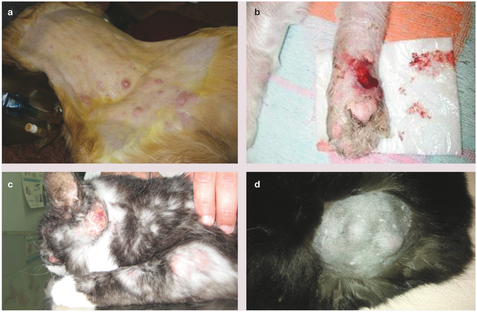
Representative photographs of cutaneous lesions caused by Candidatus ’M lepraefelis’ infection in cats. Cases 8 (a) and 5 (b) were euthanased due to persistence/progression of disease despite attempts at medical therapy. Case 16 (c) was found to have small numbers of acid-fast bacilli in the liver, spleen and lungs at necropsy. Case 20 (d) had localised disease and responded well to surgical resection and adjunctive antibiotic therapy. Image (b) courtesy of Steve Holloway


Case 16 was euthanased due to rapid clinical deterioration, despite treatment with doxycycline and enrofloxacin. Histopathological examination of tissues collected at necropsy demonstrated moderate numbers of small, multifocal granulomas in the liver parenchyma (Figure 3a) containing macrophages with intracytoplasmic acid-fast bacilli (AFB; Figure 3b) and AFB-positive macrophages circulating within the hepatic sinusoids (Figure 3c). There were also low numbers of macrophages with intracytoplasmic AFB in the pulmonary interstitium and surrounding alveolar septa and airways, and within the splenic parenchyma. Nodular granulomatous dermatitis with massive numbers of intra- and extracellular AFB was also observed, consistent with the histopathological description of previously reported cases. 3 Schwann cells around peripheral nerves in the sections of affected dermis were specifically examined for evidence of involvement and demyelination, a classic microscopic feature of Mycobacterium leprae infection in people, 15 but none was found. Likewise, endothelial invasion and thrombosis (‘Lucio’s phenomenon’), typically found in human patients infected with a related organism, Mycobacterium lepromatosis, 16 were also absent.

Photomicrograph of granulomas (arrow) within the hepatic parenchyma in a cat (case 16) that underwent necropsy (a). Macrophages containing acid-fast bacilli were found within these lesions (b) and circulating within the sinusoids (c)
Where cytology or histopathology of cutaneous lesions was available for review, a high bacterial index (BI) was observed in 21/23 cases (91%), while the remaining two had a low BI (both cats had anatomically localised disease).
Treatment and clinical outcome
Therapeutic regimens used in this study cohort varied widely, and patients were managed with variable input from the authors. As in the previous two studies in this series, options were usually influenced by owner finances and commitment, and the patient’s temperament.
Medical protocols included several months of treatment with one or more of the following drugs: rifampicin, clarithromycin, clofazimine, a fluoroquinolone (enrofloxacin, marbofloxacin or moxifloxacin) and doxycycline. Of the seven successfully treated cats where the treatment regimen was recorded, one was treated with a varying combination of clofazimine, clarithromycin, rifampicin and moxifloxacin, four were treated with clarithromycin and rifampicin (with or without surgical debulking), one received moxifloxacin and clarithromycin plus surgical resection, and one received clofazimine, doxycycline and ciprofloxacin after surgical debulking of some lesions. Three cases were euthanased or died due to disease progression despite being on protocols containing clarithromycin and/or rifampicin and/or clofazimine (although adequate compliance was not established). None of the cats treated with protocols containing doxycycline and enrofloxacin survived (five cases).
Overall, outcome was not generally favourable for cats with Candidatus ‘M lepraefelis’ infection, with resolution in only 7/20 cases (35%) where sufficient follow-up was available to make an assessment. Widespread or generalised cutaneous disease, particularly with concurrent subcutaneous oedema and/or signs of systemic illness, appeared to convey a particularly guarded prognosis.
A summary of data concerning age, gender, anatomical distribution of lesions and outcome for cats infected by Candidatus ’M lepraefelis’ is provided in Table 2.
Summary data concerning age, gender, anatomical distribution of lesions and outcome for cats infected by Candidatus ‘M lepraefelis’

BI = bacterial index
Microbiological and molecular data
Fresh tissue, methanol-fixed cytological smears and paraffin-embedded formalinfixed samples from 37 cases underwent DNA extraction, PCR amplification and sequencing. Known mutations in the rpoB gene conferring rifampicin resistance in other mycobacterial species17–19 were not found in any of the isolates in this study (data not shown). In four of the five loci examined, homogeneity of between 99.98 and 99.99% was noted (see supplementary material – Figure S1) (100% homogeneity was observed for sodA). There was no discernible geographic clustering of sequence types (see supplementary material – Table S1).
Mycobacterial DNA derived from fresh biopsy tissue from case 20 underwent draft genome sequencing during the study. The methods and results of these investigations will be published in full separately (C O’Brien et al, manuscript in preparation).
Discussion
This study greatly extends the body of knowledge concerning Candidatus ‘M lepraefelis’ infections in cats. 20
Signalment
The age range of affected cats is wider than previously reported, although most infections still occurred in adult cats over 7 years of age, and none were less than 3 years of age. Older cats may be more susceptible to this infection due to acquired defects in cell-mediated immunity or acquisition of predisposing comorbidities (early kidney disease, longstanding FIV infection, etc); or it may simply reflect increased exposure over the lifespan following a long incubation period. Male cats were predisposed, which may reflect an increased tendency to hunt potentially infected prey, or to be subject to predisposing traumatic injuries (eg, cat fights) in which there is a breach in the integrity of the skin. The ratio of domestic crossbreeds to pedigree cats in this cohort is in accord with the overall Australian cat population, suggesting that there are no breed-associated genetic predispositions towards infection.
There was a tendency for affected cats to have a higher prevalence of FIV positivity than the general population, although this may not necessarily be significant if the data were controlled for gender and age (with FIV infection being possibly more common in an older, male cohort due to the cumulative likelihood of exposure). Although none of the FIV-positive cats showed overt evidence of an AIDS-like state, this cannot be excluded as a potential risk factor for the disease, although current evidence shows that human immunodeficiency virus (HlV)-associated AIDS does not impact on the clinical progression of leprosy in HIV-positive people. 21
Clinical course
The clinical course of Candidatus ‘M lepraefelis’ infection appears to be variable, but with a tendency to progress to widespread, disseminated cutaneous involvement, sometimes after initially localised disease. The cutaneous reaction pattern of multifocal skin nodularity in these cats is strongly suggestive of haematogenous dissemination from a primary focus of infection, possibly with a pathogen with a preferred temperature range for growth that is lower than mammalian core body temperature.
The finding of mild systemic involvement was notable, as there was no evidence of this in cases infected with Candidatus ’Mycobacterium tarwinense’, 4 M lepraemurium 5 or the unnamed mycobacterium that causes leproid granulomas in dogs. It is unknown what proportion of cases are affected in this manner and at what point systemic involvement occurs. The finding of systemic disease, and indeed the presence of regional oedema, is strikingly reminiscent of Mycobacterium visibile infection reported in North American cats in the early 2000s. 22 These findings are also consistent with an early Australian report by Donnelly and colleagues, 23 in which such involvement was documented, but where no molecular diagnosis was possible as the PCR technique had not been developed. (The paraffin blocks for this early case had been discarded and were thus unavailable for retrospective PCR testing and sequence analysis.) The presence of macrophages laden with AFB in liver sinusoids is a further marker for haematogenous dissemination, and the presence of such widely disseminated disease might account for signs of inappetence, malaise and increased transaminase activities reported previously in a cat with this infection.2,3
Organism biology
Candidatus ‘M lepraefelis’ is related to M leprae, the recently characterised organism M lepromatosis, and the unnamed causative agent of bovine (and caprine 24 ) nodular thelitis based on sequence analysis of the 16S rRNA gene. 25 The phylogenetic relationship should ideally be confirmed and extended using multilocus sequence typing (MLST) analysis or, preferably, whole genome sequencing, to assess whether these organisms meet the criteria for a taxonomic species complex. A recent publication also confirms the genetic relatedness of M leprae/M lepromatosis to M haemophilum; 26 given the latter’s larger genome size, this prompts the current authors to speculate as to whether it may be the progenitor species of this group or at least possibly more closely related to the most recent common ancestor.
Analysis of five gene targets in Candidatus ‘M lepraefelis’ has demonstrated that all the clinical isolates were somewhat heterogeneous at four of the loci examined (three or four single nucleotide polymorphisms), although MLST analysis did not reveal a geographical explanation or correlation for this genetic diversity. This contrasts with the apparent genetic homogeneity of Candidatus ’M tarwinense’ 4 and M lepraemurium, 5 and at this stage it is unknown why the Candidatus ’M lepraefelis’ genome appears less stable.

Treatment and outcome
Treatment regimens were not consistent, and indeed this data set was less robust than for the other two causes of feline leprosy in the series. It can be stated with relative confidence, however, that cats with Candidatus ‘M lepraefelis’ infection responded much less favourably overall than those infected with the other organisms. Spontaneous remission was not recorded in any case. There were only a few cats in which multidrug therapy (simultaneous and/or sequential) was successful, albeit in the face of multifocal skin (and possibly internal organ) involvement.
The response to treatment was particularly poor in cats that displayed diffuse subcutaneous oedema and/or systemic signs either before or during therapy. Interestingly, peripheral oedema and systemic signs may be associated with ‘reactive’ leprosy in people with M leprae infection. 15 Studies of the lymphocyte expression of interleukin and interferon-gamma would be revealing with regards to whether a helper T-cell type 1 or type 2 response predominates in these cats, 27 and may inform whether the provision of immunomodulating agents, such as thalidomide, which has been safely used in cats, 28 could be of benefit in these cases.


Cat with a rapid-growing mycobacterial infection following one surgical intervention (debulking) and consolidation combination antimicrobial therapy. Courtesy of Dr Greg Kelman

Age distribution of cats with different forms of leproid disease
Data comparing quantitative aspects of infection with the three different agents of feline leproid disease

Kruskal-Wallis test, **Chi-square test, NSW = New South Wales
Summary of the comparative qualitative aspects of infection with the three different agents of feline leproid disease

VIC = Victoria; NSW = New South Wales
Key Points
Candidatus ‘M lepraefelis’, a fastidious mycobacterium related to the human pathogens M leprae and M lepromatosis, typically causes high BI (lepromatous) feline leprosy that may progress to generalised cutaneous and systemic mycobacteriosis.
Lesions generally consist of localised or generalised nodules, with an occasionally aggressive clinical course and a variable, but generally poor, response to therapeutic intervention.
The best management approach appears to consist of early, complete surgical resection of easily accessible lesions, and/or debulking of lesions in difficult anatomical locations, combined with two or more antimicrobial agents with known activity against slow-growing mycobacteria.
The results of draft genome analysis will likely inform further investigations into the epidemiology of this organism and explain why it cannot be grown readily using routine mycobacteriological methods.
Supplemental Material
Supplementary Figure 1
Candidatus Mycobacterium lepraefelis multilocus sequence alignments
Supplemental Material
Supplementary Table S1
Geographical distribution of Candidatus Mycobacterium lepraefelis multilocus sequence types
Footnotes
Acknowledgements
Thanks go to the staff of the Mycobacterium Reference Laboratory, VIDRL, especially Caroline Lavender, for technical assistance, and the many veterinarians and veterinary pathologists who contributed case material and other assistance to this study, especially Laura Brandt, Bronwyn Smits, Catherine Harvey, Rob Fairley, Richard McCoy and Sergio Sanchez Picado of Gribbles Pathology, and Adrienne French, Catherine Williamson, Dawn Seddon, Geoff Orbell and Cathy Harvey of New Zealand Veterinary Pathology.
Date accepted: 7 March 2017
Supplementary material
Figure S1: Candidatus ‘M lepraefelis’ multilocus sequence alignments.
Table S1: Geographical distribution of Candidatus ‘M lepraefelis’ multilocus sequence types.
Conflict of interest
The authors declared no potential conflicts of interest with respect to the research, authorship, and/or publication of this article.
Funding
This work was supported in part by a grant from the Feline Health Research Fund. Carolyn O’Brien was supported by an Australian Government Research Training Program Scholarship. Richard Malik was supported by the Valentine Charlton Bequest administered by the Centre for Veterinary Education, The University of Sydney.
References
Supplementary Material
Please find the following supplemental material available below.
For Open Access articles published under a Creative Commons License, all supplemental material carries the same license as the article it is associated with.
For non-Open Access articles published, all supplemental material carries a non-exclusive license, and permission requests for re-use of supplemental material or any part of supplemental material shall be sent directly to the copyright owner as specified in the copyright notice associated with the article.
