
Abstract
Objectives
The MRI features of the feline cerebellum and occipital bone have not previously been described in the literature. The aims of this study were three-fold. Firstly, to document variations in cerebellar shape on MRI in neurologically normal cats to support our hypothesis that crowding of the contents of the caudal fossa or herniation of the cerebellar vermis through the foramen magnum occurs frequently as an anatomical variant. Secondly, to document variations in the morphology of the occipital bone. Thirdly, to see whether these variations in shape of the feline cerebellum and occipital bone could be associated with head conformation, such as brachycephaly.
Methods
The imaging records of the small animal clinic at the Animal Health Trust between 2000 and 2013 were searched retrospectively to identify adult cats that had undergone high-field (1.5 T) MRI investigation which included the brain. Exclusion criteria included evidence of intracranial disease or the presence of cervical syringomyelia. Midline sagittal T2-weighted and transverse images were used to assess the occipital bone morphology and cerebellar shape, and to measure the width to length ratio of the cranial cavity.
Results
Fourteen different breeds were represented. A cerebellar shape consistent with crowding of the contents of the caudal fossa, or herniation through the foramen magnum was present in 40% of the entire population. Persians (recognised as a brachycephalic breed) had a higher proportion of cerebellar crowding or herniation than all other breeds. There was no significant difference in the distribution of occipital bone morphology between these breed groups.
Conclusions and relevance
It is important to recognise morphological variations of the feline cerebellum and occipital bone in order to avoid false-positive diagnoses of raised intracranial pressure and pathological herniation on MRI.
Introduction
The cerebellum lies in the caudal fossa of the cranial cavity and consists of a median vermis and cerebellar hemispheres. 1 It is bordered by the occipital bone caudally, which forms a ring, the foramen magnum, around the junction of the medulla oblongata and the spinal cord. 2 The occipital bone is composed of four parts only in dogs and cats, a dorsal supraoccipital or squamous part, two lateral exoccipital bones and the basioccipital or basilar part ventrally. The supraoccipital part of the occipital bone forms the dorsal border of the foramen magnum and partly surrounds the cerebellum. The exoccipital bones bear the occipital condyles and are part of the atlanto-occipital joints. The basioccipital bone lies ventral to the medulla oblongata and forms the ventral part of the foramen magnum. 2
Cerebellar and occipital bone morphology has been investigated in various species. Most canine studies have focused on Chiari-like malformation, mainly in the Cavalier King Charles Spaniel and Griffon Bruxellois,3–6 but studies in cats,7–9 lions10,11 and alpacas 12 have also been conducted. Several of the feline studies investigated normal morphometric features of the skull by making measurements on skull specimens.7,9 Other studies have reported the anatomy of the feline brain to provide a clinical anatomy atlas, 16 and to establish a reference when evaluating (human) neurological disease and novel therapeutics in animal models. 17
Clinically significant feline syringomyelia has also been reported,8,13–15 associated with occipital bone variations. However, the MRI features of the feline cerebellum and occipital bone have not previously been described in the literature.
In our experience, cats display a wide variability in the shape of the cerebellum and occipital bone on MRI, with varying degrees of crowding of the cerebellar vermis into the foramen magnum or herniation of the cerebellar vermis through the foramen magnum in the absence of neurological clinical signs. As this appearance in dogs may indicate either raised intracranial pressure or Chiari-like malformation, it is important to recognise it as a normal feline variant in order to avoid making false-positive diagnoses of pathology.
The aims of this study were three-fold: (1) to document variations in cerebellar shape on MRI in neurologically normal cats and to investigate the hypothesis that crowding of the contents of the caudal fossa or herniation of the cerebellar vermis through the foramen magnum occurs frequently as an anatomical variant; (2) to document variations in the morphology of the occipital bone and to determine whether cerebellar shape is related to these variations; and (3) to see whether these variations could be related to head conformation, and specifically to brachycephalic conformation, such as is seen in the Persian breed.
Materials and methods
The imaging records of the small animal clinic at the Animal Health Trust between 2000 and 2013 were searched retrospectively to identify adult cats (ie, >12 months of age) that had undergone MRI investigation which included the brain. The cats had been investigated for a variety of diseases, including nasal, middle or inner ear, thyroid, extracranial neurological and behavioural abnormalities. Inclusion criteria required a neurological examination that was normal or not localised to the brain and an absence of visible intracranial disease on MRI. Exclusion criteria included MRI findings suggesting intracranial disease such as neoplasia, inflammation or trauma, or the presence of cervical syringomyelia. All the MRI examinations included, as a minimum, sagittal and transverse T2-weighted sequences of the brain from olfactory lobes to at least the second cervical vertebra. MRI examinations were obtained using fast spin echo sequences and 3 mm slice thickness using a 1.5 T GE Signa Echospeed system and a medical extremity radiofrequency coil. All cats were anaesthetised using a premedication combination at the discretion of the anaesthetist, induced with propofol and maintained on isoflurane or sevoflurane with oxygen. All cats were positioned for scanning in dorsal recumbency with the neck gently extended and the thoracic limbs restrained caudally.
Midline sagittal T2-weighted images were used to assess the occipital bone morphology and cerebellar shape. Using freely available imaging software (ClearCanvas), the maximum width and length of the cranial cavity were also measured with electronic calipers on transverse and midline sagittal T2-weighted images.
Two radiologists, one board certified (RD) and one resident (XH), assessed each set of images, blinded to the signalment of each case and to each other’s results. Cerebellar shape and occipital bone morphology were assigned to predefined categories by each radiologist. If the categorisation differed between the two radiologists, agreement on final categorisation was made by consensus.
Cerebellar shape
From the sagittal magentic resonance (MR) images of the cerebellum, its shape in each case was allocated to one of the following three categories (Figure 1a–c):
C1 – convex caudal margin with a small area of hyperintense signal representing cerebrospinal fluid (CSF) adjacent to the caudoventral margin of the vermis, which was rounded.
C2 – flattened caudal margin with a blunt right-angle shape caudoventrally (the caudal surface could be flat or slightly irregular, depending on the shape of the adjacent cranial cortex of the occipital bone). No hyperintense signal representing CSF was seen adjacent to the caudoventral margin of the vermis.
C3 – concave caudal margin with the cerebellar vermis crowding into, or herniating through, the foramen magnum.

Representative sagittal T2-weighted images of the three cerebellar shape categories: (a) convex caudal surface and rounded caudoventral margin (category C1); (b) flattened caudal surface with caudoventral margin showing a blunt right angle (category C2); and (c) concave caudal surface with cerebellar vermis entering into or herniating through the foramen magnum (category C3)
Occipital bone morphology
From the sagittal MR images, the shape of the supraoccipital part of the occipital bone in each case was allocated to one of two categories (Figure 2a,b):
Oi, a thin occipital bone (signal-void line) lacking a medullary cavity;
Oii, a thick occipital bone containing a medullary cavity.

Representative sagittal T2-weighted images of the two occipital bone shape categories: (a) cortical bone only (category Oi); (b) occipital bone with medullary cavity (category Oii)
Intracranial cavity width-to-length ratio
A pilot study was performed to attempt to correlate head conformation with the intracranial cavity width-to-length ratio (ICCR), as it was anticipated that not all MRI studies would include the entire head on sagittal T2-weighted (T2W) images, precluding the use of other indices for assessment of head conformation.
The width of the cranial cavity at its widest point (intracranial width) and the length of the cranial cavity from the most rostral part of the cribriform plate to the dorsal margin of the foramen magnum (intracranial length) were measured in the transverse and sagittal MR images, respectively. The width of the cranial cavity was measured from the junction of bone and T2W hyperintense signal (consistent with CSF) and was performed at the widest level, usually at the level of the piriform lobes. Similarly, the length of the cranial cavity was measured from the junction of bone and T2W hyperintense signal (consistent with CSF) at the olfactory lobes to the foramen magnum (Figures 3 and 4).

Width of the cranial cavity: widest point (usually at the level of the piriform lobe)

Length of the cranial cavity: olfactory lobe to foramen magnum
The ICCR was calculated by dividing the intracranial width by the intracranial length and expressing the result as a percentage. The ICCR was used to provide information on the skull morphology; although it does not directly measure nasal shape it was anticipated that it might give an indirect indication of brachycephaly. To test this assumption, the length of the nose from cribriform plate to nares was measured on the sagittal MR images of 108 cats in which the study included the entire nose and then compared with intracranial length and the ICCR.
Statistical analysis
Initial evaluation of the continuous data suggestedthey were not normally distributed and therefore non-parametric statistical analyses were used for all evaluations. Correlation between data sets was undertaken using the Spearman rank correlation coefficient (rs) and proportions in categories assigned to groups in a contingency table were evaluated using the χ2 test. Differences between median values were evaluated using the Kruskal–Wallis (for multiple comparisons) or theMann–Whitney test (for comparison of two groups) and a P value of <0.05 was considered significant.
Results
The study population comprised 200 cats with a mean age of 7.2 years (range 1–17 years, median 7.0 years). There were three (1.5%) entire females, 87 (43.5%) neutered females, five (2.5%) entire males and 105 (52.5%) neutered males. Because of the small numbers of entire females and males, neutered and entire cats were grouped together for statistical analysis; thus, the overall study numbers were 90 (45%) female and 110 (55%) male.
Fourteen different breeds were represented: domestic shorthair (DSH, n = 129), domestic longhair (DLH, n = 15), Persian (n = 11), Siamese (n = 9), Birman (n = 7), British Shorthair (BSH, n = 7), Bengal (n = 5), Burmese (n = 5), Maine Coon (n = 5), Ragdoll (n = 2), Tonkinese (n = 2), Russian Blue (n = 1), Somali (n = 1) and Turkish Van (n = 1). For statistical analysis, breeds containing fewer than seven cats, none of which had obvious brachycephalic facial conformation, were grouped together as ‘Others’.
Cerebellar shape
As seen in Table 1, only 35 (17.5%) of the cats showed a rounded cerebellum (category C1) similar to that usually seen in dogs. This shape was seen slightly more often in male cats (23 cats, 21% of males) than in female cats (12 cats, 13% of females.) Eighty-five cats (42.5%) showed a cerebellar shape of category C2; that is, a flattened caudal margin. This shape was seen in approximately equal numbers of male and female cats (44% and 41%, respectively). The relationship between breed and cerebellar shape is shown in Figure 5. No statistically significant relationship of categories C1 and C2 with breed was detected. A cerebellar shape consistent with crowding of, or herniation through, the foramen magnum (category C3) was present in 40% of the entire population (80 cats), comprising approximately one-third of the male cats and nearly half of the female cats (35% and 46%, respectively). For this category, the median value of the cerebellar shape was significantly (P = 0.0099) different between Persian and Birman cats, Persian and DLH cats, and between Persian and Other cats, with the Persians (recognised as a brachycephalic breed) having a higher proportion of cerebellar crowding or herniation (cat-egory C3). No statistical difference in the median value of the cerebellar shape was found between Persians and DSH cats. Analysis of the data showed no significant differences in median cerebellar shape or in the distribution of cerebellar shapes between male and female cats(Table 2), and no significant correlation between age and cerebellar shape.
Relationship between breed and cerebellar shape

Data are n (%) unless otherwise indicated
DSH = domestic shorthair; DLH = domestic longhair; BSH = British Shorthair
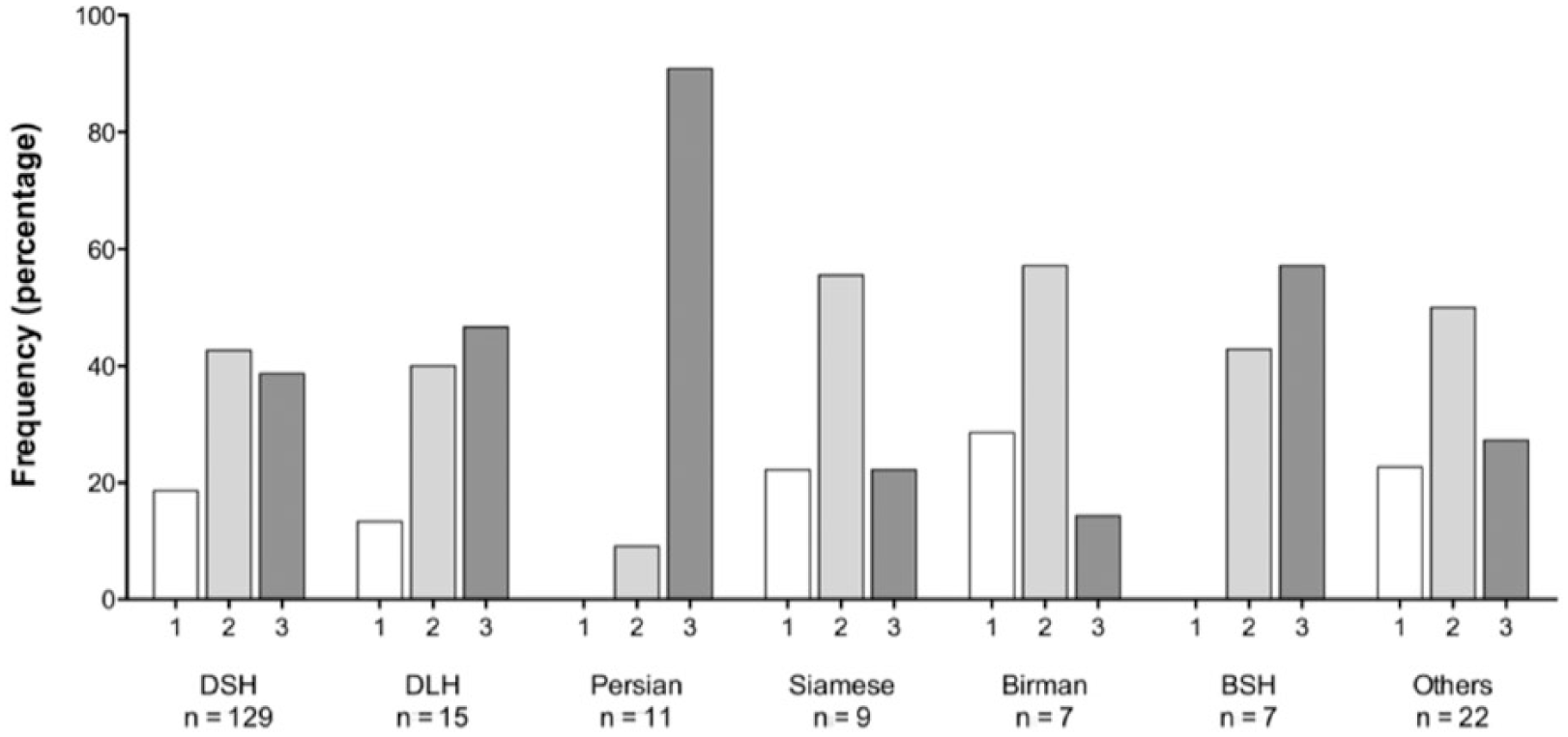
Histogram (frequency distribution) of the relationship between breed and cerebellar shape. DSH = domestic shorthair; DLH = domestic longhair; BSH = British Shorthair
Relationship between sex and cerebellar shape
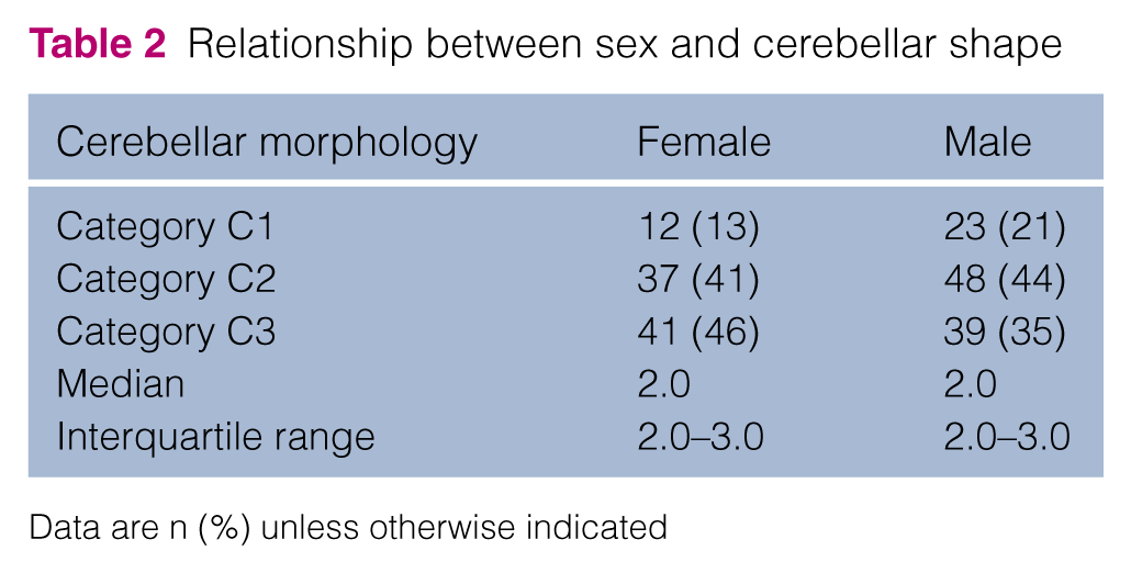
Data are n (%) unless otherwise indicated
Occipital bone morphology
A thin occipital bone (category Oi, lacking a medullary cavity) was seen in two-thirds of cats (n = 135; 67.5%), relatively slightly more often in females than in males, whereas a thick occipital bone (category Oii) was seen in one-third of cats (n = 65; 32.5%), slightly more often in males than in females (Table 3).
Relationship between sex and occipital bone morphology

Data are n (%)
The occipital bones also varied slightly in their shape ventrally. The thin bones (category Oi) were straight throughout in some cats but showed a slight caudal angulation of their ventral aspect in others. The thick bones (category Oii) sometimes ended in a blunt, rounded fashion, whereas on other occasions they showed a ventral thickening or ‘toe’ similar to that often seen in larger dogs. For valid statistical analysis of breed variation in occipital bone morphology, further combination of breed groups was necessary and four breed categories were made, comprising DSH, DLH, Persian and Others. There was no significant difference in the distribution of occipital bone morphology betweenthese breed groups. Additionally, no significant correlation was found between age or sex and occipital bone shape.
No significant differences were found when the relationship between cerebellar shape and occipital bone morphology was examined (Table 4). The three cerebellar shape categories occurred with approximately equal frequency (no statistical difference) with thin (Oi) or thick (Oii) occipital bones.
Relationship between cerebellar shape and intracranial cavity width-to-length ratio (ICCR)

ICCR
The MR scans of 108 cats included the entire nose, as well as the cranial cavity, and were included in the pilot study comparing ICCR with nasal shape in order to test the assumption that the ICCR would give an indirect indication of brachycephalic conformation. The results showed that cats with shorter nose lengths had a relatively broader cranial cavity; that is, higher ICCR. Statistically, there was a strong inverse correlation between the ICCR and the length of the nose (r2 = 0.4227, P <0.0001).
The relationship between breed and ICCR is shown in Figure 6. Statistical analysis (Kruskal–Wallis test) showed a significant difference in median values between breeds (P <0.0001). Dunn’s multiple comparison post-testanalysis revealed significant differences in median ICCR values between Persian and every other breed category except BSH cats, and between BSH and DLH cats (P <0.0001). Persian cats, and to a lesser extent BSH cats, had a significantly higher ICCR, meaning a broader, shorter cranial cavity consistent with brachycephaly.

Box and whisker plot of relationship between breed and the intracranial cavity width-to-length ratio (ICCR). A higher ICCR denotes a broader cranial cavity with respect to length. DSH = domestic shorthair; DLH = domestic longhair; BSH = British Shorthair
The relationship between the ICCR and cerebellar shape is shown in Table 4 and Figure 7, and shows that breeds having cerebellar crowding or herniation (category C3) also tended to have a shorter and broader cranial cavity (higher ICCR); that is, were more brachycephalic. A small (rs = 0.1986) but significant (P = 0.006) positive correlation was found between the ICCR and cerebellar shape, in that category C3 was correlated with a shorter and broader cranial cavity (higher ICCR); that is, in this study with Persian cats. Analysis of the data showed no significant relationship between age and ICCR. However, there was a small but significant (P = 0.0008) difference in the median ICCR between male and female cats (Table 5), with female cats having a slightly higher ICCR.

Box and whisker plot of relationship between cerebellar shape and the intracranial cavity width-to-length ratio (ICCR)
Relationship between sex and intracranial cavity width-to-length ratio (ICCR)

Discussion
Cerebellar shape
The results of our study showed that the majority of our study population were in cerebellar shape categories 2 and 3 (see Table 1 and Figure 5); that is, the caudal margin of the cerebellum was flattened or slightly concave rather than convex. A certain degree of care must be taken in comparing the appearance of the cerebellum on MRI with the anatomy. Although post-mortem examinations to confirm the shape of the cerebellum were not performed because none of these clinical cases were euthanased, it is evident that the common rounded shape of the cerebellum is much less often seen in cats on MRI. In fact, 42.5% of our population of neurologically normal cats displayed flattening of the caudal margin of the cerebellum (category C2) and a further 40% exhibited crowding of the vermis into, or herniation through, the foramen magnum (category C3), which was not previously reported in studies carried out to determine normal feline anatomy.16,17 It is possible that this variety of cerebellar shape in cats is due to Chiari-like malformation as in brachycephalic dogs, despite the fact that these cats do not generally develop syringomyelia or show clinical signs. Chiari-like malformation is an extensively studied topic in dogs, especially in Cavalier King Charles Spaniels, but the clinical relationship between an abnormal morphology of the caudal fossa (Chiari-like malformation) and the development of syringomyelia or clinical signs (eg, head scratching and neck pain), or both, remains a topic of study.
Interestingly, a recent study demonstrated that in a group of clinically normal non-Cavalier King Charles Spaniel dogs, cerebellar indentation by the occipital bone occurred in 44% and impaction into the foramen magnum occurred in 22%. This supported this study’s hypothesis that cerebellar indentation and impaction may be normal variations in dogs. 18 In this study it was also noted that an extended head position used in the acquisition of MR images significantly increased the odds of cerebellar indentation. While the head position was not investigated in this current study, it does raise an interesting question as to whether head position has an influence on the MR appearance of the cerebellum. Future studies could be considered to investigate the role of head position (that is, flexed or extended) in the conformation of the contents of the caudal fossa.
A result of this study is that, although the median value of the cerebellar shape of Persian cats was significantly different from Birman cats, DLH cats and the grouped Other cats, it was not significantly different between Persian cats and the category with the largest number, DSH cats. One explanation might be that this is a reflection of the relatively low numbers of Persian cats and therefore the inability to achieve statistical significance. However, a fact to consider is that the group of DSH cats is a diverse group with regard to morphology. Any cat without official Kennel Club papers (as stated by the owners) was grouped in this category. Hence, the facial conformation and quite possibly the cerebellar shape within this category is quite diverse. This may have been a contributing factor in decreasing the statistical difference between Persians and DSH cats. To maintain relevance in a clinical setting, it was elected primarily to compare cerebellar shape with breed. A different approach could have been to assess prospectively the facial conformation clinically and then group the cats according to the facial conformation and compare the ICCR score with the cerebellar shape. This was not possible owing to the retrospective nature of this study. It was not the intent of this study to assign brachycephalic conformation to various feline breeds, merely to document the prevalence of incidental crowding of the contents of the caudal fossa into, or herniation through, the foramen magnum. Therefore, additional studies could be recommended to investigate the morphology of various feline breeds.
Relationship to head conformation
Cerebellar shape varied significantly (P = 0.0099) between breeds, with a significantly higher incidence of category C3 shape in Persian cats compared with multiple other breeds, and between BSH and DLH cats.
One of our other hypotheses was that facial shape in the form of brachycephaly would be associated with increased prevalence of cerebellar herniation. Of the breeds evaluated, Persian cats are the most obviously brachycephalic and the data showed a higher prevalence of cerebellar shape C3 (crowding and herniation) in this breed. BSH cats have a variable head shape but a recognised trend towards brachycephaly. This is reflected in our study by the fact that although BSH cats had a more brachycephalic head conformation than DLH cats, no significant difference between BSH and other breeds could be found. Therefore, while the deduction with regard to Persian cats is evident, care must be taken when interpreting the results from the BSH cat group.
Ideally, as in human literature, a cephalic index would have been used as an indicator for brachycephaly. The cephalic index in humans is defined as a ratio between the width and length of the whole skull, 19 but this index has not been previously used in the veterinary literature. One reason might be that the skull sizes between, but also within, animal breeds vary much more than within humans. An alternative method to define brachycephaly is the craniofacial index, whereby the length of the nose is compared with the length of the cranium. 20 Unfortunately, we were unable to implement either of these indices as approximately 40% of our MRI examinations did not include the entire nose on sagittal T2W images. As we were unable to use the cephalic or craniofacial index, the ICCR was developed. Although this does not directly measure nasal shape it was anticipated that it might give an indirect indication of brachycephaly. The pilot study supported the use of the ICCR as an indirect indicator of head conformation, showing that the length of the nose and ICCR were inversely correlated, meaning short noses (brachycephalic breeds) had a higher ICCR.
Our results showed that Persian cats have a higher ICCR than every other breed and that high ICCR wassignificantly (rs = 0.1986, P = 0.006) correlated to the C3 category, which includes herniation. Thus, using the ICCR as an indication for brachycephaly, our results suggest that Persian cats have a higher incidence of cerebellar vermian crowding and herniation.
Occipital bone morphology and relationship with cerebellar shape
Our study showed that in terms of the morphology of the feline occipital bone, cats can be divided into those with a thin occipital bone and those with a thick occipital bone. A number of additional variations of the occipital bone were found, with some occipital bones (both thin and thick) not being completely straight but showing a ventral angulation or ‘toe’. In the current study, however, no significant correlation was found between the shape of the cerebellum and the morphology of the occipital bone.
Limitations of this study included the lack of correlating our MRI finding of cerebellar herniation or crowding of the contents of the caudal fossa with gross or histological confirmation. Given the soft tissue detail shown on MRI it can be assumed that the observed changes are present in situ as well. While gross or histological pathology would be a good addition, this was not possible due to the clinical nature of our study and that none of the cases displayed any pathology and therefore there was no need for histopathological testing.
Another limitation of the study is the small numbers of cats in various breed categories. This may have an effect on the statistics and mask significant differences. One example of this is the fact that BSH cats have a significantly more brachycephalic skull, but only compared with DLH cats. Four of seven BSH cats had grade C3 cerebellar shape (Figure 5 and Table 1) meaning the majority of BSH cats displayed cerebellar herniation. However, this was not statistically significant and this may have been owing to the fact that the numbers were small.
Another limiting factor in this study was the inability to use a veterinary certified method of determining brachycephaly. As discussed above, the human literature uses a craniofacial or cephalic index, and although this study provides strong evidence for breed differences and indirect evidence that this may at least be partially related to craniofacial shape, further studies are warranted to support this.
Conclusions
A high incidence of cerebellar vermian crowding into, or herniation through, the foramen magnum was observed in this population of clinically normal cats, with anincidence of 40%. There were significant breed differences in cerebellar shape, with breeds associated with brachycephaly, Persians in particular, displaying asignificantly higher incidence of cerebellar herniation. Occipital bone shape varies in cats and no correlation was found associated with breed, age or sex and, interestingly, the morphology of the occipital bone did not correlate with the shape of the feline cerebellum.
It is therefore important to recognise morphological variations of the feline cerebellum and occipital bone in order to avoid false-positive diagnoses of raised intracranial pressure and pathological herniation on MRI.
Footnotes
Conflict of interest
The authors declared no potential conflicts of interest with respect to the research, authorship, and/or publication of this article.
Funding
The authors received no financial support for the research, authorship, and/or publication of this article.