
Abstract
Objectives
Polydactyly has been reported in a number of vertebrate species, including the domestic cat. It is a common characteristic in some breeding lines of the Maine Coon. The aim of this study was to assess the limb phenotype of polydactyl cats using physical and radiographic examinations.
Methods
We used physical examination and radiography to characterise the polydactyly phenotype in a cohort of 70 Maine Coon cats, including 48 polydactyl cats from four different breeding lines from Europe, Canada and the USA.
Results
The phenotypic expression of polydactyly showed great variability, not only in digit number and conformation, but also in the structure of the carpus and tarsus. Comparison of the size of the radius in polydactyl and non-polydactyl 3-month-old kittens and adult females did not reveal any difference between polydactyl and non-polydactyl cats.
Conclusions and relevance
We conclude that polydactyly in Maine Coon cats is characterised by broad phenotypic diversity. Polydactyly not only affects digit number and conformation, but also carpus and tarsus conformation, with no apparent deleterious consequence on feline welfare.
Introduction
Polydactyly is defined as the presence of more than the usual number of digits commonly observed in the species concerned. Polydactyly has been reported in numerous species, including humans, mice, dogs, cats, horses, cattle, goats, sheep, springboks and birds.1,2 Polydactyly may be postaxial (extra digit on lateral side of the limb) or preaxial (extra digit on the medial side of the limb). It may involve a single limb, two, three or all four limbs.
Two types of postaxial polydactyly have been described in humans. In postaxial polydactyly type A, the extra digit is well formed and articulates with the fifth metacarpal bone or with an extra metacarpal bone. This type is inherited as a dominant trait with high penetrance. In postaxial polydactyly type B, also known as pedunculated postminimi, the extra digit is not well formed and is frequently present as a skin tag. Type B is unilateral in two out of three cases. It segregates as an autosomal dominant trait with reduced penetrance (OMIM #174200). 1 Four types of preaxial polydactyly have also been described in human beings. Preaxial polydactyly type I (OMIM #174400) 1 is characterised by complete or partial duplication of a normal biphalangeal thumb. It varies from an extra phalanx to a complete hypoplastic thumb and may cause broadening of the distal phalanx or a triphalangeal thumb. This type is more common in males, most often unilateral and usually not familial. It may be part of many malformation syndromes. Preaxial polydactyly type II (triphalangeal thumb, OMIM #174500) 1 is defined as an opposable thumb with three phalanges. It is usually sporadic, for example associated with prenatal exposure to hydantoin or thalidomide, but may be autosomal dominant. Preaxial polydactyly type III (polydactyly of an index finger, OMIM #174600) 1 is characterised by duplication of the index finger with or without an additional biphalangeal or triphalangeal thumb. It can be autosomal dominant or associated with genetic syndromes. In preaxial polydactyly type IV (OMIM #174700), 1 polysyndactyly (association of extra digits and webbed digits) is combined with a duplication of preaxial digits with syndactyly of the extra digits. It may lead to broadening of the thumbs or to six or seven toes and is inherited as an autosomal dominant trait with complete penetrance. Lastly, mirror-image hands and feet are defined as polydactyly of the hand or foot with the appearance of mirror duplication over the midline axis of the arm or leg in the absence of a recognisable thumb or big toe (OMIM #606850). 1
Cats usually have 18 digits, five on each forepaw and four on their hindpaws. Polydactyly has been described in some feline outbred lines, as in Hemingway’s cats. The writer Ernest Hemingway became a famous lover of polydactyl cats, after being offered a six-toed cat by a ship’s captain in the 1930s. After Hemingway’s death in 1961, his former home in Key West, Florida, became a museum and also a home for his cats. It currently houses approximately 50 descendants of these cats, about half of which are polydactyl (www.hemingwayhome.com). Two particular feline breeds also claim historical reference to polydactyly: Pixie Bob, which was derived from a polydactyl cat, and Maine Coon. The international database Poly-Trak has been set up to promote and protect this trait in Maine Coon cats (www.polytrak.net). The first reports on feline polydactyly in long haired outbred cats were published in 1947. In all reported cases, the cats showed preaxial polydactyly involving only the forelimbs or both the fore- and hindlimbs. Polydactyly of only the hindlimbs was very rarely seen. The number of extra digits varied between one and three per paw and an autosomal dominant inheritance was described.3,4
To date, polydactyl cat breeders report two morphological types of polydactyly: ‘mitten-paw’, as often seen in Pixie Bob and Hemingway’s cats, and ‘patty-foot’. Mitten-paws have an extra digit shorter than the other digits and separate from them (the distance between the extra digit and the first normal digit is greater than the distance between two normal digits). Patty-foots have extra digits of the same length and are continuous with the normal digits (same distance between extra digits and normal digits and between two extra digits). The underlying molecular mechanism leading to polydactyly in outbred cats from the UK and in Hemingway’s cats has been described. 5 However, to our knowledge, nothing has been published on polydactyly in the Maine Coon breed. Here we report the phenotypic characterisation of polydactyly, based on radiographic imaging, in four different Maine Coon breeding lines from Europe, the USA and Canada.
Materials and methods
Animals
Seventy Maine Coon cats aged from 3 months to 10 years were included in our study. They were examined in France between February 2009 and June 2010. Forty-eight had extra digits and 22 controls had 18 digits, as is usually seen in the domestic cat. The polydactyl cats came from four breeding lines: a German line (n = 2), a Canadian line (n = 31) and two lines originating from the USA named US line 1 (n = 6) and US line 2 (n = 9). All cats were included in the study at the owners’ request.
For the statistical analyses, the cats with full skeletal measurement data were split into three cohorts: 3-month-old males (n = 19), 3-month-old females (n = 8) and adult females more than 1 year of age (n = 10). Thirty-three cats were not included in the statistical analysis owing to missing skeletal measurement data or because they were between 3 months and 1 year old.
Ethical statements
As all the animals were client-owned cats and no harmful invasive procedures were performed, no animal experimentation according to the legal definition in Europe (Subject 5f of Article1, Chapter I of the Directive 2010/63/UE of the European Parliament and of the Council) was involved.
Physical and radiographic examinations
Sixty-five cats were assessed radiographically at the Toulouse School (FC), Nantes School (MF) and Alfort School (DB) of Veterinary Medicine (Canon Digital Radiography; Canon) and five cats were examined by referring clinicians. A physical examination was carried out before each radiographic assessment, focused on orthopaedic assessment. Physical and radiographic examinations were performed on awake animals without any sedation. The referring clinicians were sent a standardised protocol to ensure comparability of the radiographs. Nine views were used: dorsopalmar and mediolateral views for each distal limb and a craniocaudal view of the right radius. The radiographs obtained at the Toulouse, Nantes and Alfort Schools were recorded using the DICOM file format. All images were reviewed retrospectively by a board-certified radiologist (DB).
According to some breeders, polydactyl breeding lines are ‘bigger-boned’ than non-polydactyl ones. We wanted to verify this statement. For each cat, measurements were taken from three specific points of the radius to record radial diaphysis width, radius base width and the combined width of radius and ulna. Radial diaphysis width was calculated using the average of at least three measurements at different points of the mid radius (Figure 1a). Radius base width was measured at the base, near the physis (Figure 1b). The combined width of radius and ulna was measured at the base, near the radial physis (Figure 1c).

Measurements on radius radiographs. (A) Radius diaphysis width was measured using the average of at least three measurements at different points of the middle third radius. (B) Radius base width was measured at the base, near the physis. (C) The combined width for radius and ulna was measured at the base, near the radial physis
Statistical analyses
Results were compared by applying a non-parametric Mann–Whitney–Wilcoxon test for non-normal distributions using Statview 5.0 (SAS Institute). Statistical significance was set at P <0.01.
Results
Seventy cats from four breeding lines were examined, including 48 polydactyl cats (23 females and 25 males) and 22 non-polydactyl cats (10 females and 12 males; see supplementary material). For the 48 polydactyl cats, additional digits were observed during physical examination. The 22 control cats showed no additional digit at both physical and radiographic examinations (Figure 2). The ages of the polydactyl and control cats were not statistically different (P = 0.66), ranging from 3–55 months in polydactyl cats (median 5.5, interquartile range [IQR] 14.4) and from 3–46 months in non-polydactyl cats (median 5.0, IQR = 14.0).

Radiographic features of right forepaw and hindpaw from the same 4-year-old non-polydactyl control cat. Control cats showed 18 digits: (a) five on each forepaw and (b) four on each hindpaw. (a) The radial sesamoid bone was present and no extra carpal bone was noted. (b) No additional tarsal bone or tarsal modification was noted in control cats
One polydactyl cat in the study displayed lameness. At the age of 9 months, the cat began to display lameness in the left forelimb. Treatment with a non-steroidal anti-inflammatory drug produced improvement, but the clinical signs reappeared as soon as the treatment was stopped. On radiographs, a ‘joint mouse’ was detected in the left carpus. Surgery was performed to extract the piece of bone and the lameness subsequently disappeared. No lameness was discovered on clinical examination of the remaining 69 polydactyl and non-polydactyl cats.
Thirty-one polydactyl cats belonged to a Canadian line, six to one line bred in the USA (named US line 1), nine to another line bred in the USA (named US line 2) and two polydactyl cats belonged to a German line. The radiographs from these latter two cats and from four cats from US line 2 were adequate to evaluate the number and conformation of digits but were not technically acceptable to assess the conformation of the carpus and tarsus. The radiographs from 42 polydactyl cats were then analysed to determine the number of digits on each paw, the conformation of extra digits and the conformation of the carpus and tarsus. The 22 non-polydactyl cats were used as controls.
Control cats had a normal number of digits and normal carpal and tarsal anatomy (Figure 2). The 48 polydactyl cats had a total of 2–6 extra digits (Table 1). One cat had an incomplete extra digit on each forepaw. One cat had two extra digits on the left forepaw, one extra digit on the right forepaw, two extra digits on the left hindpaw and one extra digit on the right hindpaw. Seventeen cats had an extra digit on each forepaw and two extra digits on each hindpaw. Sixteen of them belonged to the Canadian line, and the other one to US line 2. Five cats, three from US line 1, one from US line 2 and one from the German line, were polydactyl only on their forepaws. Four cats, three from the Canadian line and one from US line 2, had extra digits only on their hindpaws, but the first digit on their forepaws was very long and the carpal structure was modified. Breeders usually describe Maine Coon cats from the Canadian line as polydactyl patty-foot, on all four paws. In this study, all four paws appeared to be patty-foot in 30 cats from the Canadian line and one cat was mitten-paw on its forepaws and patty-foot on its hindpaws (Figure 3). Various features of the forepaws were observed in this breeding line, ranging from a dewclaw that was transformed into a complete digit to two additional digits and an incomplete digit with two phalanges. The various types of additional forepaw digits included an extra claw without any phalanx (Figure 4a); a single extra phalanx with a claw (Figure 4b); two extra phalanges with a claw (Figure 4c); a small extra metacarpal bud (Figure 4d); a small, thin but complete extra digit (Figure 4e); and a complete extra digit of normal size (Figure 4f). Cats had one extra digit or a combination of these extra digit types on their forepaws.
Number of digits in the 70 Maine Coon cats, assessed by radiography

RF = right forelimb; LF = left forelimb; RH = right hindlimb; LH = left hindlimb

Clinical expression of polydactyly in the Canadian line. Most cats from the Canadian line showed patty-foot polydactyly on both (a) fore- and (b) hindpaws. Some cats were (c) mitten-paw on the forelimbs and (d) patty-foot on the hindlimbs. (a) and (b) and (c) and (d) are from the same cats, respectively

Radiographic features of forepaw polydactyly in the Canadian line. Several types of extra digit were observed in the Canadian line. (a) Left forepaw of a 3-month-old cat with a dewclaw transformed into a full-length digit and an extra claw without a phalanx (arrow). (b) Left forepaw of a 3-year-old cat with a dewclaw transformed into a full-length digit, a thin extra digit and a single extra phalanx with a claw (arrow). (c) Right forepaw of a 3-year-old cat with dewclaw transformed into a full-length digit, a thin extra digit and a biphalangeal digit with a claw (arrow). (d) Right forepaw of a 3-month-old cat with a dewclaw transformed into a full-length digit and a small metacarpal bud (arrow). (e) Right forepaw of a 3-month-old cat with a dewclaw transformed into a full-length digit and a small, thin extra digit (arrow). (f) Right forepaw of a 6-month-old cat with a dewclaw transformed into a full-length digit and a complete extra digit of normal size (arrow)
Several types of additional digits were also observed on the hindpaws: a small, thin but complete extra digit (Figure 5a); a complete extra digit of normal size (Figure 5a); two extra digits with a forked metatarsal bone and a complete extra digit (Figure 5b); two extra digits with a forked metatarsal bone and an extra metatarsal bone (Figure 5c). As observed on the forepaws, the cats had an extra digit or a combination of extra digits on their hindpaws.

Radiographic features of hindpaw polydactyly in the Canadian line. Several types of extra digits were observed in the Canadian line. (a) Right hindpaw of a 3-month-old cat with a complete extra digit of normal size (arrow) and a small, thin but complete extra digit (arrowhead). (b) Left hindpaw of a 3-month-old cat showing two extra digits with a forked metatarsal bone (arrowhead) and a complete extra digit (arrow). (c) Left hindpaw of a 1-year-old cat showing two extra digits with a forked metatarsal (arrow) and an extra metatarsal bone (arrowhead)
Cats from US line 1 have been described by breeders as showing mitten-paw polydactyly restricted to their forepaws. In this study, the six polydactyl cats from US line 1 appeared to be mitten-paw (Figure 6). Two of them had an extra digit with a forked metacarpal that was fused with the dewclaw (Figure 7a,b). This was not observed in any cat from the Canadian line. Three cats were also polydactyl on their hindpaws, which was a new discovery for this breeding line. Two of them had an extra digit only on the right hindpaw and one cat had an extra digit on each hindpaw.

Clinical expression of forepaw polydactyly in US line 1. (a,b) Cats from US line 1 usually showed mitten-paw polydactyly on their forelimbs

Radiographic features of forepaw polydactyly in US line 1. Right forepaw images of two cats from US line 1 showing fusion between an extra digit and the dewclaw and a forked metacarpal bone (arrows). (a) Two-year-old cat. (b) Four-month-old cat. (a) and (b) in Figures 6 and 7 are from the same cats, respectively
Cats from US line 2 had extra digits on their fore- and hindpaws with a mitten-paw conformation (8/9) except for one cat that had extra digits only on its forepaws. As observed in the Canadian line and US line 1, the radiographs revealed various combinations of extra digit phenotypes (Figure 8).

Radiographic features of polydactyly in US line 2. Cats from US line 2 showed several types of extra digits on their fore- and hindlimbs. They combined some types that were also observed in the Canadian line. (a) Right forepaw with a dewclaw transformed into a full-length digit (arrow). (b) Right forepaw showing a dewclaw transformed into a full-length digit and fused with a biphalangeal thin extra digit on its inner side (arrow). (c) Left forepaw with a thin extra digit (arrow), a dewclaw transformed into a full-length digit and two extra phalanges (arrowhead). (d) Right hindpaw with two extra digits. (e) Right hindpaw showing an extra incomplete metatarsal bone and two phalanges (arrow). (f) Left hindpaw with two extra digits: one thin (arrow) and one of normal size. (a) and (d) images are from the same 3-year-old cat. (b) and (e) images are from the same 2-year-old cat. (c) and (f) images are from the same 1-year-old cat
In one cat from the German line, polydactyly was restricted to the forepaws, whereas all four paws were involved in the other cat. The forepaw phenotype appeared similar to that of US line 1 cats. The hindpaw phenotype consisted of a complete extra digit that was shorter and thinner than normal.
The radiographs of 12/31 polydactyl cats from the Canadian line were clear enough to allow an assessment of any carpal modification. In all 12 cats the radial sesamoid bone (ie, abductor pollicis longus muscle sesamoid bone) was absent and the radial carpal bone was longer than in non-polydactyl cats, which could have resulted from fusion of the radial carpal and sesamoid bones, or from the loss of the radial semamoid bone and the hypertrophy of the radial carpal bone. Another hypothesis is that the radial sesamoid bone may have been present but radiographically not visible because of insufficient mineralisation (Figure 9). Additionally, 8/12 cats showed an extra carpal bone on the medial side of the distal row of the carpus, which we called carpal-0 (Figure 9a). The abnormal length of the radial carpal bone suggests that, in these eight cats, the carpal-0 bone may have fused with the radial carpal bone (Figure 9b–e). In one cat, this abnormal elongation resulted in fusion with metacarpal bone 1 (Figure 9c).

Carpal anomalies observed in polydactyl cats from the Canadian line and US line 2. (a–e) Right carpus. Carpal anomalies observed in polydactyl cats from the Canadian line and US line 2 frequently included loss of the radial sesamoid bone (ie, abductor pollicis longus muscle sesamoid bone) and presence of an extra carpal bone that we named carpal bone 0 (a, arrow). In some cats, fusion between the sesamoid bone and the radial carpal bone was suspected (a, arrowhead). This fusion could include the carpal bone 0 (b, arrow) and the metacarpal bone 1 (c, arrows). The fused radial carpal bone was often of abnormal shape (d,e, arrows). (a,c) Three-year-old cats. (b) Five-year-old cat. (d,e) Three-month-old cats
Carpal assessment was possible from the radiographs of one polydactyl cat in US line 1. In this cat, carpal bone 1 was long but no bone fusion was noted.
Carpal assessment was possible in 3/9 polydactyl cats in US line 2. The observed carpal anomalies in these cats were similar to those observed in cats from the Canadian line. In all three cats, the radial sesamoid bone was absent, the radial carpal bone was longer than in non-polydactyl cats and an extra carpal-0 bone was observed.
No assessment of carpal modification was possible from the radiographs of the two cats from the German line.
As in the carpus, several modifications of bones and joint conformation were observed in the tarsus of polydactyl cats from the Canadian line compared with non-polydactyl control cats. The central tarsal bone was enlarged in 7/12 cats examined (Figure 10). An additional tarsal bone, that we named tarsal-0, was found on the medial side of the distal row of the tarsus in 5/12 cats (Figure 10b). Tarsal bone 1 was also enlarged and was not superimposed on tarsal bone 2, as is usually observed in non-polydactyl cats. This particular configuration led to mediolateral enlargement of the joint. In 6/12 cats, we noted a lateral proliferation of the tibial tarsal bone, which could lead to a lip-like structure, overlapping the free side of the larger central tarsal bone (Figure 10c,d). In some cases (2/12), these two bony proliferations of the central tarsal and tibial tarsal bones actually fused on the medial side of the joint (Figure 10e). In contrast, in 2/12 cats we observed a smaller central tarsal bone and an enlarged tibial tarsal bone growing over its medial side, toward an enlarged tarsal bone 1 (Figure 10f).

Tarsal anomalies observed in polydactyl cats from the Canadian line. (a–f) Left tarsus. Tarsal anomalies observed in polydactyl cats from the Canadian line frequently included an enlarged central tarsal bone (a–d, arrows). Some cats had an extra tarsal bone that we named tarsal bone 0 (b, arrowhead) or a lateral proliferation of the tibial tarsal bone (c, arrowhead) that could lead to a lip-like structure overlapping the free side of the central tarsal bone (d, arrowhead). The central tarsal bone could also be fused with the tibial tarsal bone (e, arrow). In some rare cases the tibial tarsal bone was overdeveloped (f, arrowhead) and the central tarsal bone was reduced (f, arrow). (a) Five-year-old cat. (b,c) Three-year-old cats. (d) Three-month-old cat. (e) Five-month-old cat. (f) Two-year-old cat
No tarsal anomaly was observed in the one examined cat from US line 1 (n = 1/6).
Fusion between the tibial tarsal bone and the central tarsal bone was observed in one of the two examined cats from US line 2 (n = 2/9), as seen in some cats from the Canadian line.
No assessment of tarsal modification was possible from the radiographs of the two cats from the German line.
Three measurements of the radius and ulna were obtained for three cohorts of cats: 3-month-old males, 3-month-old females and adult females. No statistically significant differences between the polydactyl and non-polydactyl cats in these three cohorts were found for radius diaphysis width, radius base width or the combined width for radius and ulna (Figure 1; Table 2).
Radius and ulna measurements in 37 polydactyl and non-polydactyl control cats
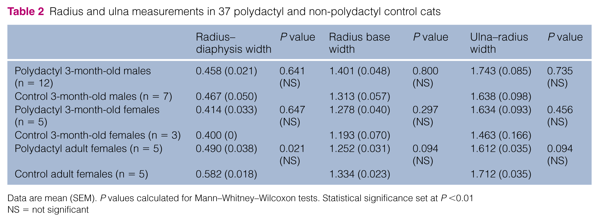
Data are mean (SEM). P values calculated for Mann–Whitney–Wilcoxon tests. Statistical significance set at P <0.01
NS = not significant
Discussion
In humans and mice, polydactyly may occur in isolation, or as part of a syndrome.1,6 In cats, polydactyly is often isolated,3,5,7,8 but some cases of feline radial hemimelia (radial agenesis) with polydactyly have been described.9,10 Cats affected by hemimelia are sometimes called ‘twisty cats’ by breeders. In mice, radial or tibial hemimelia associated with polydactyly has been described in several mutants: the Alx1 and Alx4 (aristaless-like homeobox 1 and 4) knockout mice, the dominant hemimelia spontaneous mutant, Gli3 (glioma-associated oncogene family zinc finger 3), Wnt7a (wingless-type MMTV integration site family 7A) and Shh (sonic hedgehog) mutant mice. 6 In cats, isolated polydactyly has been described in outbred cats, especially in the UK and in the USA,3,4,5,11 and in the Maine Coon and Pixie Bob breeds. The Pixie Bob breed was founded in the USA during the 1980s from a cross with a domestic shorthair female named Pixie and a polydactyl domestic shorthair male. Pixies look like bobcats and are often polydactyl on their forelimbs with a mitten-paw phenotype (www.tica.org). The Maine Coon is a natural breed originating from North America, specifically from the state of Maine, on the east coast. It has become one of the largest breeds of domestic cats and is long haired. The generally accepted history is that the breed originated from local domestic shorthair cats crossed with long haired cats brought from overseas by seafarers in the nineteenth century or earlier. Some of these seafarer cats were polydactyl. Indeed, valued as good luck charms by sea captains, polydactyl cats were frequently found along the north-eastern coast of North America. Genetic surveys dating back to the 1970s report a high frequency of the polydactyly allele in cats from coastal cities such as Halifax, Philadelphia and Boston. 11 In France, the Maine Coon breed has become particularly popular over the past 10 years. Some French breeders import numerous cats from Canada and the US to enhance the genetic diversity of their breeding lines. Some breeders have also elected to breed polydactyl cats, although polydactyly in Maine Coon cats is not recognised in some championships (www.cfa.org; www.loof.asso.fr). According to The International Cat Association, the Maine Coon Polydactyl, as part of the Maine Coon breed group (MC/MCP), was accepted into championships as of 1 May 2015 (www.tica.org). Although polydactyly has been described in outbred domestic cats,3–5 this trait has not as yet been analysed in the Maine Coon breed.
Using radiographs of polydactyl and non-polydactyl control cats, we were able to identify a considerable diversity of polydactyly phenotypes in the Maine Coon breed. However, polydactyl cats from the same breeding line seemed to share certain morphological traits. Polydactyl cats from the Canadian line investigated often had six digits on all four paws: one extra digit on the forepaws and two extra digits on the hindpaws (16/31 cats). In addition, polydactyly between the right and left forelimbs and between the right and left hindlimbs was often symmetrical (23/31 cats) and most extra digits were complete. Polydactyl cats from US line 1 had an incomplete extra digit (3/6 cats) or a complete one (3/6 cats) on their forepaws, and half of them had an incomplete digit on their hindpaws (3/6 cats). Incomplete extra digits on the forepaw were often fused with dewclaws (forked metacarpal bones). Polydactyly in US line 2 cats was heterogeneous, with many incomplete digits (5/9 cats) and asymmetry between the right and left limbs (4/9 cats). Polydactyly in Maine Coon cats also affected carpal and tarsal conformation. The common changes identified included loss of the radial sesamoid bone (ie, abductor pollicis longus muscle sesamoid bone), extra carpal and tarsal bones and fusion between bones. Finally, no difference in radius bone size between polydactyl and non-polydactyl control cats could be found at 3 months of age in either sex or in adult females (males were not analysed), which implies that there is no bony evidence to support the common affirmation by breeders that polydactyl cats have larger limbs than non-polydactyl cats.
Lameness was detected in only one out of 48 polydactyl cats, but there was no proof that it was related to polydactyly. It can therefore be assumed that polydactyly has no impact on cat welfare.
The largest study of feline polydactyly to date was published in 1947.3,4 It described polydactyl outbred cats from a line derived from two polydactyl females born in California. Our findings in Maine Coon cats are in agreement with these earlier observations on outbred cats. Most of these outbred cats had an extra digit on their forepaws and one extra digit (24/97 cats) or two (12/97 cats) on their hindpaws. Many of them were also polydactyl on their forepaws only (22/97 cats). The conformations of extra digits were as heterogeneous as those observed in our cohort of Maine Coon cats. We detected one difference from the study of outbred cats in that 4/48 cats in our study had extra digits only on their hindpaws. This was not reported in the previous study. 4 However, these cats exhibited abnormal carpal conformation, as observed in forelimb polydactyl cats, and very long first digits of the forepaws. The author also noted that extra digits included veins, muscles and bones as observed in other digits. As an exception, the radial sesamoid bone was missing, as also observed in our Maine Coon polydactyl cats. 3
Conclusions
Polydactyly in Maine Coon cats is characterised by broad phenotypic diversity, as previously reported in outbred cats in the 1940s. Polydactyly not only affects digit number and conformation, but also carpus and tarsus conformation, with no apparent deleterious consequence on feline welfare. Additional studies involving cats aged more than 5 years will be necessary to characterise fully the possible evolution of the polydactyl phenotype during the aging process.
Supplemental Material
Click here for Supplementary Material
Table to show the breeding line, sex, age and phenotype of the 70 Maine Coon cats
Footnotes
Acknowledgements
We wish to thank the practitioners who supplied the radiographs, the radiology technicians from the Alfort, Toulouse and Nantes veterinary schools, the Maine Coon breeders and owners who contributed, and Diana Warwick and Marc Chodkiewicz for editing the manuscript.
Conflict of interest
The authors declared no potential conflicts of interest with respect to the research, authorship, and/or publication of this article.
Funding
This work was supported by the French Felinotechnic Society (Société Française de Félinotechnie [SFF]).
Supplementary material
Table to show the breeding line, sex, age and phenotype of the 70 Maine Coon cats.
References
Supplementary Material
Please find the following supplemental material available below.
For Open Access articles published under a Creative Commons License, all supplemental material carries the same license as the article it is associated with.
For non-Open Access articles published, all supplemental material carries a non-exclusive license, and permission requests for re-use of supplemental material or any part of supplemental material shall be sent directly to the copyright owner as specified in the copyright notice associated with the article.