
Abstract
Objectives
From May 2009 to January 2015, 208 Maine Coon cats presented to the Tierklinik Hollabrunn – a small animal referral and first-opinion centre – and 17 (8.17%) cats were diagnosed with a slipped capital femoral epiphysis (SCFE). Over the same time period, 29 (0.67%) of 4348 cats (all breeds) were diagnosed with SCFE.
Methods
Clinical and orthopaedic examinations and diagnostic imaging were performed on all affected Maine Coons. Age at first presentation, sex, body weight, body condition score (BCS), unilateral or bilateral manifestation of the disease, activity level and duration of lameness, age at neutering and known family history of disease were recorded. Sixteen of 17 Maine Coons were surgically treated. Surgically removed femoral tissue samples were histologically examined in 13 cases.
Results
The mean age at first presentation was 21.47 months; male to female ratio was 16:1; mean body weight was 7.5 kg (range 5.3–9.3 kg); and mean BCS was 5.06/9.0. Seven cats were bilaterally affected; the median duration of decreased activity level and lameness was 2 weeks; mean age at neutering was 7.7 months (range 3.0–12.0 months); and four cats were littermates. Fourteen femoral head and neck ostectomies, eight total hip replacements and one primary fixation were performed. All 13 histologically available samples confirmed the diagnosis of SCFE.
Conclusions and relevance
To date, SCFE has been reported only occasionally in Maine Coon cats. However, the results of this study showed that Maine Coons were approximately 12-fold more likely to develop SCFE than the overall population of cats presenting to the Tierklinik Hollabrunn over the same time period. Male sex, neutering, delayed physeal closure and breed-specific high body weight may play an important role in the pathogenesis of SCFE in Maine Coon cats.
Introduction
Slipped capital femoral epiphysis (SCFE), also referred to as feline capital physeal dysplasia syndrome and spontaneous femoral capital physeal fracture, is a unilateral or bilateral progressive displacement of the capital femoral epiphysis from the proximal femoral metaphysis through the growth plate, without a history of significant trauma.1–3 SCFE is differentiated from the traumatically induced Salter–Harris type I fracture of the femoral capital epiphysis. 4 According to Queen et al, 3 the radiological diagnosis of SCFE is based on typical signs that include radiolucent areas within the proximal femoral metaphysis consistent with bone necrosis and bone loss. Physeal widening is considered one of the first radiological signs of SCFE. 4 As the disease progresses, the dysplastic physis is no longer able to resist mechanical stress, which results in femoral neck displacement and separation. Other changes commonly seen include metaphyseal bone remodelling and metaphyseal and epiphyseal sclerosis.1–3,5 Histologically, SCFE is characterised by widening of the physeal cartilage and the appearance of irregular chondrocyte clusters in an abundant extracellular matrix on the metaphyseal and epiphyseal sides of the physis. 2 In contrast, samples of patients with a Salter–Harris type I fracture exhibit a linear arrangement of chondrocytes on both sides of the physis. 6 Various factors, including genetics, obesity, endocrine imbalances, neutering and sex, are believed to play a role in the development of the disease.1–3 SCFE is mainly reported in Siamese and domestic shorthair cats and only sporadically in other breeds such as domestic longhair cats, Siamese cross, British Blue, Birman and Colour Point cats.2,3,5,7,8 Mature, neutered, overweight male cats are primarily affected.1–3 In Maine Coons, this condition has only been reported in the literature in four cases so far.1,9
The aims of this study were to examine a possible over-representation for SCFE in Maine Coon cats in the study population, to detect possible risk factors and to compare our findings with the results of previous studies.1–3
Materials and methods
Animals
For this study, all records of Maine Coons with radiological signs of SCFE in the absence of a history of trauma that presented to the Tierklinik Hollabrunn from May 2009 to January 2015 were reviewed. For each of the Maine Coon cats, age at presentation, sex, body weight, body condition score (BCS) 10 and unilateral or bilateral manifestation of the disease were recorded.
To rule in or out a possible breed predisposition for SCFE in Maine Coon cats, all cats with no history of trauma and radiographic signs of SCFE were also recorded. The percentage of Maine Coons affected by SCFE in the Maine Coon population was compared with the percentage of overall SCFE-affected cats in the overall cat population that presented to the hospital during the study period.
History and preoperative examinations
The following data were retrospectively collected from the owners: age at neutering, activity level, duration of lameness and known family history of disease.
A full physical examination, as well as an orthopaedic and neurological examination, was performed in all cats with SCFE.
Lameness scoring was based on a five-point scale: 0 = no detectable lameness, 1 = mild weightbearing lameness, 2 = moderate weightbearing lameness, 3 = marked weightbearing lameness and 4 = non-weight-bearing lameness.
Pain scoring was based on a four-point scale: 0 = no signs of pain during manipulation, 1 = mild pain (eg, cat turns head towards manipulated site), 2 = moderate pain (eg, cat pulls limb away during manipulation) and 3 = severe pain (eg, cat vocalises or is aggressive during manipulation).
Radiographic assessment
Pelvic radiographs of the cats were taken in a ventrodorsal direction with fully extended hips and lateral open leg views (Figure 1). In some cases, frog leg views were also taken to assess the physeal widening and femoral neck displacement (Figure 2). This view depicts the hips with less tension on the joint capsule, thus allowing a confirmation of metaphyseal separation. 11

Pelvic radiograph in a ventrodorsal direction with fully extended hips (cat 10). Note the more prominent osteolysis of the left femoral neck

Pelvic radiograph in a frog-leg view (cat 10). There is bilateral physeal widening without (right) and with (left) minimal metaphyseal displacement
In cats with confirmed SCFE on one side with clinically or radiographically suspicious contralateral findings, magnetic resonance imaging (MRI) (n = 1) or repeat radiographs (n = 3) were performed.
To determine the closure of the growth plates in the proximal femur, radiographs of the distal femur and proximal tibia were taken in a craniocaudal and mediolateral direction. One DECVS (GS) and one RECVS (DB) independently investigated radiographs of the distal femur and proximal tibia in the affected Maine Coons. If there was a radiolucent line visible within the physis in craniocaudal and/or mediolateral views the physis was scored as open. The third person (MB) compared the scores of both investigators and found no difference between them. The age of cats with open physes was compared with the age of cats with physiologically normal physeal closure. 12
Surgery
According to the financial ability of the owners, either a femoral head and neck ostectomy (FHNO) or a total hip replacement was performed. In one cat with the clinical suspicion of SCFE, absence of femoral neck osteolysis and femoral neck displacement on radiographs, but MRI findings indicative for SCFE, two Kirschner wires were placed to stabilise the epiphysis.
Histopathological examination
For histological examination, tissue samples obtained during ostectomies were fixed in 4% buffered formalin and decalcified as long as necessary by Decal (Quartett Immunodiagnostika und Biotechnologie Vertriebs GmbH), embedded in paraffin wax, sectioned and stained with haematoxylin and eosin. Histological samples of the femoral neck from an age-matched control cat, which had died for reasons unrelated to this study, were used for comparison.
Results
Animals
Seventeen SCFE-affected Maine Coons (16 castrated males, one spayed female [male to female ratio 16:1]) were included in this study. Mean ± SD age at first presentation was 21.47 ± 3.13 months (range 17.00–27.00 months). Mean ± SD body weight was 7.50 ± 0.45 kg (median 7 kg; range 5.3–9.3 kg). BCS in four cats was 4/9, in eight cats it was 5/9, in four cats it was 6/9 and in one cat it was 7/9 (mean ± SD BCS 5.06 ± 0.78/9.00) (Table 1). Fourteen right and 10 left femora were affected. Seven of the 17 Maine Coons (41.18%) were affected bilaterally. Overall, 29 cats were diagnosed with SCFE during the study period, irrespective of breed, including 17 (58.62%) Maine Coon cats. The remaining SCFE cats included six domestic shorthair cats, four British Shorthairs, one Persian and one British Longhair cat. A total of 208 (4.78%) Maine Coon cats presented to the hospital in the overall population of 4348 cats of different breeds.
Cat characteristics
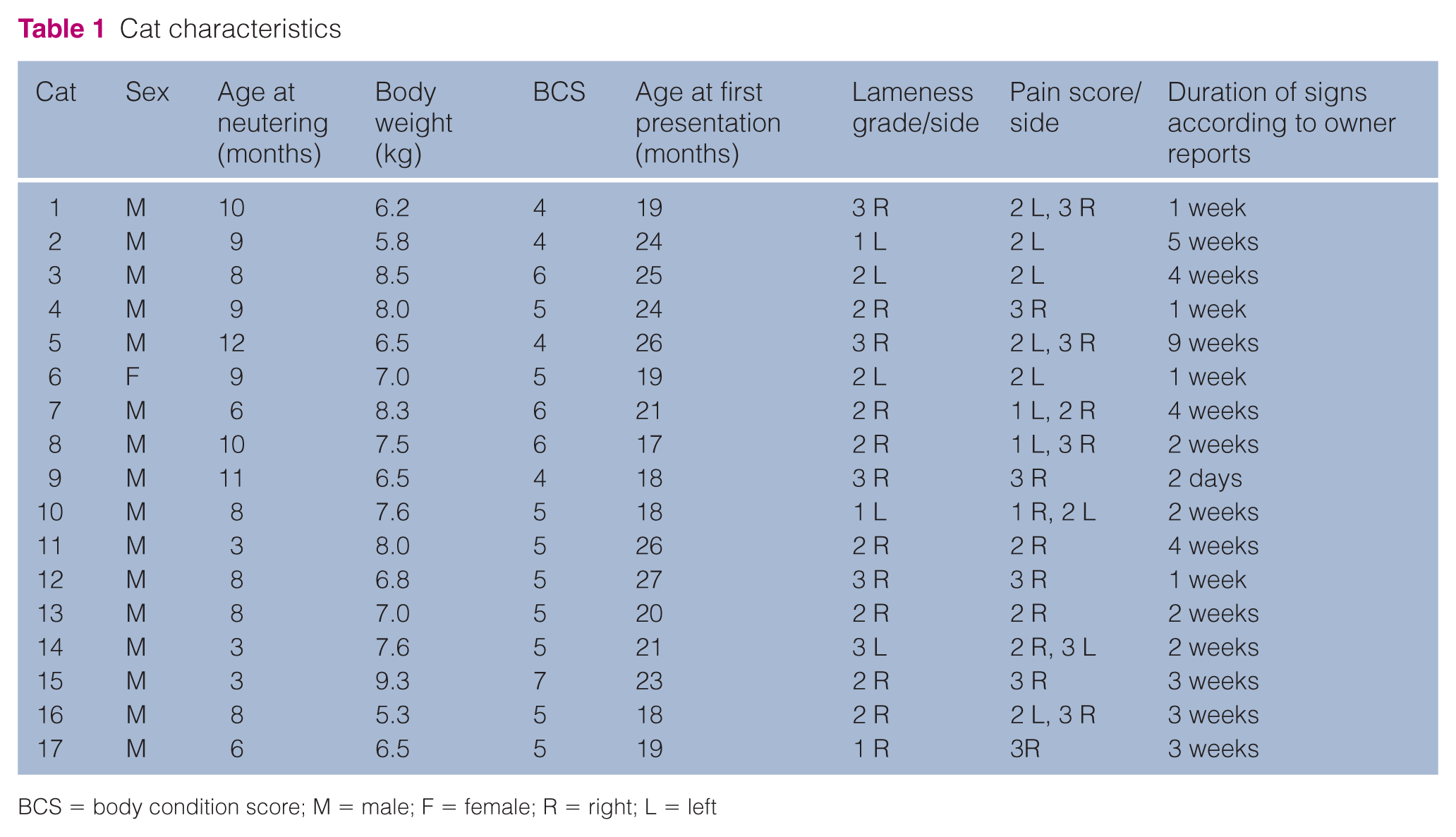
BCS = body condition score; M = male; F = female; R = right; L = left
Seventeen out of the 208 (8.17%) Maine Coons were affected and represented 58.62% of all cats diagnosed with SCFE. This indicates a more than 12-fold greater incidence relative to the 0.67% incidence of SCFE in the general clinic population (29/4348).
History and preoperative examination
Neutering of all Maine Coons with SCFE had been performed on the cats at a mean ± SD age of 7.70 ± 2.55 months (median 8 months, range 3.0–12.0 months). Five (29.41%) out of 17 cats were neutered before sexual maturation (prior to 7 months) (Table 1). 13
Cats 4 and 6 and cats 14 and 15 were from the same litter, and a littermate of cat 7 was diagnosed with SCFE in another clinic. None of the cats exhibited a history of noteworthy trauma. The owners reported decreased mobility or lameness between 2 days (cat 9) and 9 weeks (cat 5) (median 2 weeks) prior to presentation (Table 1).
Two cats exhibited grade 1 hindlimb lameness, and nine and six cats exhibited grade 2 and grade 3 hindlimb lameness, respectively (Table 1). All cats exhibited pain upon manipulation of the affected hip joint (Table 1), especially with full extension and abduction.
Neurological examination was unremarkable in all cats.
Radiographic assessment
Nineteen femora of 17 cats exhibited different severities of femoral neck osteolysis and sclerosis, epiphyseal sclerosis and obvious metaphyseal displacement in the craniolateral direction as previously described.1,3 In cat 10, physeal widening was visible bilaterally in the frog leg position, and minimal metaphyseal displacement was visible on the left side; therefore, an MRI was performed, which confirmed early SCFE on the right side. The owners of three clinically suspicious cases (cat 8L, cat 7L, and cat 14R [Figure 3]), with SCFE already confirmed on the contralateral side, refused MRI examination for financial reasons. When repeat radiographs at 4 weeks (cat 8L), 5 weeks (cat 14R [Figure 4]) and 6 months (cat 7L: in another clinic) were performed, the diagnosis of contralateral SCFE in all three patients was confirmed.

Cat 14 at first presentation with hindlimb lameness on the left side and bilaterally painful hips. Note the mild osteolysis on the dorsal side of the right femoral neck. There is prominent osteolysis and sclerosis of the left femoral neck with separation and metaphyseal displacement

Cat 14 at 35 days after first presentation and total hip replacement (CFX Micro Hip; BIOMEDTRIX) on the left side. There is osteolysis and sclerosis development with separation and metaphyseal displacement on the right side
Physeal closure of the distal femur and proximal tibia was evident in four (23.53%) Maine Coons (2, 3, 5 and 11) with a median age of 25.5 months (range 24.0–26.0 months). Physeal closure was not completed in 13 (76.47%) cats (1, 4, 6–10, 12–17) with a median age of 19 months (range 17–27 months).
Surgery
FHNO was performed in 14/24 affected Maine Coon femora (two in other small animal clinics). Total hip replacement was performed in 8/24 femora. In one case, with minimal femoral neck osteolysis and no femoral neck displacement, the epiphysis was stabilised with two Kirschner wires. One unilaterally affected Maine Coon was lost to follow-up because the owner refused surgical treatment.
Histological examination
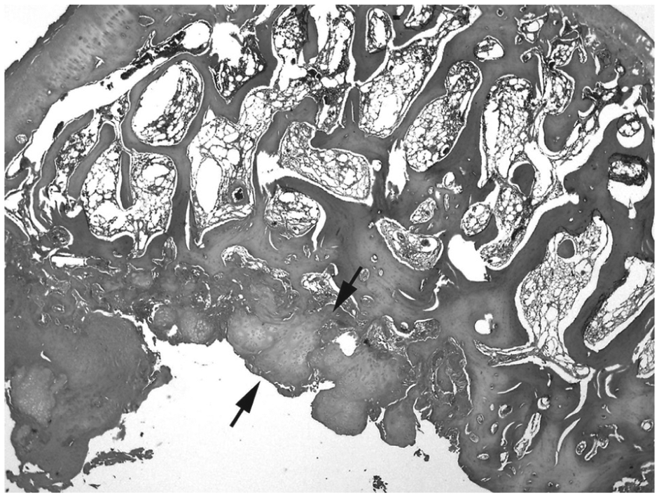
Histological examinations of 13 excised femoral heads or their fragments from 11 Maine Coon cats were available for review. The specimens revealed a broad zone of hypertrophic, irregularly arranged, sometimes necrotic, cartilage at the site of the capital femoral epiphysis (Figure 5). Chondrocytes were separated by an abundant matrix and typically arranged in clusters (Figure 6). The physis of all cases was thickened relative to that of an age-matched control cat (Figure 7). In some cases, discrete focal haemorrhage was evident. Additionally, in three cases (cats 10, 11 and 14) in which the femoral necks were available for histological examination (Figure 8), necrotic areas at the dorsal femoral neck region were clearly visible. These areas were filled with proliferating fibroblasts or granulation tissue. Mild inflammatory reactive infiltrates and fibrin exudation were noted in two other cases.
Slipped femoral capital epiphysis with hypertrophic cartilage (arrows) at the cleavage (cat 5). Haematoxylin and eosin stain (×12)

Clusters of irregularly arranged chondrocytes embedded in abundant extracellular matrix (cat 5). Haematoxylin and eosin stain (×100)

Femoral capital epiphysis of an age-matched control cat with closed growth plate. Haematoxylin and eosin stain (×12)

Extensive osteolytic process at the dorsal femoral neck region with some osteoclasts (arrows) (cat 5). Haematoxylin and eosin stain (× 200)
Discussion
Although the proximal femoral metaphysis is indeed displacing from capital femoral epiphysis, ‘slipped capital femoral epiphysis’ is still the most used term regarding this disease in cats.2,4,7,9
One aim of this study was to evaluate a possible breed predisposition for SCFE in Maine Coon cats. In the study period of May 2009 to January 2015, Maine Coons comprised 4.78% of the overall cat population presenting to the Tierklinik Hollabrunn, and 58.62% of the cats diagnosed with SCFE. A comparison of the 8.17% incidence of SCFE in Maine Coons with the 0.67% incidence in the general population indicates a more than 12-fold higher incidence in Maine Coons relative to the overall cat population presenting to the clinic in the same time period. Because of the relatively small number of SCFE cases in other breeds compared with the 17 Maine Coons in this study, the disposition of other breeds to SCFE was not investigated further.
Previous reports of SCFE in Maine coons have been published for four cats.1,9 To investigate possible risk factors for SCFE in Maine Coons, the results of this study were compared with the two published studies concerning Maine Coon cats,1,9 as well as with all other studies describing SCFE in different breeds of cat.2,3 At first presentation, the median age of the Maine Coon cats of the study population was 21 months, which correlates with previous studies describing SCFE in Maine Coons.1,9 The male to female ratio in our group was 16:1. Previously,1,9 SCFE in Maine Coons has only been described in males. Studies involving different breeds of cats reported a significant over-representation of males, but females were also occasionally affected.1–3,8 According to body weight documentation in over 460 Maine Coons, 14 male cats generally gain higher body weights than females. With respect to SCFE in humans, 15 overweight boys at an average age of 13 years are predominantly affected. African Americans and Pacific Islanders also exhibit a higher risk of developing SCFE. Relative femoral retroversion,16–19 increased physeal slope, 20 and growth, sex and thyroid hormone imbalances are also considered risk factors in humans. 21 Heritability has previously been suggested as a possible risk factor in humans,22,23 as well as in cats. Four cats in this study were littermates. Further investigations, especially in collaboration with breeders, could provide more information about possible predisposing factors for SCFE.
Physeal closure of the distal femur and the proximal tibia in cats occurs at 54–76 weeks and 50–76 weeks of age, respectively. 12 In our study, radiographically incomplete physeal closure of the distal femur and proximal tibia was observed in 13/17 Maine Coons (76.47%). The median age of these patients was 19 months (range 17–27 months), suggesting that growth in the Maine Coons in our study was prolonged relative to those cats in the aforementioned study. 12 Further studies are necessary to investigate the factors contributing to delayed physeal closure in Maine Coons and to clarify if SCFE is part of a generalised growth disturbance or if the disease is exclusively limited to the physis of the proximal femur. Delayed physeal closure was also recorded in previous reports regarding SCFE.1,3,8 We are not aware of reports specifically defining the time point of physiological physeal closure in Maine Coons. A breed-specific delayed physeal closure may suggest a breed predisposition for SCFE in Maine Coon cats.
All four Maine Coons in the previous two studies,1,9 as well as the Maine Coons in this study, were castrated or spayed. Five of 17 cats (29.41%) were neutered early, prior to 7 months of age. 13 Previous studies have suggested that early neutering may be one of the predisposing factors for delayed physeal closure.1,2,7 Root and Olson did not find any difference in physeal closure between cats neutered at 7 weeks or at 7 months of age. 13 However, the same study reported that gonadectomy retards radial physeal closure and significantly increases radial length compared with intact female and male cats. 13 Another recent study reported that physeal closure occurs later in neutered than in intact males for the greater trochanter, distal femur and tibial tuberosity. However, no effect of neutering has been demonstrated for the proximal tibial or the proximal femoral physes. 24 None of these studies indicates delayed physeal closure of the proximal femur related to gonadectomy. The results of this study, which included only Maine Coons, raises questions concerning the effect of delayed physeal closure of other bones on overall growth and body weight, thus potentiating the incidence of SCFE.
The median body weight of cats in our study was 7 kg, which correlates well with the documented average body weight in Maine Coons, according to Diehl. 14 In contrast to previous studies,1,2,8 there were only three slightly (BCS 6/9) and one mildly overweight (BCS 7/9) cats out of 17 Maine Coons in this study (median BCS 5/9). This finding suggests that obesity alone may not contribute to SCFE in Maine Coon cats, but the comparatively high body weight and mechanical overload could promote the development of SCFE.
Fourteen right femora and 10 left femora were affected. According to the small number of patients it is not possible to confirm a side predilection for the right hindlimb. All cats were lame at first presentation and exhibited pain upon femoral abduction and full extension of the hip on the affected side. In this study, 41.18% of cats (7/17) were bilaterally affected. This finding is consistent with other studies, which also reported a high incidence of bilateral occurrence of SCFE.1–3,5,8All bilaterally affected cats presented with clinical signs of SCFE bilaterally, but MRI (n = 1) and repeat radiographs (n = 3) were necessary to confirm diagnosis. In cat 14, Figures 3 and 4 show development of necrosis in 35 days. Cat 7 was operated on (FHNO) after 6 months in another small animal clinic because of obvious separation (personal communication). In humans, 25 MRI is a valuable tool for the early diagnosis of SCFE but, for financial reasons, was only performed in one case in this study. Further investigation is needed to assess whether MRI can improve the early diagnosis of SCFE in cats and therefore provide better prognosis regarding primary fixation in cats as in humans.26,27 In other cases with more advanced osteolysis, we consider total hip replacement to be a better surgical option than FHNO,28,29 but, for financial reasons, more owners favour FHNO.
Histological examination confirmed SCFE in the 13 femora of the 11 Maine Coons that had been radiographically diagnosed with SCFE. In humans, SCFE is characterised by proliferative and hypertrophic zones of slipped growth plates with diminished cellularity, marked distortion of the architecture, and disarray of cartilage columns. 30 Additionally, deficiencies and abnormalities exist in the supporting collagenous framework of the slipped plates. 30 Chondrocyte degeneration and death are seen at all levels of the proliferative and hypertrophic zones, suggesting a disturbance in the life cycle of chondrocytes. 30 Similar changes have been seen in cats with SCFE. 2 These are characterised by a distinctive lesion in the physeal cartilage, consisting of irregular clusters of chondrocytes that are separated by abundant matrix on both the epiphyseal and metaphyseal sides of the cleavage site. 2 Despite the extended duration of clinical signs and progressed radiographic changes, including marked femoral neck remodelling and sclerosis of the metaphyseal an epiphyseal region, histological examination showed no difference concerning the characteristic histological appearance of SCFE in cat 5. Results of our histological examinations from the 13 femora are identical to those findings previously reported for cats.1,2
One of the limitations of this study is the small number of patients. Lameness examination was only performed visually and is therefore less objective then force plate and gait analysis. 31 However, force plate gait analysis is not a well established method for diagnosing lameness in cats. Finally, owing to financial limitations, not all excised femoral heads and necks could be histologically evaluated.
Conclusions
The results of this study indicate that Maine Coons were approximately 12-fold more likely to develop SCFE than the overall cat population that presented to the Tierklinik Hollabrunn from 2009 to 2015. Sex (male), neutering, delayed physeal closure and breed-specific high body weight may play important roles in the pathogenesis of SCFE in Maine Coon cats. Whether early neutering and heritability contribute to SCFE remains undetermined. In general, SCFE should be considered an important differential diagnosis in male Maine Coons aged between 1.0 and 2.5 years that exhibit hindlimb lameness and a reduced activity level.
Footnotes
Acknowledgements
We thank Karin Fragner, Klaus Bittermann and Tijana Borak for providing technical assistance.
Conflict of interest
The authors declared no potential conflicts of interest with respect to the research, authorship, and/or publication of this article.
Funding
This research received no specific grant from any funding agency in the public, commercial or not-for-profit sectors.