
Abstract
Objectives
We aimed to (1) evaluate how frequently the uterus and ovaries of healthy, non-pregnant queens are visible; (2) describe their appearance; (3) take their measurements; and (4) determine intra- and inter-observer variabilities in their measurements. We hypothesised that, using a high-frequency linear probe, the uterus and ovaries could be ultrasonographically visualised during any period of the sexual cycle and with any level of operator expertise.
Methods
Eight queens were enrolled in the study and the ultrasonographical appearance of their uterus and ovaries assessed with a high-frequency linear probe of 15–19 MHz. The diameter of the uterine horns, body and cervix in transverse and longitudinal sections, and the length of the ovaries were recorded. Three observers of different expertise level participated in the study, and the differences between the separate measurements made per queen were evaluated.
Results
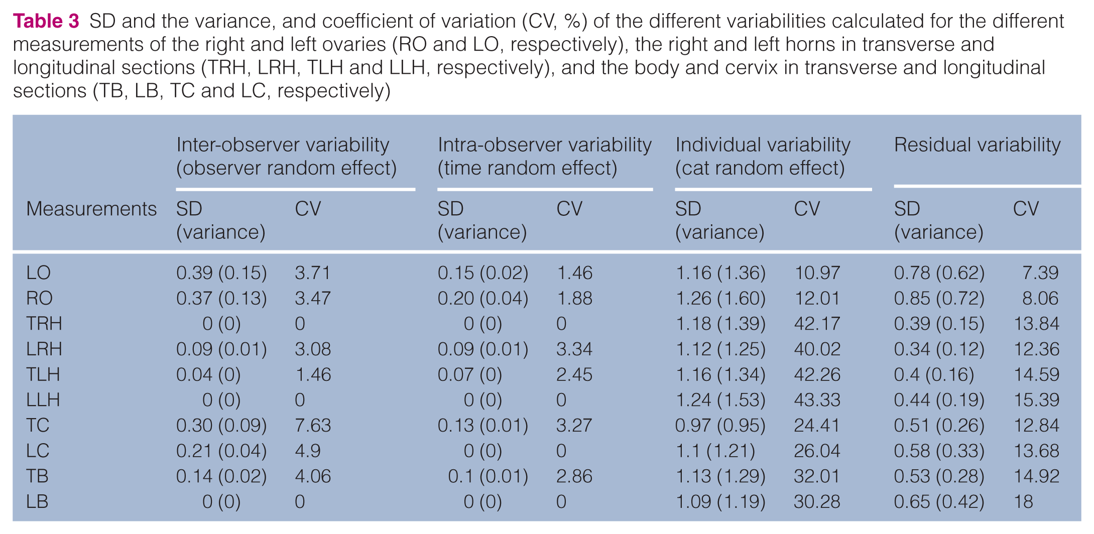
The ovaries and the entire uterus were visualised in every queen. The ovaries were ovoid structures with submillimetric follicles during anoestrus and additional larger follicles depending on the stage of the cycle. An ovarian pattern suggesting cortex and medulla was observed in half the cases. In the uterus, the serosa was a thin hyperechoic outer rim, and layering was observed in half the cases. The cervix was difficult to identify. The intra- and inter-observer variabilities in the uterine horns and the ovaries were minimal (coefficient of variation [CV] 1.4–4.1%) compared with the differences within the queens (CV 10.9–43.4%). The longitudinal and transverse measurements of the horns and the uterine body were the same.
Conclusions and relevance
The ovaries and uterine horns in queens are accessible ultrasonographically at any stage of their cycle, and can be measured with low intra- and inter-observer variabilities.
Introduction
Ultrasound (US) imaging of the feline genital tract has been poorly reported in the literature. Pregnancy diagnosis, prediction of gestational age and parturition time, prediction of kittens’ birth weight, postpartum involvement and some pathological conditions have been described.1–12 The normal, non-gravid uterus in cats is usually described as not visible; 13 to our knowledge, the normal ultrasonographical anatomy of the uterus in queens has not been described. In one study of uterine width in gross specimens, no statistical differences were found between the normal group of queens, including queens in anoestrus, oestrus and within 2 months postpartum (mean = 0.39 cm, 0.56 cm and 0.46 cm, respectively), whereas the pathological group had a significantly higher measurement (0.88 cm). 14 However, neither the values of the ultrasonographical measurements of the uterus, nor the reliability, repeatability and variability of these measurements in healthy queens is known.
Ultrasonographical descriptions of the ovaries are also poorly documented. A recent study that described the follicular dynamics by transabdominal ultrasonography of the anovulatory and mechanically induced ovulatory cycles demonstrated that ovarian ultrasonography via the transabdominal route is a safe, reliable, non-invasive technique for monitoring follicular growth in female cats. 15 However, ovarian ultrasonography required high-resolution equipment, a cooperative queen and an experienced operator. Neither the appearance nor the length of the ovaries in a non-oestrous period is known on US. The gross lengths of ovaries reported in anatomy textbooks range from 8–10 mm.16,17
According to published reports, measurements can be divided into two parameters: actual value and noise.18–20 To evaluate the performance of a technique, the noise must be divided into several components as inter-observer, intra-observer and inter-individual variability, as well as some residual variation. Ultrasonography is highly dependent on the observer and measurements can differ according to biological variations.
This study aimed to visualise, describe and measure the uterus and ovaries in non-gravid queens, and to evaluate the frequency of the observations. The variability of the intra- and inter-observers’ measurements were determined.
Materials and methods
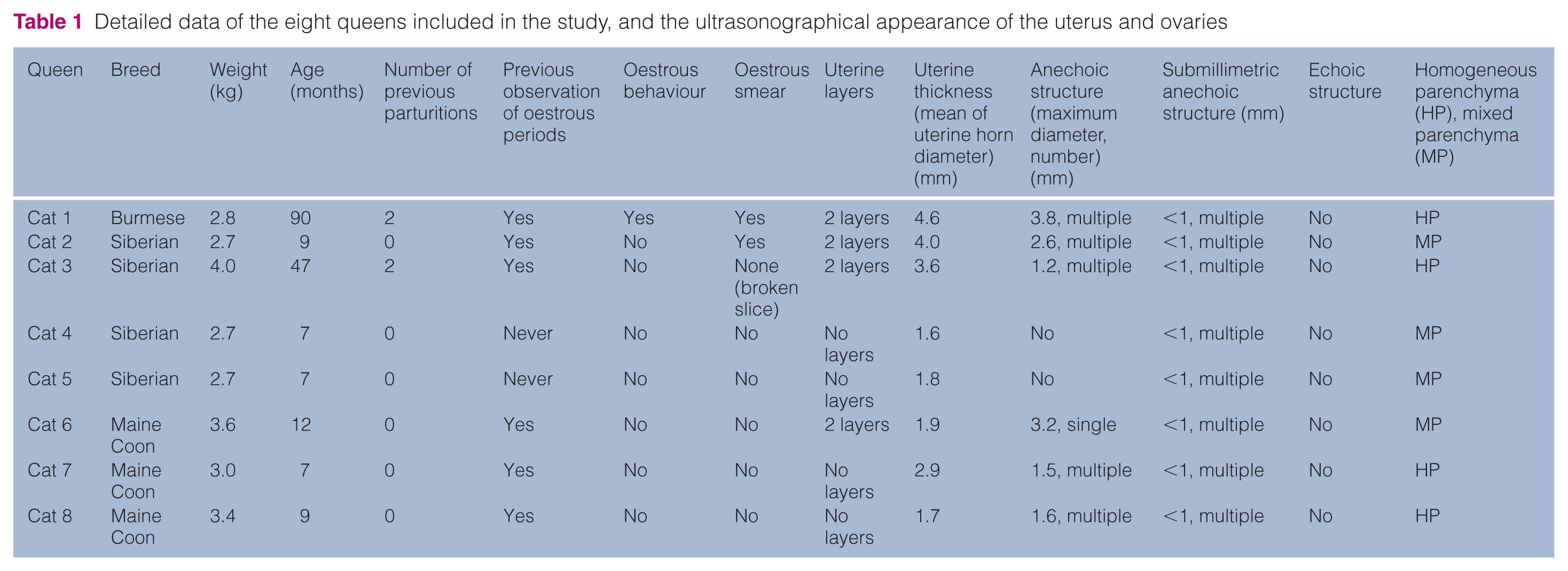
Eight intact, non-pregnant queens (age 7–90 months; weight 2.7–4.0 kg) were enrolled in this study (Table 1): Burmese (n = 1); Siberians (n = 4); Maine Coons (n = 3). Two queens had previously been pregnant, but not in the year prior to the study. No previous uterine diseases or infertility had been recorded. The queens were clinically healthy, as determined by the absence of clinical signs and a normal clinical examination. The sexual behaviour observed by the owner was recorded. No blood tests were performed. A vaginal cytology was performed in all of the queens and coloured with a Diff-Quik method to stage the oestrous cycle.
Detailed data of the eight queens included in the study, and the ultrasonographical appearance of the uterus and ovaries
Hair from the abdominal wall was clipped and coupling gel applied. The queens were scanned in dorsal recumbency without sedation for no longer than 7 mins per examination. Three observers were enrolled in this study: an ECVDI diplomate (obs 1) with queen genital US experience; a second-year ECVDI resident (obs 2); and an intern in medical imaging (obs 3). All used the same machine settings and a high-frequency linear probe of 15–19 MHz (Aplio400; Toshiba). The ultrasonographical examinations were performed on three consecutive days, with three queens scanned the first and third days, and two queens the second day. The ultrasonographical examinations of the same queen by the same observer were performed at least 15 mins apart. All queens were examined by US three times per operator, giving a total of nine examinations per queen.
The entire uterine horn was scanned to ensure that its diameter and appearance were similar throughout. The observers evaluated the presence or absence of layering within the uterine horns and body. The presence of round anechoic structures, their largest diameter and heterogeneities of the parenchyma were recorded for the ovaries.
Measurements were made on frozen images. For this study, we considered the uterine horns as tubular in shape. Longitudinal and transverse sections of the two horns were scanned halfway between the ovary and the body of the uterus, and measurements of the horn (diameters of the right horn in transverse section, the right horn in longitudinal section, the left horn in transverse section and the left horn in longitudinal section) were taken. Then, longitudinal and transverse sections of the body and cervix of the uterus were measured (diameters of the body in transverse section, the body in longitudinal section, the cervix in transverse section and the cervix in longitudinal section). Longitudinal images of the ovaries were taken and the maximum length recorded (lengths of the right ovary and left ovary). The cursors were positioned using the edge shadowing at the cranial and caudal poles of the ovaries.
A software program (R version 2.10.0; R Foundation for Statistical Computing) and a linear mixed-effects model were used to perform the analysis. 20 The time (scan periods 1, 2 or 3 for the same observer), observers (obs 1, obs 2 and obs 3) and cats were considered random effects. The SD of time, observer and cat were calculated to appreciate the intra-observer, inter-observer and individual variability, respectively. The coefficient variation (SD divided by measurement mean) was also calculated. Transverse and longitudinal measurements of the horns, body of the uterus and the cervix were compared by the paired Student’s t-test.
Results
On cytology, oestrus was recognised when a clear background, a mild amount of cellular debris and a majority of superficial cells were observed.14,21 Two vaginal cytological examinations were compatible with oestrus (cats 1 and 2) (Table 1). Cat 1 was presented with oestrous behaviour and cat 2 had a silent oestrus. In cats 4 and 5, which had poor cellular smears, oestrous behaviours were never observed, most likely because they were young and prepubescent. The other five queens, which had smears with moderate cellularity composed of a mixed cell population, were neither in oestrus nor anoestrus, so an interestrous period was suspected.
All three observers scanned the genital tract from caudal to cranial, starting from the uterine body between the urinary bladder and the colon in the caudal abdomen. They found the ovaries by following the uterine horns or at the caudal aspect of the kidneys. They all had difficulty recognising the cervix because a mild enlargement of the caudal uterine body and the typical ‘bull’s eye’ aspect described in other species were absent. 13 The observers measured the cervix at the most caudal, widest part of the uterine body. They all visualised the uterine horns, although that was occasionally complicated by bowel loops, which crossed the field of view. The most experienced observer (obs 1) had no difficulty assessing the ovaries, but the other two found it more time consuming.
The uterine horns and body in all cats were similar in appearance (Table 1; Figures 1 and 2). In each case, the uterus presented a thin hyperechoic outer rim that was more clearly seen in the longitudinal scan than the transverse. Two layers were distinguished in 4/8 queens. The observers saw no liquid in the lumen of the uterus. They did see a thin hyperechoic line at the level of the virtual lumen. Submillimetric anechoic structures could be appreciated in all the ovaries (Figure 3). Supramillimetric anechoic cavities were also detected in 6/8 queens but not in the two (cats 4 and 5) that were suspected to be prepubescent cats. Cats 1 and 2, which were in oestrus, had multiple cavities >2 mm. Cats 3, 7 and 8, which were suspected of proestrus based on vaginal smears, had multiple cavities <2 mm. Cat 6 had a single anechoic large cavity of 3.2 mm, which, based on the vaginal smear, was considered likely to be a cyst. In seven cats, the follicles were distributed subjectively closer to the ovary’s periphery, while in cat 1, the large follicles occupied the major part of the ovaries. In cats 2, 4, 5 and 6, the central section of the ovary (without anechoic structures) appeared more hyperechoic. The measurements of both uterine horns and the body (P >0.05) showed no significant difference in the transverse or longitudinal sections; however, the longitudinal and transverse diameters of the cervix (P <0.05) did show a significant difference (Tables 2 and 3). The variability between individual queens was highest. The intra-observer variability was lower than the inter-observer. Both were less significant than the individual variability and the residual effect. No inter- and intra-observer variability was observed for the uterine horn, uterine body and ovary measurements.

Longitudinal (a, a’) and transverse (b, b’) planes of the left uterine horn in cat 1 (Burmese, aged 7.5 years, multiparous). Layers can be seen. The diameter of the uterus is 5.3 mm. The serosa appears as a thin hyperechoic line (represented by a solid line on [a’] and [b’]). Two other layers are detected corresponding to the endometrium and the myometrium (separated by a dashed line on [a’] and [b’]). On the longitudinal plane, a thin hyperechoic line (dotted line on [a’]), is detected and corresponds to the lumen of the uterine horn
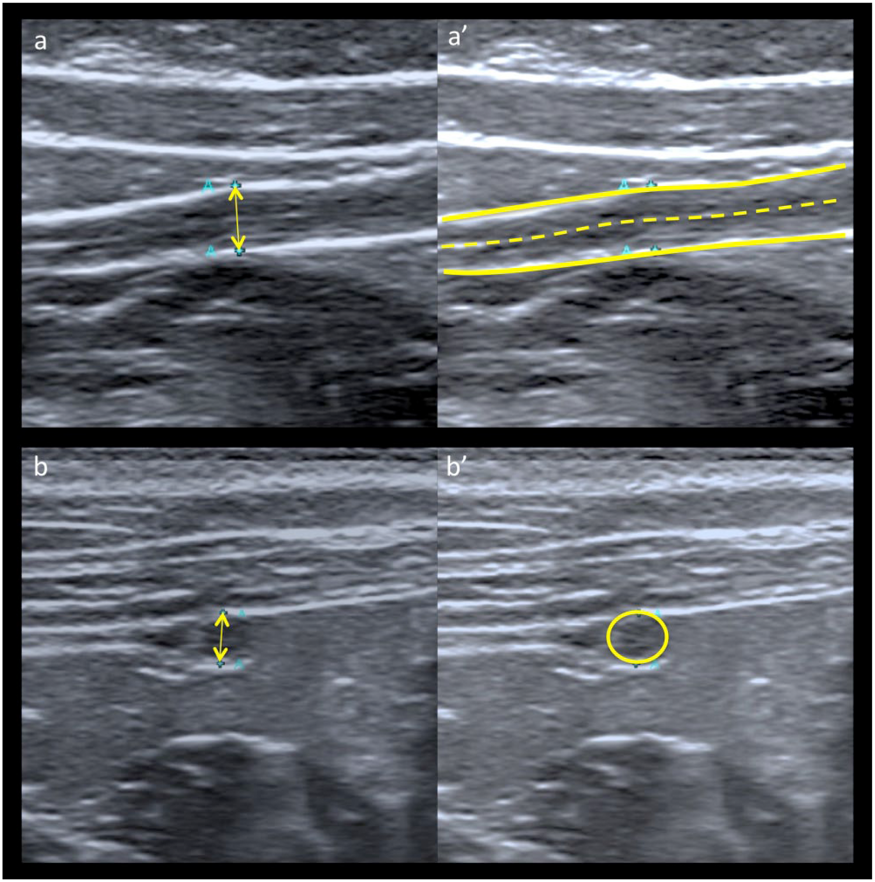
Longitudinal (a, a’) and transverse (b, b’) planes of the left uterine horn of cat 5 (Siberian, aged 7 months, nulliparous). No layers can be detected. The diameter of the uterus is 1.9 mm. The serosa appears as a thin hyperechoic line (represented by a solid line on [a’] and [b’]). On the longitudinal plane, a thin hyperechoic line (dashed line on [a’]) is detected and corresponds to the lumen of the uterine horn

Appearance of the ovaries. The outline of the ovary is highlighted by a solid line (a’, b’, c’, d’). (a, a’) Long-axis images of the left ovary of cat 5, which was prepubescent. The ovary measures 9.6 mm. Submillimetric follicles only can be detected (arrows on [a’]). The ovary presented a mixed parenchyma with a hyperechoic medulla (surrounded by a dashed line on [a’]) compared with the cortex. (b, b’) Long-axis images of the right ovary of cat 7, which was considered to be in proestrus. The ovary measures 9.9 mm. Infra- and supramillimetric follicles are visualised (<2 mm) (circles and arrows on [b’]) and the parenchyma of the ovary is homogeneous. (c, c’) Long-axis images of the right ovary of cat 2, which was in oestrus. The ovary measures 11.9 mm. A submillimetric follicle can also be visualised (red dashed arrow on [c’]). Three large supramillimetric follicles are detected within a homogeneous parenchyma (arrows on [c’]) and are >2 mm in diameter. (d, d’) Long-axis images of the right ovary of cat 6. The ovary measures 12.9 mm. Submillimetric follicles can be visualised. A single large supramillimetric structure is visualised (>2 mm), which is compatible with an ovarian cyst. The ovary has a mixed parenchyma with a hyperechoic medulla (surrounding by a dashed line on [d’]) compared with the cortex
Mean ± SD ultrasound measurements (mm) of the right and left ovaries (RO and LO, respectively), the right and left horns in transverse and longitudinal sections (TRH, LRH, TLH and LLH, respectively), and the body and cervix in transverse and longitudinal sections (TB, LB, TC and LC, respectively) obtained by each observer (obs 1, obs 2 and obs 3)

SD and the variance, and coefficient of variation (CV, %) of the different variabilities calculated for the different measurements of the right and left ovaries (RO and LO, respectively), the right and left horns in transverse and longitudinal sections (TRH, LRH, TLH and LLH, respectively), and the body and cervix in transverse and longitudinal sections (TB, LB, TC and LC, respectively)
Discussion
The estimated sexual status (according to behaviour and vaginal cytology) for seven cats was consistent with the ultrasonographical appearance of the ovaries. Measurements of progesterone and oestradiol would have been valuable in determining the stage of oestrus and potentially the significance of the cystic structure in cat 6. The structure in the ovary may have been a cyst because the vaginal smear did not suggest oestrus; however, its diameter was <4.1 mm, the maximum size of a follicle in a cycling queen.15,21
Compared with a previous study conducted by Axnér on seven queens, the ovaries and uterus could be visualised in every queen and both horns were followed entirely, despite the observers’ different levels of expertise in genital US. 7 However, in the Axnér study, the transducer resolution was lower than the resolution used in the present study (8–5 MHz vs 15–19 MHz), which may explain the better visualisation.
When looking at the uterus, described in postpartum involution in the queen, bitch, gilt and woman, we always found an hyperechoic outer rim, which was considered most likely the serosa.3,22–25 Cats 1, 2, 3 and 7 showed layers in the uterine wall, which have been described during postpartum involution up to 24 days after parturition in queens and during dioestrus, pregnancy and pseudopregnancy in bitches.1,3,4 Layering of the uterus at the time of oestrus has also been observed in cows, gilts, mares, bitches and women.11,26–28 The endometrium has been described as a hypoechoic layer; in our study, the two visible layers were interpreted as the endometrium (inner) and the myometrium (middle). Layering has also been associated with a uterus with a large diameter, which may correlate with our study, where three queens were older. In human medicine, a positive correlation exists between the size of the uterus and the woman’s age.29,30 No correlation was observed between the layering and stage of oestrus in the current study. Owing to low numbers, the effect of the age of the cat was not considered in this study, and further studies are required to assess an age effect.
The hyperechoic line visible in the lumen of the uterus could be explained by the reflection of the echoes on the opposing layers of the endometrium, which lies in close apposition in the empty uterus and projects as an echogenic spot, according to a study in horses. 26
In this study, multiple submillimetric anechoic structures were observed in the ovaries. These images have been described in prepubescent girls and correspond to primordial follicles,24,31 which were not described in previous reports on the ultrasonographical appearance of the feline ovary, where the smallest structure was 2 mm in diameter during oestrus. 15 However, as mentioned previously, we used a transducer with a higher frequency that allowed a higher resolution and visualisation of smaller structures. The preferential localisation of the follicles at the ovary’s periphery is compatible with a previous histological description of queen ovaries where the follicles were located in the cortex, and the medulla contained the vasculature and nerves. 32 The relative hyperechogenicity of the medulla in four cases could not be related to age or stage of cycle, and further investigation of this is needed.
With the measurements, the values obtained by US were higher than the description on gross evaluation,16,17 which could be explained by the presence of the suspensory ligament of the ovary at the cranial pole of the ovary that could extend the length of the ovary compared with gross evaluation.
The measurements obtained of the uterine horns were compatible with the macroscopic measurements in a group of normal cats. 14 No intra- or inter-observer effects (P >0.05) were seen on the uterine diameter measurements, which suggests that the level of expertise did not produce significant variability, and thus obtaining those measurements is not highly challenging when a high-frequency linear probe is used. Also, no significant differences were observed between the measurements in longitudinal and transverse sections, so both can be used equally in practice. The high variability of the cervix measurements can be explained by the difficulty of clearly identifying the cervix. The inter- and intra-observer variabilities on the uterus were minimal compared with the variability between the queens, but the diameters were very small. The low inter- and intra-observer variability can also be explained by the consistent observation of the thin hyperechoic serosa and the position of the cursor could therefore be more reliable.
This study was limited by the absence of post-ovariohysterectomy gross examination or histologic examination, as well as the use of a high frequency linear probe, which is not commonly available in general practices. Other limitations related to the appearance of the ovaries and the uterus are the low number of queens, and the breed, age and stage of cycle variation.
Conclusions
According to this study, the ovaries and uterus in queens are accessible ultrasonographically, with the use of a high-frequency linear probe, at any stage of their cycle. Further studies are needed to determine the normal appearance and measurements of the ovaries and uterus during the entire cycle, as well as the correlation between the ultrasonographical images and the histological findings, in order to facilitate early diagnosis of abnormalities of these structures.
Footnotes
Acknowledgements
We thank Ms Zannini for her assistance, and all her marvellous cats.
Conflict of interest
The authors do not have any potential conflicts of interest to declare.
Funding
This research received no specific grant from any funding agency in the public, commercial, or not-for-profit sectors