
Abstract
Eleven pregnancies in six queens were monitored daily from day 7 to day 28, corresponding to the end of the embryonic period, using ultrasonography with a 12.5 MHz probe. The first mating was considered as the presumed start of gestation, as has been described to be the case in 92.3% of pregnancies. The embryonic vesicles were identified on day 11, while the embryo appeared on day 15 or 16. The stage of pregnancy could be evaluated approximately by measuring the length of the embryonic vesicle or the crown–rump length of the embryo from days 11 and 17, respectively, up until the end of the embryonic phase of gestation. The visualisation of certain organs could also be used to date gestation; for example, the limbs, neural tube and stomach were visible from days 19, 20 and 26, respectively. The 12.5 MHz probe did not enable the diagnosis of gestation to be performed any earlier than with 7.5 and 10 MHz probes. However, there was a significant difference in comparison with a 5 MHz probe.
Introduction
Pregnancy diagnosis in the cat has already been studied extensively. Probes with a frequency of ⩽10 MHz are usually employed. This is the first study to use a 12.5 MHz probe.
The objective was to ascertain whether an earlier diagnosis could be attained with this higher-frequency probe, and to define new landmarks for determining the precise stage of gestation during the embryonic period, that is up to day 28.
Materials and methods
The animals used for the study belonged to the feline unit of the reproductive pathology department (Table 1) of the National Veterinary School of Nantes, France. They were all fed the same diet, and were regularly vaccinated and dewormed.
Queens monitored during gestation
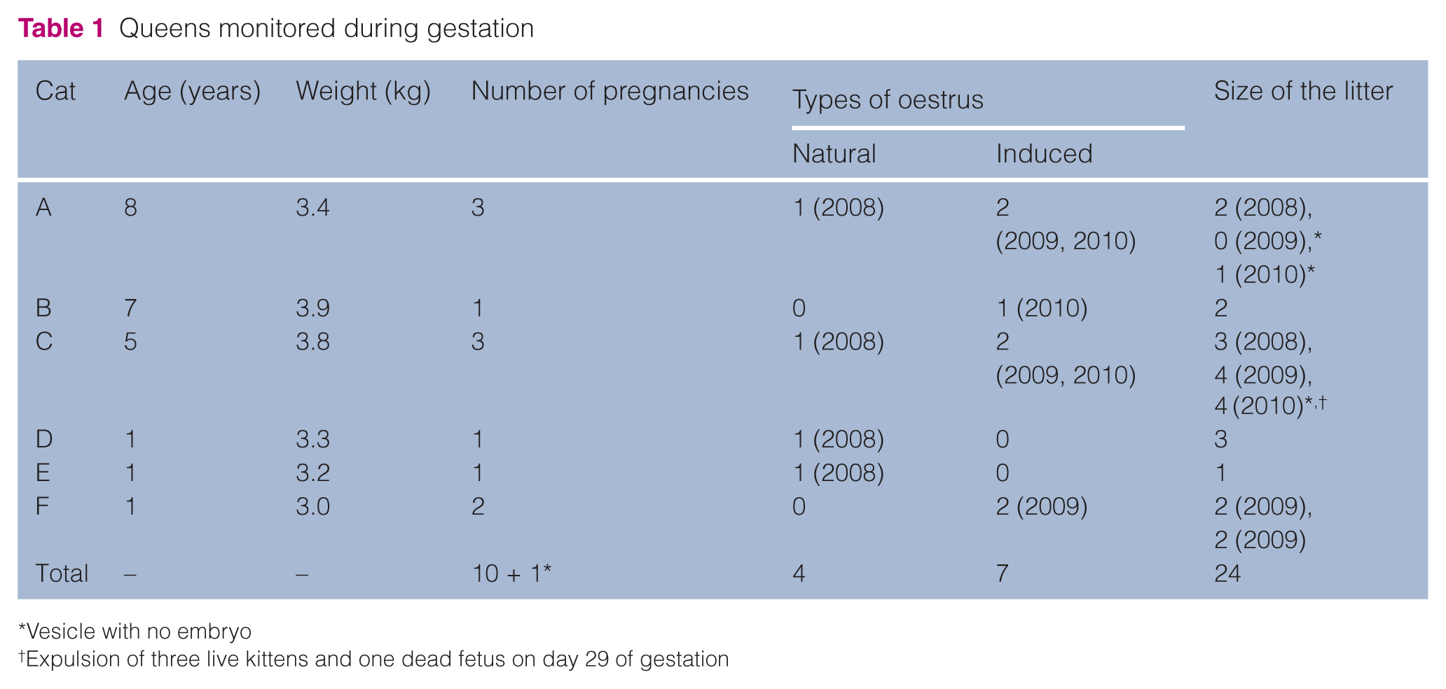
Vesicle with no embryo
Expulsion of three live kittens and one dead fetus on day 29 of gestation
The oestrous cycle of the cats in the study was followed during their reproductive cycle, both within the reproductive season from February to September, and also during the off-season. Oestrus was detected every 2 days by observing the behaviour of females in the presence of the male. Off-season, artificial lighting was provided for 14 h a day and oestrus was detected as described above after 3 weeks of artificial lighting. When a queen accepted the male, she was isolated for 48 h with the tomcat, a 6-year-old domestic shorthair cat, weighing 4.8 kg. The first mating was always observed until the end (including postmating behaviour). Four gestations followed natural oestrus and seven followed induced oestrus (artificial lighting), but during one of the latter gestations, only one empty vesicle was observed. The oldest queens were examined by ultrasound using light restraint (cats A–C), enabling three gestations to be monitored in two of them. Restraint of the youngest queens (cats D–F) was harder, which limited pregnancy monitoring (with the exception of cat F, 2) (Table 1).
We used an ‘IXOS Vet’ (Esaote, Pie Medical) ultrasound machine on a mobile trolley with a super video graphics array monitor enabling direct recording of the ultrasound images on Imagelab software. The machine was equipped with two-dimensional and time-motion modes, and colour Doppler. It enabled the image to be frozen on the screen and had a 15-s memory, which allowed replay to visualise the interesting sequence and precise selection of the best image. A linear LA 523 probe was used, with an adjustable frequency of 7.5, 10, or 12.5 MHz. Only the latter frequency was used during pregnancy examinations.
The queens were clipped over their entire ventral abdomens, from the xiphisternum to the pubis, to around 5 cm on either side of the mammary glands. Clipping was repeated over the course of gestation as necessary. The animal was usually placed in dorsal decubitus, held by two assistants, and always examined by the same experienced operator, who is a diplomate of the European College of Animal Reproduction. The examinations were occasionally performed with the queen standing if they did not tolerate being held in decubitus.
The date of the first observed mating was taken to be the start of gestation. The ultrasound examination was performed daily from day 7 of gestation until parturition. Each day, each pregnant cat was subjected to ultrasound for between 15 and 45 mins as a function of the stage of gestation and their patience. The ultrasound probe was placed in front of the pubis lateral to the mammary chain. Once the uterus was located next to the bladder, the horns were followed one after the other up to the ovaries. Two gestations could not be monitored through to parturition, as the cats did not tolerate the ultrasound examination (cats D and F, 2009). Ten gestations were monitored, with six being monitored through to parturition (cat A, 2010; cat B, 2010; cat C, 2009 and 2010; cat E, 2008; cat F, 2009 [first gestation]). The last four were monitored intermittently (cat A, 2008; cat C, 2008; cat D, 2008; cat F, 2009 [second gestation]) owing to the unavailability of operators or occasionally difficult restraint of certain queens (cats D and F).
Results
In this study, the mean duration of pregnancy was 65.2 ± 0.7 days (64–67 days).
On day 8 of gestation, one suspicious image of an embryonic vesicle was observed in one queen, which was suggestive of gestation. However, this image was not found on days 9 and 10 of gestation, whereas on day 11, the embryonic vesicle was clearly visible. It appeared as a small anechoic cavity in the lumen of the uterus, 1.2 mm (± 0.1) in diameter, next to the bladder (Figure 1). At least one embryonic vesicle was observed at this stage in all the queens confirmed to be pregnant.

Embryonic vesicle (EV) next to the bladder (B) on day 11 of gestation
The embryonic vesicle grew without changing its appearance or shape up until day 15, at which point it measured 5.3 mm (± 0.6) in diameter, on average. The vesicles observed within a same litter were of different sizes on any given date.
The embryo usually appeared on day 16 (6/10), more rarely on day 15 (2/10) or day 17 (2/10), as a hyperechoic element jutting out into the anechoic embryonic vesicle (Figure 2).

Embryo (E) in an embryonic vesicle (EV) and trophoblast (T) on day 16 of gestation
The heart was visible from day 16 of gestation (3/10), but more commonly from day 17 (6/10) and sometimes day 18 (1/10). Visualisation of the heartbeat confirmed the viability of the embryo.
From day 19 of gestation, the head could be distinguished from the rest of the body by its characteristic anechoic cavity. The fore- and hindlimb buds also appeared on day 19 of gestation (Figure 3). The head was easier to see from day 20.

Embryo on day 19 of gestation. Head (H), forelimb bud (FB), hindlimb bud (HB) and allantois (A) are visible
The neural tube and liver were observed from day 20 of gestation. The latter appeared as a hyperechoic mark just caudal to the heart, which was easy to locate by visualisation of the heartbeat (Figures 4 and 5).

Embryo and neural tube (NT) on day 20 of gestation

Embryo on day 21 of gestation. Head (Hd), heart (Ht), liver (L), allantois (A) and yolk sac (YS) are obvious
On day 22 of gestation, the embryo took on a figure-of-eight shape. The head and body were clearly demarcated and of roughly the same size. The head was almost entirely filled with fluid, which was seen on the ultrasound as a significant zone of anechogenicity (Figure 6).

Embryo on day 22 of gestation. Head (H) and body (B) have the same size
The fore- and hindlimb buds were clearly visible on day 24 of gestation. However, the development of the forelimbs seemed to be one day ahead of the hindlimbs. The tail became visible (Figure 7).

Embryo on day 24 of gestation. Forelimb (Fl), hindlimb (Hl), tail (T), liver (L), heart (Ht) and head (Hd) are visible
The stomach appeared on day 26 of gestation and became easily identifiable from day 27 of gestation (Figure 8).

Embryo on day 27 of gestation. Forelimb (Fl), head (Hd), stomach (S) and tail (T) are visible
The genital bud appeared anterior to the tail on day 27 of gestation, but it was impossible to sex the embryo at this stage.
The left–right symmetry of the head started to become clearly apparent on the ultrasound examination of days 27 and 28 of gestation, with the development of the cerebral hemispheres—slightly hyperechoic structures, reducing the size of the anechoic zone. The heart, liver, and stomach were clearly visible at this stage, and the embryo had a similar shape to that which it would have at birth (Figure 9).
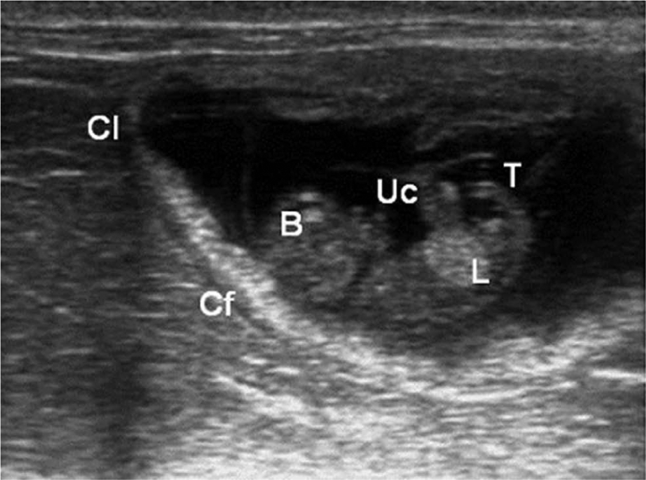
Embryo on day 28 of gestation. Chorion laeve (non-placental) (Cl), chorion frondosum (placental) (Cf), umbilical cord (Uc), tail (T), liver (L) and brain (B) are visible
During embryo development, the stage of gestation could be estimated by measuring the length of the vesicle from day 11 of gestation and the crown–rump length of the embryo from day 17 (Table 2).
Lengths of the embryonic vesicle and the embryo as a function of the stage of gestation

The size of the litter could only be assessed after the embryo appeared on day 17 of gestation; each uterine horn was examined up to the ovary, passing lateral to the mammary chain. It should be confirmed a few days later; embryonic resorption was possible and was observed during this study (cat A, 2008). In this case, the embryonic vesicle became deformed, and lost its spherical shape with a crinkled and less well-defined wall. The vesicle then regressed over a few days.
Discussion
The embryonic period was arbitrarily defined from a purely embryological point of view as ending on day 28, as established by Knospe 1 by dissecting embryos.
Some pregnancies were obtained off-season as a result of artificial lighting. No difference regarding oestrus, pregnancy or litter size was observed between natural or induced oestrus; as a result, this variable was dismissed when interpreting the results.
The queens used for the ultrasound examinations were experimental domestic shorthair cats, of varying ages, but of a similar body condition score. They were subject to the same environmental factors, were housed together and mated to the same male. However, this posed a problem for transposing the results to a more random feline population, as might be encountered in practice.
In this study, all of the ultrasound examinations were conducted by the same operator on a daily basis from day 7 of gestation, with the exception of four, which could not be monitored in the same fashion owing to problems with the availability of the operators or the lack of patience of certain queens. The older cats were trained to accept the ultrasound examinations and could stay on their backs for long periods, while the younger cats only tolerated this position for a few minutes. The precocity of the pregnancy diagnosis was influenced by how long the cat tolerated the ultrasound examination.
The timing of the examination differed from one day to the next, which could have a slight influence on the measurements and observations. The first mating was considered as the presumed start of gestation, as has been described to be the case in 92.3% of cases, according to Georgiev et al, 2 but ovulation is often triggered by repeated matings, which is why we left the female with the male for 2 days. Malandain et al 3 have shown that ovulation was spread over a 10 h period in nulliparous queens, over nine ovulatory cycles. This period was longer in multiparous queens as the litters were often bigger, which explained why the vesicles were of different sizes in the early stages of pregnancy diagnosis. Size variation of embryonic vesicles was not observed by Georgiev et al, 2 who diagnosed pregnancy on day 25 after the beginning of oestrus—a stage in which no variation regarding the size of embryonic vesicles was observed in our study. A possible difference between nulliparous and multiparous queens for size variation of vesicles was not researched in our study because there was no apparent difference in the size of the litters and a low number of queens.
The first embryonic vesicle was observed on day 11 of gestation, even if a suspicious image of an embryonic vesicle was seen on day 8. This concurred with the results obtained with a 7.5 MHz probe.4,5 However, with a 10 MHz probe, the embryonic vesicle was observed on day 10 of gestation. 6 In this last study by Zambelli et al, 6 mating was observed by the owners of the queens, who had not recorded the dates of oestrus precisely or the number of matings, which rendered the estimation of the start of gestation imprecise and certainly at least 24 h later than the actual start of gestation. Moreover, the fact that no vesicle was observed in our study on days 9 and 10 could be related to the small size of the litters. The results obtained with a 12.5 MHz probe were better than those obtained with a 5 MHz probe;7,8 with the latter, the embryonic vesicle was only visible from day 16 or 17. The various embryonic vesicles within a same litter did not all appear at the same time. 9
The embryo was observed from day 15 of gestation, and regularly on day 16. It appeared as a hyperechoic element within an anechoic cavity. It was visible from day 20 day with a 5 MHz probe,7,8 and from day 14 with a 10 MHz probe. 10 In the latter case, it was adherent to the wall and only protruded into the cavity of the embryonic vesicle from day 16. 10 It was clearly separated from the wall on day 18 of gestation. 6
The heart was observed with the 12.5 MHz probe from day 16 of gestation, and more generally on day 17. This concurred with the results obtained with 7.5 and 10 MHz probes. 10 However, with a 5 MHz probe, the heart was only visible from day 22.7,8
The use of a 12.5 MHz probe did not improve the precocity of pregnancy diagnosis in comparison with a 7.5 or 10-MHz probe, but enabled significant progress in comparison with a 5 MHz probe, which is still commonly used in practice. Indeed, vesicles and embryos could be seen 5 days earlier with a 12.5 MHz probe than with a 5 MHz one (day 11 VS day 16 for the vesicle and day 15 VS day 20 for the embryo).
With a 12.5 MHz probe, the stomach was visible from day 26 of gestation, while it was only visible from day 29 or 30 with 7.5 and 5 MHz probes, respectively.7,8,11 While one could assume that the observation of the stomach signified that the gestation was older than 30 days with a 5 MHz probe, 8 this was not true with a 12.5 MHz probe. Similarly, the neural tube appeared on day 20 of gestation with a 12.5 MHz probe compared with day 30 with a 7.5 MHz probe 11 and day 34 with a 5 MHz one.7,8
The genital bud was visible from day 27 of gestation; it was observed from day 38 of gestation with a 10 MHz probe and was never observed with a 5 MHz one.7,8,10
The comparison between the probes is summarised in Table 3.
First appearance (days of pregnancy) of different structures depending on the probe frequency

The estimation of litter size was more or less reliable as a function of the stage of gestation. The best period started after 17 days of gestation, with the appearance of the embryo in the embryonic vesicle. Some vesicles may be reabsorbed before day 18 of gestation. Litter size could only be estimated with any certainty with a methodical and rigorous exploration of the uterine horns after this date.
The estimation of the stage of gestation, based on the size of the embryonic vesicles, was more reliable before day 25. After this date, it was too easy to deform the vesicles under the pressure of the ultrasound probe. The measurements were slightly lower than those obtained at the same stage of gestation with a 5 MHz or 10 MHz probe.6–8 This could be explained by the superior resolution of the 12.5 MHz probe. The results could also be influenced by the relative uncertainty about the beginning of pregnancy or by the experience of the operator. The crown–rump length of the embryos was also a good indicator. The results obtained with a 12.5 MHz probe were similar to those obtained with a 7.5 MHz probe. 6 If Table 2 was used to predict the term of pregnancies followed in this study, there may be up to 2 days of error. This deviation may be related to the variability of the duration of pregnancy, which, in turn, may be influenced by the difficulty in determining the precise time of fertilisation. These results were similar to those obtained by Beccaglia and Luvoni, 12 who found out that at the fourth week of pregnancy the mean accuracy (± 1 day) of the prediction of parturition date obtained with the inner diameter of chorionic cavity in queens was 81%.
Finally, the apparition of the various organs could be used as landmarks to estimate the stage of gestation. However, this required a certain amount of experience and good knowledge of the equipment, as the images were more or less precise, and the organs appeared more or less early depending on the frequency of the probe used. Thus, with a 12.5 MHz probe, pregnancy diagnosis could be performed from day 11 (5 days earlier compared with a 5 MHz probe), with the observation of a vesicle, and confirmed on day 16 with the appearance of the embryo. The embryo took on a ‘figure-of-eight’ shape at around day 22. Gestation was more than 26 days along if the stomach was visible.
Conclusions
The 12.5 MHz probe did not enable the diagnosis of gestation to be performed any earlier than use of the 7.5 and 10 MHz probes. However, there was a significant difference in comparison with a 5 MHz probe.
Footnotes
Conflict of interest
The authors do not have any potential conflicts of interest to declare.
Funding
This research received no specific grant from any funding agency in the public, commercial or not-for-profit sectors.