
Abstract
Practical relevance:
Diagnostic and therapeutic cystourethroscopy has become very popular in the diagnosis and treatment of canine lower and upper urinary tract disease in the past 5–7 years. As expertise is expanding, the use of similar techniques in feline patients is growing.
Outline:
This is a brief overview of the indications, equipment needed and procedure for performing cystourethroscopy in feline patients. The principal focus is normal feline lower urinary tract anatomy. Some examples of abnormalities that might be seen are also provided, and therapeutic cystourethroscopy is touched upon.

Indications for cystourethroscopy
Signs of lower urinary tract disease in feline patients are among the most common presenting complaints by owners attending small animal veterinary practice.1,2 These signs include dysuria, hematuria, pollakiuria, nocturia, periuria, stranguria, vulvar or prepucial discharge and incontinence. Other indications for endoscopic evaluation are abnormal findings on ultrasound examination or contrast studies, recurrent urinary tract infections and the presence of urinary calculi. Imaging of the vestibule, vagina, urethra, bladder and ureterovesicular junctions can be accomplished with diagnostic cystourethroscopy to confirm normal anatomy and/or the presence of tumors, strictures, stones, foreign bodies, bladder wall lesions and upper tract renal/ ureteral bleeding from the ureterovesicular junction, among other abnormalities.3,4
In addition to the diagnostic utility of cystourethroscopy, numerous therapeutic interventions can be performed simultaneously, including stone removal (basketing, voiding, laser lithotripsy), cystoscopic-guided laser ablation of ectopic ureters, polyp resection, stricture balloon dilation or stenting, malignant obstruction biopsy and/or stenting, injection of bulking agents for incontinence, cauterization for bleeding, and endoscopic retrograde ureteral stenting for ureteral obstructions.3 –16
Equipment
Various flexible, rigid and semi-rigid endoscopes can be used for cystourethroscopy in cats (Figure 1):
A
A
A

Equipment needed for cystourethroscopy, including (a) light box, (b) light guide cable, (c) camera and camera box and (d) various cystoscopes. The five cystoscopes displayed are: (1) semi-rigid male/feline endoscope, (2) 1.9 mm 30° rigid cystoscope, (3) 2.7 mm 30° rigid cystoscope, (4) 4 mm 30° rigid cystoscope and (5) 2.5 mm flexible fiberoptic cystoureteroscope
Room set-up and use of good quality monitors are both important factors when performing cystourethroscopy in order that everything can be visualized appropriately. The monitor should ideally be positioned directly in line with the scope so that the operator is always looking forwards (Figure 2a,b). If this is not possible, the monitor can also be positioned to the left of the operator (Figure 2c,d).

Room set-up for routine cystourethroscopy. (a,b) Clinicians at the Animal Medical Center have the advantage of a high-tech set-up enabling them to face forwards during the procedure. While in practice this is seldom possible (c,d), good visualization remains key and is achievable. Images (c) and (d) courtesy of Christiane Stengel, Tierklinik Hofheim, Germany
A fluid bag of warm sterile saline (1 l) should be hanging. The author prefers to use a pressure bag to maintain good fluid flow through the narrow working channel of the endoscope, and typically warms the fluid in a fluid warmer. Care must be taken to ensure the bladder is never overdistended, as this could result in excessive bleeding and, potentially, bladder rupture. The feline bladder can typically hold between 5 and 25 ml/kg of fluid and care must be taken to ensure that the bladder is not overfilled.
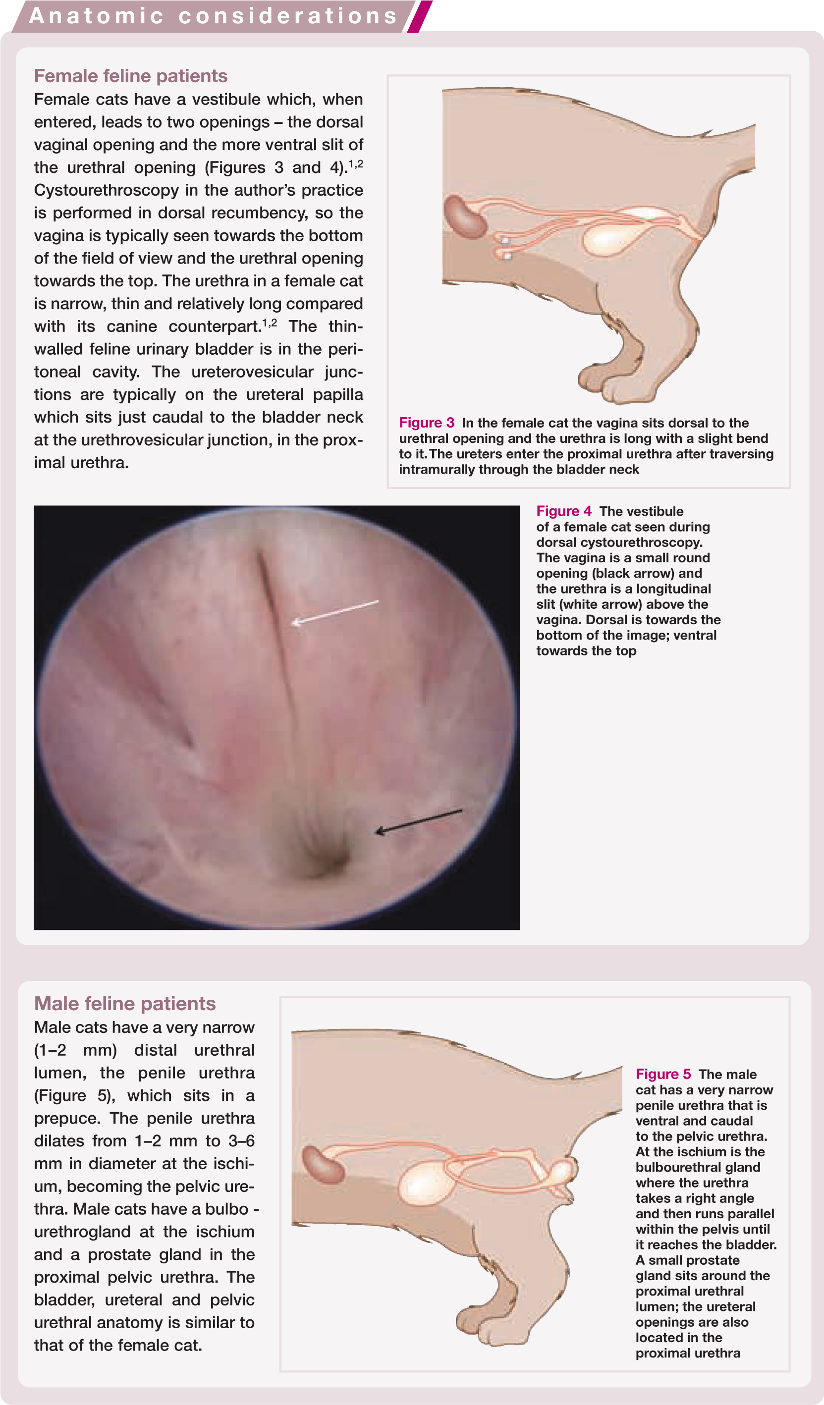

In the female cat the vagina sits dorsal to the urethral opening and the urethra is long with a slight bend to it. The ureters enter the proximal urethra after traversing intramurally through the bladder neck

The vestibule of a female cat seen during dorsal cystourethroscopy. The vagina is a small round opening (black arrow) and the urethra is a longitudinal slit (white arrow) above the vagina. Dorsal is towards the bottom of the image; ventral towards the top

The male cat has a very narrow penile urethra that is ventral and caudal to the pelvic urethra. At the ischium is the bulbourethral gland where the urethra takes a right angle and then runs parallel within the pelvis until it reaches the bladder. A small prostate gland sits around the proximal urethral lumen; the ureteral openings are also located in the proximal urethra
The cystoscope typically has three ports: a working channel, an ingress and an egress port. Fluids should be attached to one port for ingress, and an extension set to one port for egress. The working channel port needs a good quality cap to prevent fluids from running out of the channel during interventions requiring the channel to be open (Figure 6). An eyepiece attaches to the camera head and the rigid scope has a 30° angle to facilitate visualization at different angles throughout the urethra and bladder (Figure 6).

(a) A 2.7 mm 30° rigid cystoscope. Yellow arrow indicates the eyepiece that attaches to the camera; red arrow indicates the working channel of the endoscope; black and white arrows indicate the ports for ingress or egress of irrigation fluids; blue arrow indicates the adaptor for the light guide cable; purple arrow indicates the end of the endoscope, demonstrating the 30° angle. (b) The same endoscope attached to the camera head (yellow arrow), showing the fluid line for irrigation (black arrow), working channel cap (red arrow) and light guide cable (blue arrow)
Additional equipment needed for therapeutic cystourethroscopy is described in more detail in the accompanying article in this Special Issue series.
Cystourethroscopy procedure
For both male and female cats the author positions the patient in dorsal recumbency with the perineum aligned with the end of the table, and the tail hanging straight down. Placing the patient in a small trough will help to keep it upright and secure. The entire area of the vulva/prepuce and perineum is clipped and aseptically prepared. The vulva or prepuce is flushed with dilute chlorhexidine solution. For male and smaller female cats the abdomen is clipped and scrubbed as well, in case antegrade cystourethroscopy is needed. Ideally, the patient is then covered with a sterile patient drape, with a hole made in the area of the vulva/prepuce, using towel clamps to hold it in place. The author always gowns and gloves for the procedure.
Female cats
Using a 1.9 mm 30° rigid cystoscope the vulva is entered and the endoscope is aimed down towards the vestibule (Figure 4). Once the urethral opening, which typically appears as a vertical slit, is visualized the vaginal opening is inspected but the vagina is not entered, as this is considered more contaminated than the urethra. The endoscope is then gently passed into the urethral lumen, being careful to avoid overfilling the urethra and bladder with aggressive fluid irrigation but using enough irrigation to allow the urethra to distend for safe endoscope passage. If the endoscope feels tight or is hitting resistance the procedure should be aborted, as the smallest rigid endoscope can be too large for smaller female cats. In such cases, exchanging for a flexible ureteroscope or semi-rigid male cat cystoscope should be considered. If this is not possible then antegrade cystourethroscopy can be performed (see later).
Once the urethra is entered care should be taken to maintain the lumen of the urethra at the bottom of the screen to keep the endoscope straight and the 30° angle of the cystoscope aiming up (Figure 7). The most distal aspect of the urethra is often the most narrow and is lighter in color than the proximal urethra. As the mid-urethra is entered, a dorsal ridge will be seen at the bottom of the screen (Figure 7b,c); this is a normal feature in cats. At the most proximal aspect of this ridge are the ureteral papillae (Figure 7d) that sit in the proximal urethra. Each ureteral opening (ureterovesicular junction) appears as a slit on the top of each papilla (Figure 8).
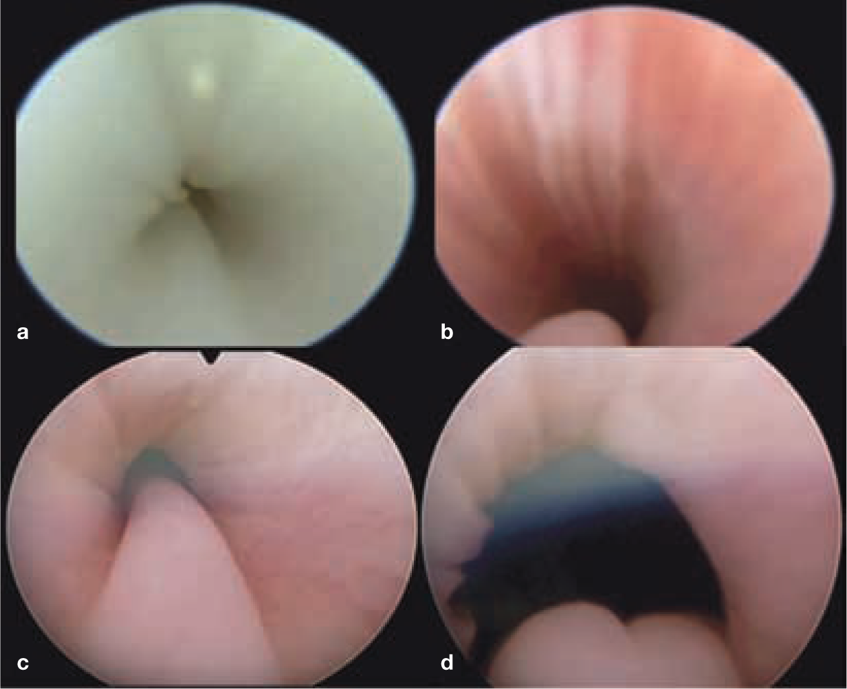
Endoscopic images of a female cat during urethroscopy in dorsal recumbency. (a) The distal urethra is very narrow and often more pale than the mid-proximal urethra. The folds of the urethral tissue are seen and small stones are visualized in this image. (b) The pelvic urethra has a dorsal bulge of tissue (bottom) that extends as far as the ureteral papillae. Note the parallel folds of urethral tissue ventrally. Note also that the lumen of the urethra is maintained towards the bottom of the screen to retain the 30° angle, keeping the endoscope straight in the urethral lumen to prevent urethral trauma. (c) The proximal urethra, with a viewof the urethrovesicular junction/trigone. (d) The ureteral papilla at the urethrovesicular junction prior to entering the urinary bladder

Endoscopic images of a female cat during dorsal cystourethroscopy showing (a) the ureteral papilla (arrow) in the proximal urethra. (b) The ureterovesicular junction is seen as a small hole on the top of the papilla (arrow). (c) Close-up image of the ureteral opening (arrow)
On entering the bladder, the irrigation fluid should be turned off immediately and the bladder drained through the egress port. The entire bladder from apex to trigone should be carefully evaluated for vascularity (Figure 9), thickness, nodules, smoothness, urachus and size. Finally the endoscope should be turned with the 30° angle facing down (towards the floor), keeping the camera in the same position, so that the dorsal (dependent) aspect of the bladder and urethra can be carefully visualized. This allows the ureteral openings to be investigated (Figure 8). The ureteral openings sit in the proximal urethra, just caudal to the bladder neck; the openings appear as a small slit on each ureteral papilla. Urine should be visualized coming from each orifice to be sure there is no blood and that each ureter is patent. If stones are present they may be seen on the bladder floor as they will fall to the dependent portion of the bladder (Figure 10).

Cystoscopic images of the bladder wall in two cats. (a) Normal vasculature of the bladder wall. (b) Thickened pale bladder wall with minimal vasculature seen in a cat with chronic cystitis and stone disease

Bladder stones on the dependent surface of the urinary bladder seen during cystourethroscopy. Note the pale bladder wall


Schematic of the PCCL procedure (discussed further in the accompanying article)
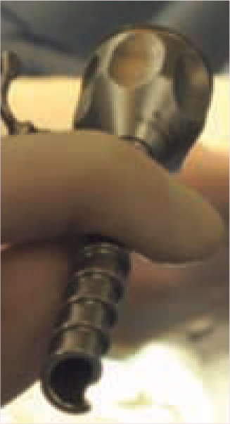
Screw trocar used to maintain irrigation and distension during antegrade cystourethroscopy (PCCL procedure)

Antegrade cystourethroscopy (PCCL procedure) in a male cat with bladder stones. (a) Normal bladder wall. (b) Stones (calcium oxalate) on the dependent surface of the bladder wall. (c) Proximal urethra showing the ureteral papilla and the slits of the ureterovesicular junction. (d) Mid-urethral lumen viewed with the rigid cystoscope
Male cats
In male cats cystourethroscopy can either be performed in a retrograde manner using a semi-rigid endoscope or in an antegrade manner using a 1.9 mm rigid cystoscope (see page 39). The semi-rigid scope allows visualization of the penile urethra but the image quality and ability to irrigate, biopsy or perform therapeutic interventions is limited due to the absence of, or only a very small, working channel (Figure 14).

Urethroscopic images of a male cat during semi-rigid cystourethroscopy. The image quality is not as good as that provided by a rigid endoscope. This is a 0° endoscope so the image does not need to be angled and the lumen should remain in the center of the screen. (a) Distal penile urethra, (b) mid-pelvic urethra, (c) proximal pelvic urethra and (d) ureteral papilla in the proximal urethra
Abnormalities
The most common abnormalities seen in the urethra in the author’s experience are strictures, stones and tumors (Figure 15). Interestingly, strictures are most commonly seen by the author in the pelvic urethra after trauma or congenitally in the very proximal urethra at the ureterovesicular junction. Ectopic ureteral openings are not commonly seen in cats and the operator should realize that the normal feline ureteral openings are found in the proximal urethra, rather than the bladder, and thus these are not ectopic.

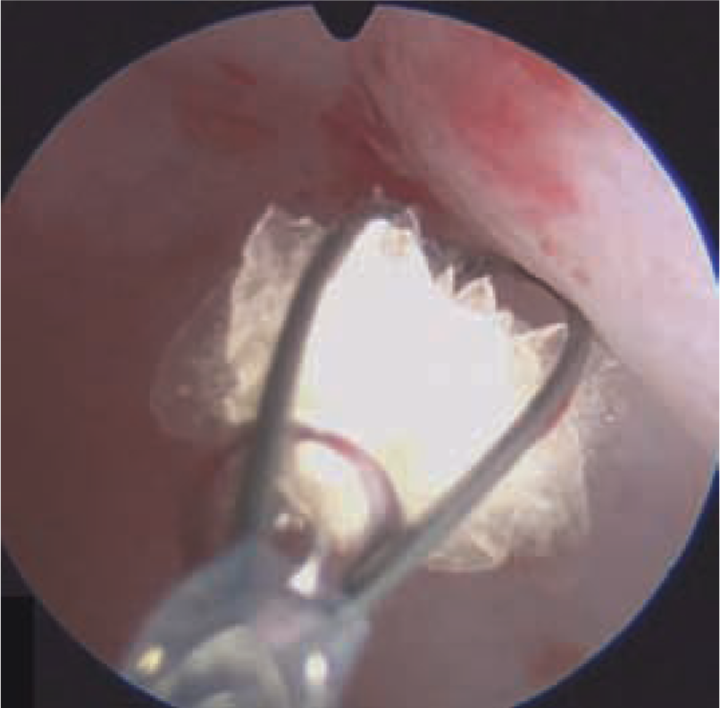
Endoscopic biopsy cup forceps being used to sample a bladder tumor
In the urinary bladder, tumors, stones, polyps and glomerulations, associated with feline idiopathic cystitis, can be seen endoscopically. A diagnosis can be obtained using various instruments/biopsy methods (cup biopsy forceps, basket, snare) (Figure 16). Therapeutically, wires, catheters, snares, lasers and stents can be employed (Figure 17), and this is expanded on in the accompanying article.
Bladder stone being retrieved with a stone retrieval basket during PCCL in a male cat

Urethral abnormalities seen during urethroscopy in three different female cats. (a) Congenital urethral stricture located between the ureteral openings and the bladder trigone. Note that the ureteral openings (black arrow) are on the distal side of the stricture. The narrow urethral opening is shown with a red arrow. (b) Bladder stones within the urethral lumen. (c) Proximal urethral transitional cell carcinoma causing an obstruction
Key points
Cystourethroscopy is a valuable diagnostic tool in the evaluation of feline patients with lower urinary tract disease.
Care should be taken as the feline urethra and bladder are very fragile and can be easily traumatized during cystourethroscopy.
Therapeutic cystourethroscopy is being more commonly employed in feline patients for the treatment of stone disease, incontinence, neoplasia and ureteral obstructions/disease.
To perform this technique safely and effectively suitable training should be undertaken to ensure appropriate technique is used and the best instruments are available.
Knowledge of the anatomical normals is vital in the diagnosis of disease.
Footnotes
Funding
The author received no specific grant from any funding agency in the public, commercial or not-for-profit sectors for the preparation of this article.
Conflict of interest
The author is an advisor for Infiniti Medical and Norfolk Vet Products.