
Abstract
Overview:
Mycobacterial infections are important in humans and animals. Cats can be infected by several Mycobacterium species, which may cause different syndromes, mainly tuberculosis, atypical or non-tuberculous mycobacteriosis and leprosy. In recent years, awareness has increased about how to recognise and confirm these infections. More cases are diagnosed today, which probably means that the disease has escaped detection in the past.
Infection:
Most cases in cats are cutaneous, presenting as nodules in the skin and draining tracts, ulceration and local lymphadenopathy; however, systemic dissemination may also occur.
Diagnosis:
Definitive diagnosis is difficult when the bacterium cannot be detected by histology or culture. However, species confirmation is essential for treatment and prognosis, so material for culture and polymerase chain reaction should be submitted in every suspected case.
Treatment:
Treatment is challenging. A combination of two or three antibiotics is needed, and treatment must be continued for some months, which makes owner compliance especially difficult in cats.
Zoonotic risk:
There is a zoonotic risk associated with some mycobacterial species. Concerns should be communicated in every case of an immunocompromised owner in contact with an infected cat.
Bacterial properties
Mycobacteria are intracellular, acid-fast, slow-growing bacilliform bacteria, highly resistant to environmental conditions.1,2 Mycobacterial taxonomy is complex and many species can infect the cat and induce a variety of clinical presentations. Different classifications have been made in the past based on features and ability to grow in culture, as well as biochemical properties. 2 The use of molecular techniques has led to taxonomic changes, and some species have been or will be classified into different groups. 2
For practical purposes, we here classify mycobacteria on the basis of their biological behaviour, including aspects of clinical presentation, diagnosis and culture, their response to treatment and on zoonotic aspects.
Tuberculosis (TB) complex group
Mycobacterium tuberculosis (mainly infecting humans and dogs, rarely cats and other species), M bovis (infecting cattle, dogs and cats) and M microti (infecting small rodents like voles, shrews and cats) are bacteria that can be grown only in specific culture media. Tuberculosis in cats is a systemic disease with disseminated internal lesions,3,4 but cats with M microti infection may also present with localised or disseminated cutaneous disease. 5
Non-tuberculous mycobacteria (NTM) group
This group includes a large number of slow-growing species like M mageritense, M genavese and M malmoense and rapidly growing species like M fortuitum, M chelonae-abscessus, M avium, M smegmatis, M flavescens and the M avium–intracellulare complex (MAC) among others. NTM infections in cats are typically subcutaneous (local or disseminated), rarely progressing to systemic disease. 6 However, MAC infections, which in some classifications are included within the TB complex group, are frequently sytemic.3,7
Feline leprosy
The M lepraemurium and several other species cannot be grown in culture. Infection in cats is restricted to the skin where it produces mainly localised and rarely disseminated cutaneous nodules. 8
Epidemiology
The prevalence of mycobacterial infections in cats is unknown. They are considered rare, but case series or case reports from the United States, Australia, New Zealand and several European countries have been published. In recent years, more cases have been recognised, probably meaning that the infection was under-diagnosed previously.7,9,10 A 2009 survey from diagnostic laboratories in the UK evaluating tissue samples with a histological diagnosis of mycobacterial infection showed a significant incidence of around 1%. 10
Data on the prevalence of the different mycobacterial species are also lacking. However, a recent retrospective study from the UK evaluating 339 cases of mycobacterial disease in cats found that 53% could not be identified through culture, 19% were M microti, 15% M bovis, 7% MAC and 6% of the NTM group. 5 Most cats with mycobacterial infections have an outdoor lifestyle;5,8 living in a non-urban area seems to increase the risk. 10 Adult tom cats seem to be predisposed to developing disease,3,5 as are Siamese and Abyssinian breeds.5,7,11
TB complex group
M microti infection is mainly related to direct contact with small rodents like voles and mice. 12
M tuberculosis infection is rare in cats, probably due to their natural resistance. 13 M tuberculosis and M bovis are directly transmitted to cats by ingestion of milk from infected cattle and by direct or environmental contact with badgers (M bovis). 14
NTM group
The main risk to cats is wound contamination by mycobacteria present in the environment (soil, water and decaying vegetation).6,9,15,16
Feline leprosy
The main risk comes from direct contact or rodent bites, or from wound contamination by mycobacteria present in the soil or on plants.7,17
Pathogenesis
Mycobacteria infect the macrophage and induce granulomatous and pyogranulomatous inflammatory responses in the organs involved. 18 The mycobacterial species, route of infection and immune responses determine the extent, location and severity of the lesions.
TB complex group
The primary site of infection by M tuberculosis and M bovis may be the alimentary tract, lungs or skin.2,15 From these sites dissemination and systemic infection may occur. Occasionally, the infection is primarily systemic.
In M microti infection, the portal of entry is the skin, in locations commonly affected by wild rodent bites (the face and legs). 5
NTM group
The primary site of infection is the skin, with mainly traumatic or surgical wounds becoming contaminated with mycobacteria.6,8 Some fast-growing mycobacteria show a predilection to replicate in lipid-rich tissues, like the ventral abdominal and inguinal areas, particularly after a surgical wound contamination (Figure 1). A lipoid pneumonia case caused by mycobacterial infection has also been reported. 19 Dissemination from the skin and systemic infections are not common for bacteria of this group, with the exception of MAC infections, which are easily disseminated.6,11,20–22

Mycobacterial infection in the ventral abdomen. Courtesy of Richard Malik, University of Sydney Veterinary School
Feline leprosy
The primary site of infection is the skin, with localised subcutaneous granulomas and, less commonly, disseminated skin granulomas. 7
Clinical presentation
Most mycobacterial infections occur in immunocompetent animals.3,5 Cases in cats with primary or acquired immunodeficiency have been documented. Two feline immunodeficiency virus-positive cats with atypical mycobacterial infection have been reported.23,24 One case has been documented of an atypical mycobacterial infection in a cat with an idiopathic CD4+ lymphopenia. 25 Two cases (MAC disseminated infection and mycobacterial osteomyelitis) have been reported after renal transplantation and long-term immunosuppressive therapy with ciclosporin.26,27

Cutaneous forms
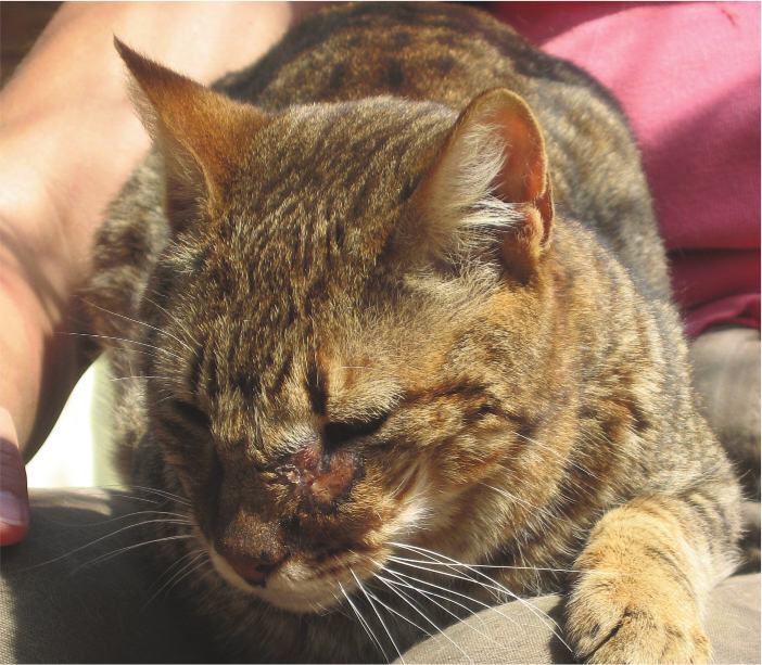
M microti, the NTM group and feline leprosy species are the most common mycobacteria producing skin lesions. These typically consist of dermal nodules, non-healing wounds with draining tracts and ulceration (Figures 2–4).4,5,8–10 Common locations are the facial area, extremities, tail base, perineum, ventral thorax and abdomen. Lesions may be solitary or multiple.5,9 Multiple skin lesions may result from local spread or haematogenous dissemination. Local or generalised lymphadenopathy is present in about half of the cases and may be the only clinical sign (especially submandibular and prescapular). 5

Ulcerated skin nodule in M microti infection. Courtesy of Richard Malik, University of Sydney Veterinary School
Subcutaneous skin nodule in NTM infection. Courtesy of Albert Lloret, Veterinary Teaching Hospital, Universitat Autonoma, Barcelona, Spain

(a,b) Subcutaneous nodules in a cat with leprosy. Courtesy of Richard Malik, University of Sydney Veterinary School
Visceral (digestive or respiratory) or systemic forms
The TB complex and MAC species are the most common mycobacteria producing visceral or systemic lesions.3,20–22 NTM infections rarely produce disseminated disease. 19 Common clinical signs and abnormalities are digestive (weight loss, mesenteric lymphadenopathy) or respiratory (pneumonia, hilar lymphadenopathy, pneumothorax, pleural or perdicardial effusions), and may be accompanied by signs of systemic dissemination like fever, ocular signs, splenomegaly, hepatomegaly, generalised lymphadenopathy, bone lesions and central nervous system signs.3,6,11,20–22,27
Diagnosis
Diagnosis may be difficult, especially when skin lesions are absent. It is based on a clinical suspicion when the presentation is indicative and other diseases are ruled out. In these cases, appropriate samples should be obtained for cytology and/or histology (including acid-fast staining), culture and polymerase chain reaction (PCR).
Haematology and biochemistry changes are non-specific, suggesting a chronic inflammatory condition. Hypercalcaemia due to granulomatous disease has been reported in systemic MAC 6 and M microti infections. 4 Cats infected with mycobacteria may show reduced levels of vitamin D when compared with healthy cats, as occurs in humans. 28
Thoracic radiographic changes are variable and non-specific, ranging from no abnormalities to bronchial, alveolar or interstitial nodular mixed patterns, pleural effusion and/or mediastinal and perihilar lymphadenopathy (Figure 5). 29 Appendicular radiographs may show bone osteolytic lesions, and less frequently osteoproliferative changes, associated with systemic infections.27,29 Abdominal ultrasound may be useful for identifying mesenteric lymphadenopathy or granulomatous lesions and as a guide to obtain fine needle aspirates. 26

Mixed bronchial–interstitial pattern in the lungs of a cat with TB complex group infection. Courtesy of Richard Malik, University of Sydney Veterinary School
Cytology
Fine needle aspirates or smears from skin lesions (nodules, ulcers, draining tracts) or from granulomatous lymph nodes should always be stained for acid-fast bacteria using, for example, the Ziehl-Neelsen (ZN) procedure. Sensitivity is variable, as the number of bacteria within macrophages varies depending on the mycobacterial species and on the host’s immune response.
2
A negative cytology result does not rule out mycobacterial infection [
Histology
Histology is useful in the diagnosis of mycobacterial infections. It allows the inflammatory pattern (pyogranulomatous or granulomatous) to be assessed and special acid-fast staining, like ZN, to be performed (Figure 6).18,30 However, only a few bacteria may be present and pass undetected by staining (M microti and some NTM species), although culture or PCR may give a positive result [

Large numbers of acid-fast bacteria revealed using the Ziehl–Neelsen stain. Courtesy of Richard Malik, University of Sydney Veterinary School
Culture
Culture from a fresh tissue sample is useful to confirm mycobacterial infection and to identify the species involved, which has implications for treatment, prognosis and assessment of zoonotic risk. This needs to be done in a specialised laboratory and, even still, many mycobacterial species are slow growing (2 to 3 months) or fail to grow [
PCR
PCR (eventually followed by sequencing) is the recommended test for diagnosing mycobacterial infections [
Interferon gamma test and other immunoassays
These tests are currently commercially unavailable, but they are promising for the diagnosis of TB complex group infections.31,32 A recent study evaluating a cell-based interferon gamma test and serum antibody tests showed excellent specificity and a variable, moderate sensitivity [

Treatment
Treatment of mycobacterial infections is generally difficult and challenging. There are no prospective controlled clinical trials, and recommendations are based on case reports or retrospective studies. Good outcomes have been published after identification of the mycobacterial species and treatment with a long (several months) course of an appropriate antibiotic combination [
Some important considerations before embarking on treatment are outlined in the box above.
TB complex group and NTM group
Currently, double or triple therapy is recommended: rifampicin (10–15 mg/kg q24h) plus a quinolone (marbofloxacin 2 mg/kg q24h) plus a macrolide (clarithromycin 125 mg/cat q24h or azithromycin 5–15 mg/kg q24h) for 6–9 months. Ideally, the three drugs should be giving during an initial phase of 2 months, followed by two of the drugs for 4–7 months [
Short courses of antibiotic and/or monotherapy (eg, quinolones or beta-lactams) have been associated with clinical responses and remissions, but also with a high risk of relapse, which may be followed by systemic spread. 30 It is recommended always to start complete treatment while waiting for diagnostic confirmation and species identification.
MAC infections
Disseminated MAC infections usually respond poorly to treatment, and quinolones are not very effective [
Feline leprosy
Most cats with leprosy can be cured by surgery (small lesions) and applying combinations of rifampicin, clofazimine and clarithromycin for several months [
Prevention
Keeping cats indoors and avoiding contact with wild rodents are the only measures for preventing mycobacterial infection.
Prognosis
The prognosis generally must be considered guarded, but depends on the mycobacterial species and the extent and severity of the disease. Disseminated infections (TB complex and MAC) are associated with a poor prognosis.3,9,20–22 Localised skin disease (NTM infection), M microti infection and leprosy may have a good prognosis if treated appropriately.7,8,30

Footnotes
Key Points
Funding
The authors received no specific grant from any funding agency in the public, commercial or not-for-profit sectors for the preparation of this article. The ABCD is supported by Merial, but is a scientifically independent body.
Conflict of interest
The authors do not have any potential conflicts of interest to declare.