
Abstract
A spayed female domestic shorthair cat was first examined at the age of 16 months because of persistent licking of the perineal area. The cat had a grossly enlarged and oedematous vulva with pronounced superficial pyoderma of the perivulvar area, which responded favourably to systemic antibiotics, analgesics and local corticosteroids. A month after the initial examination, the cat was re-presented owing to pollakiuria, stranguria and dyschesia. The oedema of the vulva had disappeared and the vulvar labia were fused together; there was only a fistulous tract with a diameter of 1 mm present in the area of the vulva, and the cat strained to urinate through that opening. A contrast study revealed normal transit through the lower urinary tract, but labial adhesions resulted in the development of dilation cranially, where the vaginal vestibule was supposed to be. Vaginoplasty was subsequently performed, the cat recovered normally and, 10 months after the procedure, the lumen of the vaginostoma is preserved and the cat is urinating without difficulty.
Case Report
Labial fusion, also termed labial adhesion, labial agglutination and synechia vulvae, is an acquired condition caused by midline adherence of the vulvar labia minora. 1 It is not rare in human medicine and has been reported to occur in up to 1.8% of female prepubertal patients, with a peak incidence around the age of 13–23 months. 2 It also occurs in menopausal women, but is quite uncommon in reproductive-age women.2 –4 The cause of the condition is a predisposition of the patient and chronic irritation of the perivulvar skin due to poor hygiene or dirty diapers, 1 senile vaginitis, lack of sexual activity, female circumcision, local trauma or vaginal laceration following childbirth.3,4 As the disease typically starts at the age of 6 months, when the influence of maternal oestrogen in a baby gradually disappears, oestrogen is also suspected of playing a role in the occurrence of the disease. 1 Most patients are asymptomatic and about 80% of labial fusions resolve spontaneously within 1 year. If treatment is needed it consists of topical use of oestrogen or corticosteroid ointments,2,5,6 and surgical treatment in severe cases and cases resistant to conservative treatment.3,4
We suspect that a similar pathology occurs occasionally in companion animals. A literature search revealed one case of vaginoperineal fistula after a bite wound and consecutive scarring and contracture of the tissue in the perineal region in a cat. 7
In this report, we describe labial fusion in an adult domestic shorthair cat.
A 16-month-old, spayed female domestic shorthair cat was presented to the Clinic for Small Animal Medicine and Surgery, Veterinary Faculty Ljubljana, with persistent licking of the perineal region for several days. First examination revealed a very enlarged and swollen vulva with pronounced superficial pyoderma of the perivulvar region. A small amount of sero-haemorrhagic vaginal discharge was noticed.
Urinalysis and complete blood count were without clinically relevant changes, and ultrasonographic examination of the abdomen also revealed no changes. Symptomatic treatment was initiated with an antibiotic (single application of cefovecin, Convenia; Pfizer, 0.1 ml/kg SC), analgesic (meloxicam, Meloxidyl; Ceva, 0.1 mg/kg PO once a day for 3 days, then twice further, one dose q48h), local treatment with corticosteroid ointment (betamethasone in combination with gentamicin, Kuterid genta; Lek) and prevention of licking with an Elizabethan collar.
At recheck 2 days later, the oedema had subsided and the cat had tolerated the treatment very well. A week later, the condition was significantly improved, but the pyoderma had not yet resolved completely, so the treatment was continued.
One month after the initial examination, the cat was re-presented owing to pollakiuria, stranguria, mucous diarrhoea and dyschesia. The owners had removed the Elizabethan collar, contrary to our instructions, before the perivulvar pyoderma had resolved completely.
Clinical examination revealed total resolution of the perivulvar oedema, but the vulva did not have a normal morphological structure. The vulvar labia were fused together, with only a pinpoint opening located dorsolaterally to the vulva, 1 cm below the anus (Figure 1). Manual bladder expression was extremely painful and resulted in leakage of urine through the opening.
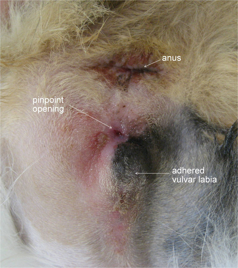
Vulvar labia are adhered, and a pinpoint opening near the vulva is seen
With the cat under general anaesthesia, we performed a contrast radiographic examination of the vagina, urethra and the urinary bladder. Positive contrast medium (3 ml, Omnipaque; Higiea) was applied retrogradely with a feline urinary catheter (Buster cat catheter 1.0 × 130 mm; Buster), which was introduced through the opening into the urinary bladder in order to achieve a good orientation point for the further study. As the contrast-filled urinary catheter was slowly extracted, the urethra was outlined, demonstrating direct communication between the fistula and urinary bladder, with a normal position and morphology of both structures. Application of an additional 2 ml of the contrast medium in the vaginal vestibule revealed saccular dilation of the vestibule in the dorsocaudal direction (Figure 2). The vestibule communicated with the surroundings through the fistula. Owing to contrast medium spillage through the fistula, only partial presentation of the vagina could be seen. Standard retrograde vaginourethrography 8 was considered impossible owing to the small fistula opening. Other abnormal adhesions were not noticed.
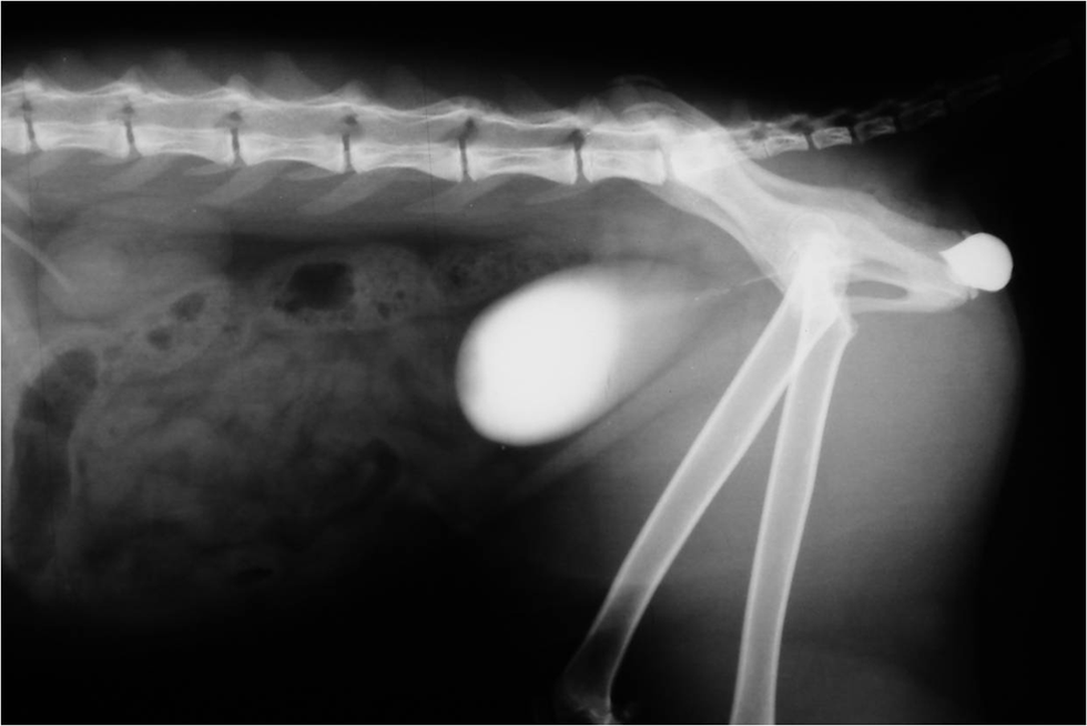
Positive contrast medium, applied retrogradely in the urinary bladder, urethra and saccular dilation of the vaginal vestibule
Owing to severe pain and straining during urination and defecation, the cat’s quality of life was diminished, so surgical correction of the problem with vaginoplasty was proposed.
On the day of the surgical procedure, cystocentesis was performed and routine microbiology urine culture was negative.
With the cat under general anaesthesia, an intravenous catheter was introduced initially through the opening into the urethra, which was later replaced by a urinary catheter. An incision was made dorsally and ventrally from the visible opening to expose the lumen of vaginal dilation. Immediately after the incision, the wall of the vaginal vestibule was exposed and sutured to the skin with single sutures with polypropilene 5/0 (Surgipro II; Covidien) ensuring a bigger opening, morphologically similar to the vulva (Figure 3). During the procedure, the external urethral orifice was examined, revealing its normal position and patency.

With an intravenous catheter inserted in the urethra (a), an incision was made dorsally and ventrally from the visible opening to expose the lumen of the vaginal dilation (b). The wall of the vaginal vestibule was exposed and sutured to the skin with single sutures (c)
At suture removal, the wound was healed completely, and the cat urinated and defecated normally, without any signs of pain. Ten months after the procedure, the lumen of the vaginostoma was maintained and the cat was behaving normally.
In humans, labial fusion is an acquired condition occurring mostly in hypoestrogenic women with irritation or local trauma of the perivulvar skin.1–4,9 Owing to the rarity of such cases, a similar aetiology in cats can only be speculated. Ovariohysterectomised queens have an oestradiol concentration <9 pmol/l. 10 In intact queens, the oestradiol concentration varies during the oestrous cycle from approximately 91.8 pmol/l to >183.55 pmol/l and returns to basal levels of <55.1 pmol/l between cycles and during seasonal anoestrous. 11 Although the oestradiol concentration was not measured in this case, we suspect a very low level, as the cat had been spayed 8 months previously. However, the main causative factor for labial fusion in the presented case was probably chronic inflammation of the vulvar and perivulvar tissue. The condition was further aggravated by self-injury due to the cat’s constant licking of the affected area. The cause of the vulvitis and pyoderma of the perivulvar region was not identified, but urinary tract infection, as one of the most likely causes, was eliminated. Similarly, chronic irritation, inflammation or extensive damage to the vulvar and perivulvar tissue play an important role in the aetiology of labial fusion in women.3,12
Human patients are usually asymptomatic, some experience dysuria, urinary tract infection, 2 incontinence, colpitis 5 and impossible intercourse.3,12 Our patient had similar clinical signs, pollakiuria, stranguria and dyschesia, although urinary tract infection, which develops in 14% of human patients, 2 was not proven at any time during the treatment. These signs were not expected to resolve spontaneously and, owing to the cat’s clinical status, in our opinion, prolonged topical therapy was not acceptable, 2 especially as a newly-published study has reported only a 15.4% and 15.6% success rate of oestrogen and betamethasone topical treatment in women, respectively, and only slightly better results with a combination of the two medications. 6 However, some authors report that betamethasone may separate fusion quicker with less recurrence and fewer side effects than topical oestrogen therapy. 5 However, in our case, betamethasone cream, used at the beginning of the cat’s treatment, did not prevent the formation of labial fusion.
Conclusions
The only other veterinary patient with similar pathology was first treated with prepubic urethrostomy but was re-presented 11 months later with a stricture of the vulvar stoma and accumulation of vaginal secretions, and the creation of a fistulous tract. Vaginoplasty was performed at this point. 7 This case proves that preservation of the vulvar opening for the drainage of vaginal secretions in female cats is necessary, even if the cat is spayed and urine is diverted. We thus decided on surgical separation of the labial fusion and vaginoplasty as the first-line treatment. Such an approach is also recommended in human medicine in symptomatic patients resistant to conservative management and those with urinary retention. 2 The cat resumed normal micturition immediately after the procedure and remained recurrence free at the time of writing, 10 months after the final treatment.
Footnotes
Funding
This research received no specific grant from any funding agency in the public, commercial or not-for-profit sectors.
Conflict of interest
The authors do not have any potential conflicts of interest to declare.