
Abstract
Medical records of 1407 cats with dermatologic diagnoses made at Cornell University teaching hospital from 1988 to 2003 were tabulated. We expressed the diagnoses as counts, percentages of the cats with dermatologic disease (1407) and percentages of all cats seen at the university hospital (22,135) during the same period. A total of 1887 diagnoses were made in the 1407 cats. We compared the age, sex and breed group of our cases with all those 22,135 cats in (‘1-by-c’) χ2 tests in which the hospital population was considered a standard (rather than a ‘sample’). The 10 most common dermatoses, their counts, and the proportions of dermatologic diagnoses and of the total cat population that the cats with these dermatoses represented were: allergy (298; 15.8%; 1.35%), atopic dermatitis (194; 10.3%; 0.88%), bacterial folliculitis/furunculosis (189; 10.0%; 0.85%), otodectic mange (115; 6.1%; 0.52%), flea infestation (99; 5.2%; 0.45%), feline acne (74; 3.9%; 0.33%), flea-bite allergy (70; 3.7%; 0.32%), cutaneous adverse drug reaction (56; 3.0%; 0.25%), idiopathic eosinophilic-granuloma complex (55; 2.9%; 0.25%) and abscess (51; 2.7%; 0.23%). Allergies of all types, combined, accounted for 32.7% of all the feline dermatoses. Relative to the standard of the total hospital population, cats <2 years old and females (both intact and spayed) were significantly under-represented (all P≤0.001) in the dermatologic case series. In contrast, Himalayans (compared with domestic short- or longhair, Persian, Siamese and other breeds) and males (both intact and neutered) were significantly over-represented (all P ≤0.001).
Introduction
Only a few peer-reviewed publications address the prevalence and types of feline dermatologic conditions seen in private or specialty veterinary practice. In a study commissioned by the British Small Animal Veterinary Association (BSAVA) Scientific Committee in 1972–1973, data were collected from 61 veterinarians representing all regions of the BSAVA throughout England, Scotland and Wales. 1 Disorders of the skin and ears accounted for 12.5% of the feline caseload. In the USA, the National Companion Animal Study collected data in 1995 on all diseases diagnosed in 52 private practices in 31 states. 2 The most common dermatologic diagnoses in cats were flea infestation (9.2% of all diagnoses), Otodectes species infestation (7.4%), abscess (6.5%), ‘miliary dermatitis’ (2.3%), ‘otitis externa’ (2.3%) and ‘dermatitis’ (1.7%). (The latter three conditions are, of course, cutaneous reaction patterns rather than ‘diagnoses’.) A survey of dermatologic conditions seen in 154 cats in 20 private practices in the UK was conducted in 1998–2001. 3 Cats with disorders of the skin and ears accounted for 13% of all cats examined.
Scott and Paradis summarized data on 111 cats with dermatologic disorders seen over a 1-year period (1987–1988) in a university hospital in Canada. 4 Cats with skin and ear conditions accounted for 15.2% of all cats examined during the study period.
The purpose of this article is to report the results of a retrospective study of 1407 cats examined over a 15-year period by the dermatology service at a university hospital in northeast USA.
Materials and methods
A retrospective review was conducted on the medical records of all 1407 individual cats examined by the dermatology service of the Cornell University Hospital for Animals (CUHA) over a 15-year period (1988–2003). Signalment (age, breed, sex) data were compared with that of the general CUHA cat population for the same time period. All diagnoses had been confirmed by standard clinical, laboratory, histopathologic and therapeutic criteria. 5 A diagnosis of ‘allergy’ was made when a differential diagnosis of atopic dermatitis, food allergy or both of these was achieved, but definitive allergy testing could not be performed. These cats had non-seasonal dermatoses characterized by one or more of the classic cutaneous reaction patterns seen in feline skin: miliary dermatitis, eosinophilic granuloma complex, symmetrical self-induced hypotrichosis and symmetrical pruritus with excoriation. 5
Statistical analysis for age, breed and sex
The CUHA age data were recorded in 1-year increments (<1 year, 1, 2, …,18 years) and our case series was categorized similarly. The breeds were categorized as domestic shorthair (DSH) or longhair (DLH), Himalayan, Persian, Siamese and other. Sex was a four-level variable for intact and non-intact males and females. The CUHA data categories provided the expected proportions of our cases that would have been each age category, breed category or sex category, if age, breed and sex were not related to having dermatologic diagnoses. These expected numbers were compared with the observed counts in ‘1-by-c’ χ2 tests. (There were 19 columns — ‘c’ — for age, six for breed and four for sex.) We interpreted that any one χ2 test was significant at P≤0.0167 (an overall P value of 0.05, divided by 3 for three formal tests to make a Bonferroni adjustment for multiple tests). We then identified the cell-χ2 values that were significant at P≤0.001 for a χ2 with 1 degree of freedom (a small P value to account for multiple implicit ‘1-by-2’ comparisons) and interpreted those categories as ones that ‘contributed importantly to the risk of dermatologic diagnoses’ (alternatively, ‘protection from’ — if fewer cats were in that category than expected).
Results
Cats examined were the following states or Canadian provinces: New York (1315 cats), Pennsylvania (76), Connecticut (three), Massachusetts (three), New Jersey (three), Ontario (two), Delaware (one), Maryland (one), Vermont (one), Wisconsin (one) and West Virginia (one). Seven hundred and fifty-one cats (53.4%) were first-opinion cases, 627 cats (44.6%) had been examined previously by one to eight veterinarians; this information was missing for 29 cats (2.0%). Cats examined by the dermatology service accounted for 1407 (6.4%) of the 22,135 cats examined at the CUHA during the study period.
Breed, age and sex
Relative to the standard of the total cat hospital population, cats <2 years old and females (both intact and spayed) were significantly under-represented (all P ≤0.001) in the dermatologic case series. In contrast, Himalayans and males (both intact and neutered) were significantly over-represented (all P≤0.001). Only six (0.4%) of the 1407 cats with dermatologic disease had feline leukemia virus or feline immunodeficiency virus infection and all six cats had different dermatoses.
Three hundred and seventy-one cats (26.4%) had two or more concurrent diagnoses. The number of diagnoses per cat was as follows: one (994 cats; 70.6% of total), two (311; 22.1%), three (78; 5.5%), four (18; 1.3%), five (4; 0.3%) and six (2; 0.1%). Hence, a total of 1887 diagnoses were made in the 1407 cats. One hundred and forty-two different diagnoses were rendered (Tables 1–9). The 10 most common diagnoses were allergy (298 cats; 15.8% of total), atopic dermatitis (194; 10.3%), bacterial folliculitis/furunculosis (189; 10.0%), otodectic mange (115; 6.1%), flea infestation (99; 5.2%), feline acne (74; 3.9%), flea-bite allergy (70; 3.7%), idiopathic eosinophilic granuloma complex (55; 2.9%), cutaneous adverse drug reaction (56; 2.9%) and abscess (51; 2.7%). These ‘top 10’ diagnoses are listed alongside the findings from two other studies in Table 1. The proportions of the dermatologic diseases compared to the total cat hospital population is presented in Tables 2–9.
The ‘top 10’ feline dermatologic diagnoses from three studies (ranked from highest prevalence in descending order)

CUHA study; 1407 cats
Scott and Paradis; 4 111 cats
Hill et al; 3 154 cats
Six conditions tied for tenth place: atopic dermatitis, cyst, aural hematoma, polyp, dermatophytosis, plasma cell pododermatitis
Allergic dermatoses diagnosed at the Cornell University Hospital for Animals (n = 617 diagnoses)

These cats had atopic dermatitis, food allergy or both. Allergy testing was not performed
Nine of these cats had concurrent food allergy
Nine of these cats had concurrent atopic dermatitis
Bacterial dermatoses diagnosed at the Cornell University Hospital for Animals (n = 291 diagnoses)

Parasitic dermatoses diagnosed at the Cornell University Hospital for Animals (n = 266 diagnoses)

Seven of these cats had Demodex cati, two had Demodex gatoi
Dermatoses of the external ear diagnosed at the Cornell University Hospital for Animals (n = 185 diagnoses)

Nineteen of the cats (44.2%) had allergy or atopic dermatitis
These cats had allergy, atopic dermatitis or food allergy. Symmetrical otic pruritus was the only reaction pattern
All cats had Demodex cati
No diagnostics were performed
Cutaneous neoplasms, cysts, keratoses, cutaneous horns and hamartomas diagnosed at the Cornell University Hospital for Animals (n = 157 diagnoses)

One of these cats was reported previously in Scott et al 10
One cat had a metastatic pancreatic carcinoma, the other had a metastatic biliary carcinoma
This cat had a metastatic pancreatic carcinoma
Fungal dermatoses diagnosed at the Cornell University Hospital for Animals (n = 112 diagnoses)

One of these cats also had multiple dermatophytic pseudomycetomas
Immune-mediated dermatoses diagnosed at the Cornell University Hospital for Animals (n = 104 diagnoses)

Fourteen of these cases were reported previously in Scott et al. 11 None of these reactions was believed to be a simple irritant reaction or an expected side effect
One of these cases was previously reported in Kalaher and Scott. 12 ‡One of these cases was reported previously in Hickford et al 13
One of these cases was reported previously in Scott et al 14
This cat had either bullous pemphigoid or mucous membrane pemphigoid. Discriminating immunodiagnostic testing was not available
Miscellaneous conditions diagnosed at the Cornell University Hospital for Animals (n = 296 diagnoses)

These three cats had thin, hypotonic, dry skin with dull, dry, brittle hair coats. All had concurrent systemic diseases
These three cats had environmental stressors that preceded the behavior, and the cats did not respond to parasiticides, novel diets and antipruritic medications. ‡No diagnostic tests or treatments were allowed in these three cats
This cat was reported previously in Scott and Miller 15
The most common group of diseases were allergic in origin (Table 2). Together, allergy, atopic dermatitis, flea-bite allergy, food allergy and mosquito-bite allergy accounted for 32.7% of all diagnoses. Nine cats had concurrent atopic dermatitis and food allergy. Cats with allergy or atopic dermatitis had secondary bacterial dermatitis and/or bacterial otitis externa in 10.4% and 17%, respectively, of the cases. Cats with allergy or atopic dermatitis had secondary malassezia dermatitis or malassezia otitis externa in 4.4% and 6.1%, respectively, of the cases.
Bacterial infections accounted for 15.4% (291 cases) of all dermatologic diagnoses (Table 3). Bacterial folliculitis/furunculosis was the most common entity in this group, and was secondary to another disease — especially feline acne, allergy and atopic dermatitis — in 134 (70.9%) of the cases. Parasitic diseases accounted for 14% (266 cases) of all dermatoses (Table 4). Otodectic mange, flea infestation and cheyletiellosis accounted for the majority of cases. Otodectic mange was the only diagnosis in 58 cases (3.1%) and in 26/115 cases (22.6%) the infestation was ectopic (other areas of the skin affected in addition to the ear canals). Flea infestation was the only diagnosis in 27 cases (1.4%). Demodicosis was associated with Demodex cati in 7/9 cases, and Demodex gatoi in 2/9 cases. In 6/7 cases associated with D cati the infestation was confined to the external ear canals. The case of generalized demodicosis caused by D cati was associated with chronic glucocorticoid administration.
Diseases of the external ear accounted for 9.8% (185 cases) of all dermatoses (Table 5). The most common conditions in this group were malassezia otitis externa, bacterial otitis externa and allergies. Idiopathic excessive cerumen production was diagnosed in nine cats. All cats had had the condition since the owners acquired them (8 weeks to 1 year of age). All cats were DSH and two were littermates. The condition was bilaterally symmetrical, non-pruritic and not associated with secondary otic infections.
A grouping of neoplasms, cysts, keratoses, cutaneous horns and hamartomas accounted for 8.3% (157 cases) of all diagnoses (Table 6). Squamous cell carcinoma, mast cell tumor and epitrichial sweat gland cyst were the most common entities in this grouping. Most squamous cell carcinomas involved the pinna or nasal planum.
Fungal diseases accounted for 5.9% (112 cases) of all dermatoses (Table 7). Dermatophytosis (the most common fungal disease) was caused by Microsporum canis (44/45 cases) or Trichophyton mentagrophytes (1/45 cases). malassezia otitis and/or dermatitis were secondary to another disease — especially allergy and atopic dermatitis — in 37 (62%) of the cases.
Immune-mediated diseases accounted for 5.5% (104 cases) of all dermatoses (Table 8). Nineteen (34%) of the cutaneous adverse drug reactions were associated with the topical application of otic preparations. Nine (16.1%) of the cutaneous adverse drug reactions were characterized by allergy-like symmetrical pruritus of the face and ears: four in association with methimazole, two with amoxicillin-clavulanate, and one each with amoxicillin, spirinolactone and diltiazem/aspirin. Two DSH cats developed contact dermatitis (eosinophilic plaque) at the site of application of an imidacloprid-containing spot-on product (Advantage; Bayer Health Care), and one of these cats also developed an identical reaction at the site of application of a fipronil-containing spot-on product (Frontline; Merial). None of the cases of pemphigus foliaceus were known to be precipitated by drug or vaccine administration. Vasculitis was idiopathic in 6/8 cases, and associated with Pseudomonas species infection or herpesvirus respiratory disease in the other two cases.
A number of miscellaneous dermatoses was seen in 296 cats (Table 9), accounting for 15.7% of all dermatoses. Feline acne (comedones on the chin) was diagnosed in 74 cats (3.9%), was the sole diagnosis in 18/74 cats (24.3%) and was the subject of a previous report. 6 Feline acne was complicated by secondary bacterial folliculitis/furunculosis in 31/74 cats (41.9%). Cats with idiopathic eosinophilic granuloma (2.9% of all diagnoses) was the subject of a separate report. 7
The cats with impacted anal sacs, hyperthyroidism and cystitis presented for self-induced alopecia, predominantly in the ventral abdominal and rump/tailhead regions. The two cats with dacryocystitis with nasolacrimal duct obstruction presented for allergy-like facial pruritus.
The six cats with telogen defluxion were postsurgical (three) or had feline infectious peritonitis (one), intestinal lymphoma being treated with prednisolone (one), and idiopathic nasal discharge and weight loss. The two cats with anagen defluxion were being treated with prednisolone and cyclosporine for pure red cell aplasia.
The cats with idiopathic transient pruritus presented for bilaterally symmetrical, lesionless pruritus. Three of these cats were younger than 1 year old and had more-or-less generalized pruritus. The other two were 5 and 12 years old and had pruritus of both ears or chin, respectively. The cats were otherwise healthy and no triggering factor (vaccinations, medications, dietary changes, environmental stressors, etc.) could be elucidated. The cats received no treatment and all recovered spontaneously after a total course of 8 weeks to 4 months. No recurrences were reported during 2–4-year follow-up periods.
Congenital pawpad hyperkeratosis was diagnosed in three cats: a female DSH, a spayed female DLH and a castrated male Russian Blue. The condition was characterized by asymptomatic villous hyperkeratosis of all pawpads. The cats were otherwise healthy. No treatment was rendered and the cats remained asymptomatic.
Both cats with iatrogenic hyperglucocorticoidism had acquired skin fragility. The cat with spontaneous hyperadrenocorticism had an adrenocortical adenoma.
Two cats (listed as ‘normal’ in Table 9) were presented having been exposed to cats with dermatophytosis. Both cats were clinically normal and Wood’s light examination and toothbrush cultures were negative.
Follow-up periods of 8 weeks to 12 years were available for 1166 cats (82.9%). Follow-up periods of at least 1 year and 3 years were available for 593 cats (42.1%) and 241 cats (17.1%), respectively. No follow-up information was available for 101 first-opinion cases and 140 multiple-opinion cases. Of the 241 cats with no available follow-up information, 85 (35.3%) had received treatment and 156 (64.7%) had been given various options to consider. The most common diagnosis in both groups — first-opinion versus multiple-opinion, treatment versus options — was allergy.
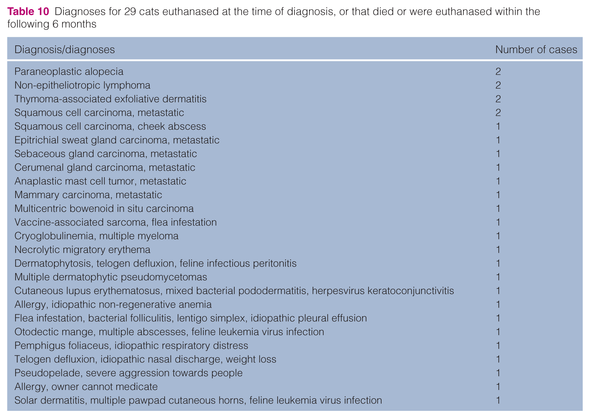
Thirty-seven cats (2.6% of the cats with dermatoses) were euthanased or died during the 15-year study period. Twenty-nine of these cats were euthanased at the time of diagnosis or shortly thereafter (Table 10). Eight cats were euthanased or died 6 months to 3 years after initial examination at the CUHA. For six of these cats, the cause for euthanasia or death was not related to their dermatologic diagnosis/diagnoses. Two Persian cats were euthanased owing to recurring dermatophytosis (M canis) 6 months after initial examination.
Diagnoses for 29 cats euthanased at the time of diagnosis, or that died or were euthanased within the following 6 months
Discussion
To our knowledge, our university-hospital study is the largest detailed and peer-reviewed article on the prevalence and types of dermatologic disorders in cats in publication. University-based studies might not be representative of the general population of cats examined at private veterinary practices, but might, rather, represent a subset of patients with disorders that are difficult to diagnose and to treat. 2 In this regard, our study consisted predominantly (53.4%) of first-opinion cases.
Cats in our study accounted for 6.4% of all cats examined during the study period. This percentage is definitely low. The dermatology service consults on many cats with skin and ear disorders that are presented to other hospital services: community practice, internal medicine, soft tissue surgery, orthopedic surgery, oncology and ophthalmology. Many of these consultations are not recorded on medical-record problem lists or discharge sheets and the dermatology service receives no follow-up information. Hence, we limited our retrospective study to cases examined and followed by the dermatology service. This may, in part, explain why cats with dermatologic disorders accounted for a larger percentage of the total cat population samples in other studies.1,3,4
In our study, 26.4% of the cats had two or more concurrent dermatologic diagnoses. This multiplicity of diagnoses has not been reported previously. We also report for the first time a significantly increased risk for Himalayans and males, and a significantly decreased risk for cats <2 years old and females. The decreased risk for cats <2 years old could be due to referral bias.
Allergic disorders were the most common diagnoses in our study, which is different from previous reports (Table 1).3,4 Bacterial folliculitis/furunculosis and bacterial otitis externa occurred as secondary infections in over 10% of the cats with allergy or atopic dermatitis in our study. This has not been reported previously. Over 4% of the cats with allergy or atopic dermatitis in our study had secondary malassezia otitis externa or malassezia dermatitis. This has not been reported previously.
Bacterial infections were the second most common diagnoses in our study, which is similar to previous reports (Table 1).3,4 Bacterial folliculitis/furunculosis accounted for 10% of all diagnoses and were secondary to other diseases — especially allergies or feline acne — in 70.9% of the cases. In other sources, bacterial folliculitis/furunculosis are reported to be rare. 5
Parasitic disorders were the third most common diagnoses in our study, which is similar to previous reports (Table 1).3,4 Otodectic mange was the most common parasitic disorder and was ectopic in over 20% of the cases. Previous studies3–5 have not evaluated the prevalence of this phenomenon.
Diseases of the external ear were the fourth most common group of diagnoses in our study, which is similar to a previous report (Table 1). 3 Dermatophytosis accounted for 2.4% of the diagnoses in our study. This is in line with previous reports. 5 Malassezia dermatitis and otitis externa accounted for 3.2% of our diagnoses. Other sources consider dermatologic disorders associated with Malassezia species to be rare in cats.5,8
Conclusions
We report for the first time three different feline dermatoses. The first is a syndrome of idiopathic excessive cerumen production. According to the owners, these cats had ‘always’ (since acquisition) had the disorder. Although cosmetically distasteful to the owners, the condition did not seem to bother the affected cats. No cats were known to be cured of this apparently unusual disorder. The second is a syndrome of idiopathic transient pruritus. The cats had symmetrical, lesionless pruritus, resembling allergy. However, (i) no triggering agents were identified, (ii) all cats recovered spontaneously within 4 months and (iii) no recurrences were reported over 2–4-year follow-up periods. The third is a congenital pawpad hyperkeratosis in three cats.
Psychogenic alopecia was diagnosed in only three cats in our study. We agree with other sources that conclude that psychogenic alopecia is a rare cause of allergy-like behavior in cats.5,9 Most of the cats in our study with apparent symmetrical pruritus were allergic (allergy, atopic dermatitis, food allergy), with other cases explained by cutaneous adverse drug reaction, impacted anal sacs, hyperthyroidism, D gatoi infestation, dacryocystitis, cystitis and idiopathic transient pruritus.
Follow-up periods are not reported in other publications,2 –4 but were available for 82.9% of the cats in our study. It is commonly believed that owners of animals that are referred to specialists are more highly motivated. However, in our study, follow-up information was available for 86.7% and 77.7%, respectively, of the first- opinion and referral cases.
Only 2.6% of the cats in our 15-year study were known to have been euthanased (Table 10) or to have died. Although this is not surprising in a specialty like dermatology, such information has not been reported previously.
Footnotes
Funding
This research received no specific grant from any funding agency in the public, commercial, or not-for-profit sectors.
Conflict of interest
The authors do not have any potential conflicts of interest to declare.