
Abstract
The thymidine kinases are enzymes that convert deoxythymidine to deoxythymidine monophosphate and have a function in DNA synthesis. Rapidly proliferating cells will have higher levels of thymidine kinase. Serum thymidine kinase activity (sTK) is a useful tumour marker in humans and dogs, with utility as a prognostic indicator in lymphoma. In the current study serum samples were collected from 49 clinically healthy cats, 33 with lymphoma, 55 with inflammatory disease and 34 with non-haematopoietic neoplasia (NHPN). sTK was measured using a radioenzyme assay and a reference interval (1.96 × SD) was established from the clinically healthy cats (<5.5 U/l). Mean sTK activity for healthy cats was 2.2 U/l (range 0.8–8.4, ± SD 1.7). Mean sTK activity for cats with lymphoma was 17.5 U/l (range 1.0–100.0 SD ± 27.4). Mean sTK activity for cats with NHPN was 4.2 U/l (range 1.0–45.0, SD ± 8.6). Mean sTK activity for the inflammatory group was 3.4 U/l (range 1.0–19.6, SD 3.9). Cats with lymphoma had significantly higher sTK activity than healthy cats or cats with inflammatory disease (P <0.0001) and cats with NHPN (P <0.0002). sTK activity is a potentially useful biomarker for feline lymphoma and further study is required to assess its utility as a prognostic indicator.
Introduction
Lymphoma is the most common malignancy in cats, accounting for approximately 33% of all tumours in this species. 1 Multiple lymphoma anatomical locations have been described and a variety of chemotherapeutic protocols used in the management of this disease.2–4 Diagnosis is generally achieved via cytology or histopathology. Reliable prognostic indicators or diagnostic markers seem elusive in this species. Factors proven prognostic in dogs seem not so in cats, including immunophenotyping 5 and measurement of serum alpha 1 acid glycoprotein. 6 Clinical response to treatment, feline leukaemia virus (FeLV) status, location and clinical stage remain the only consistent prognostic factors in cats.4,5,7 Given the paucity of information on prognosis, a marker to assist in the diagnosis and prognosis of this disease would be clinically useful.
Thymidine kinase (TK) is a cytoplasmic enzyme involved in phosphorylation of deoxythymidine to deoxythymidine monophosphate as part of the important one-step salvage pathway of pyrimidine synthesis. TK exists in both cytosolic (TK1) and mitochondrial forms (TK2) (Figure 1). TK1 is associated closely with cellular proliferation, and its activity increases markedly after the G1-S transition in the cell cycle and then declines markedly in G2. 8 Therefore, increases in TK1 activity extracellularly may be associated with the overall degree of DNA synthesis and thus correlated to the rate of cell proliferation, which is particularly high in haematopoietic malignancies.
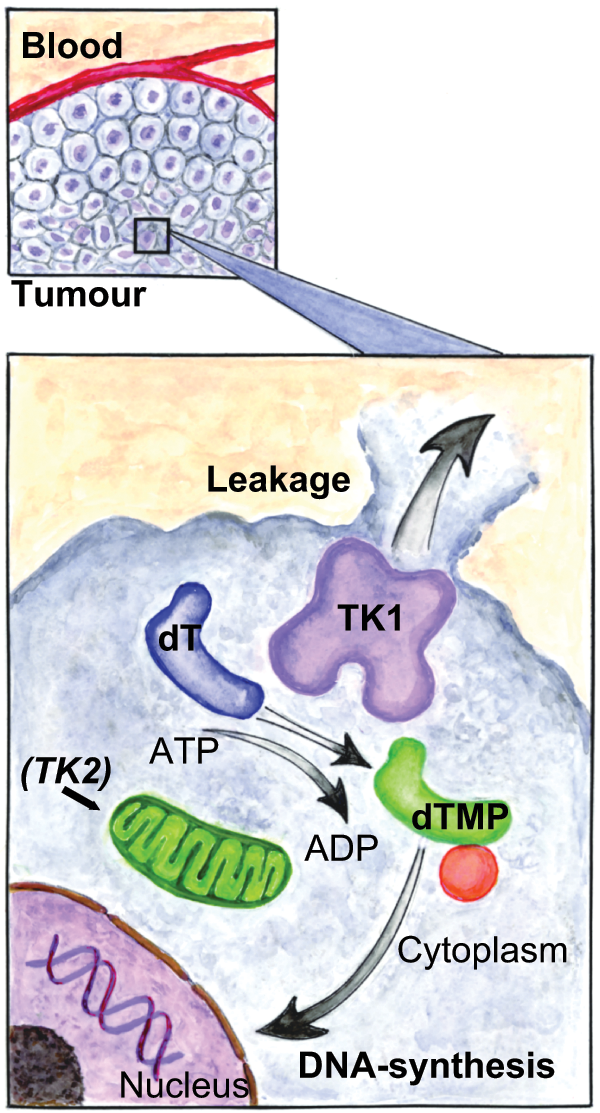
Illustration of the role of thymidine kinase (TK) in the salvage pathway of pyrimidine synthesis. Two isoenzymes of TK exist. TK2 is expressed in the mitochondrion and is present during the entire cell cycle. Leakage of TK1 through the cell membrane reflects either the overall degree of DNA synthesis or the number of cells dying in the replicative stage. In the presence of adenosine triphosphate (ATP), TK1 catalyses the conversion of deoxythymidine (dT) to deoxythymidine monophosphate (dTMP). Deoxythymidine monophosphate is subsequently phosphorylated to its triphosphate analogue (dTTP) before being a substrate for DNA synthesis. Reproduced from Von Euler et al with kind permission from Wiley-Blackwell Publishing.11 Illustration by Eva Hall
Measurement of serum TK1 (sTK) activity has multiple applications in human clinical oncology, including the early detection of malignancy, monitoring of response to treatment, prognostic information and early detection of relapse. This has been shown to apply to multiple malignancies, including non-Hodgkin’s lymphoma, leukaemia, as well as non-haematopoietic neoplasia.9,10 TK activity has also been studied in canine oncology where levels have been shown to reflect clinical stage of lymphoma, as well as prognosis, and have utility in the prediction of relapse before the development of clinically detectable disease. 11
Experience of measurement of sTK activity in cats is limited to a small pilot study which indicated the potential of this marker in feline lymphoma and justified further study. 12 Therefore, the first aim of the current study was to measure sTK activity in a group of clinically healthy cats to create a reference interval. The second aim was to evaluate the potential clinical utility of sTK activity as a marker for lymphoma by measuring sTK activity in cats with inflammatory disease, non-haematopoietic neoplasia (NHPN) and lymphoma.
Materials and methods
Assay of TK activity
sTK activity was measured using a radioenzyme technique (Prolifigen TK-REA; DiaSorin) in which the substrate analogue 125I-iododeoxyuridine is converted to 125I-iododexoxyuridine monophosphate by TK. The phosphorylated product is absorbed onto a separator tablet (aluminium hydroxide) and radioactivity measured after several washings. TK activity is then reported as units per litre (U/l), where one unit corresponds to a substrate turnover of 1.2 × 10⁻¹² katal (mol/s). The linear range of the assay was 1–100 U/l. All assays were performed in the UK by the same operator (HE) and the same operator analysed all the Swedish samples.
Clinically healthy cats
Excess stored serum samples from clinically healthy cats were used in the study. Twenty-seven samples had been collected from cats as part of the Pet Travel Scheme in the UK. 13 Twenty-two samples were collected from cats at the Swedish University of Agricultural Science (SLU) during a health check prior to neutering with owner consent, and meeting criteria from the Swedish Ethical Committee and the Swedish Board of Agriculture. All cats were examined by a veterinarian at the time of sampling and reported to be clinically healthy.
Clinical cases
Clinical cases were identified at three Veterinary Hospitals (Bristol, Cambridge and Uppsala) following a search of clinical records for cats with the following diagnoses: lymphoma, inflammatory disease and NHPN. Cases that met the following inclusion criteria were included in the study: age greater than 6 months (owing to potential effects of age on TK activity levels), 11 availability of non-haemolysed stored excess serum, complete case history and follow-up information according to specific group requirements as detailed below.
Cases of lymphoma
Cases of lymphoma were included in the study if they were diagnosed with lymphoma after histopathological or cytological examination of samples collected either via biopsy or at post mortem. Cases receiving chemotherapeutic treatment (including corticosteroids) in the 3 months prior to the diagnosis were excluded. Lymphoma anatomical locations were extracted from the clinical notes and described as gastrointestinal (including hepatic), extranodal (nasal, renal, central nervous system, ocular, other), multicentric or mediastinal. 2 FeLV and feline immunodeficiency virus (FIV) status were recorded in each case where available.
Cases of inflammatory disease
Cases were classified as inflammatory and included in the study if they had a confirmed diagnosis of inflammatory disease based on a combination of clinical findings, diagnostic imaging, clinicopathological results, bacterial culture, cytology and histopathology. In addition, inclusion in this category required follow-up information available for the 3 months post-sampling, during which time the diagnosis had not changed and haematopoietic or other neoplasia had not developed. Follow-up information was obtained from updated case records or via contact with the veterinarians in charge of the case. Cases receiving corticosteroid treatment or other immunosuppressive medications in the 3 months before sample collection were excluded.
Cases of NHPN
Cases with NHPN were included in the study if they had not received chemotherapeutic treatment (including corticosteroids) in the 3 months before sample collection and the diagnosis was confirmed after histopathological or cytological examination of samples collected either via biopsy or at post mortem.
Statistical analysis
All data analysis was performed using JMP version 9 (SAS Institute). For the purpose of statistical analysis only, samples with sTK activity results that fell outside the linear range of the assay (<1 or >100 U/l) were assigned values of 1 or 100, respectively. The upper limit of the reference interval for normal cats was calculated as the 1.96 × SD. The Tukey–Kramer test was used to identify differences between groups (based on an assumption of normality in larger populations) and Dunn’s multiple comparison test was employed for post hoc analysis. Sensitivity and specificity values for the lymphoma group were calculated using receiver–operator characteristic (ROC) analysis. Significance was taken as P <0.05 throughout.
Results
Clinically healthy cats
This group consisted of 49 cats with a median age of 49 months (range 6 months to 198 months). The group included 24 males (18 neutered, six entire) and 25 females (13 neutered and 12 entire). The majority were non-pedigree cats [43 domestic shorthair (DSH), 87.8%] with two Persians, one Tonkinese, one Burmese, one Abyssinian and one Russian-blue.
Clinical cases
One hundred and twenty-two cases met the inclusion criteria, including 85 cats seen at The University of Bristol, 31 at The Swedish University of Agricultural Science and six at The University of Cambridge. Cases included 55 cats with inflammatory disease, 34 with non-haematopoietic neoplasia and 33 with lymphoma.
Lymphoma group
This group consisted of 33 cats with a median age of 9 years (range 12–208 months). The group included 17 males (16 neutered, one entire) and 16 females (15 neutered and one entire). The majority of cats were non-pedigree [20 DSH, one domestic longhair (DLH)]. Of the remaining cats two were Persians, two were Oriental, two were Siamese, one was Birman, one was Abyssinian, one was Cornish Rex, one was Tonkinese, one was Somali and one was Bengal.
Based on the anatomical location of the lymphoma 17 cats were diagnosed with gastrointestinal lymphoma, 10 with extranodal lymphoma (five with nasal, two with renal, one ocular, one dermal, one with both nasal and renal), three with multicentric, two with mediastinal and one cat with intravascular lymphoma (malignant angioendotheliomatosis). All cats tested negative for FIV. Two cats were positive for FeLV, one cat had multicentric lymphoma and one had gastrointestinal lymphoma.
Inflammatory disease group
This group consisted of 55 cats with a median age of 7 years (range 12–156 months). The group included 40 males (33 neutered, seven entire) and 15 females (13 neutered and two entire). The majority of cats were non-pedigree (60.0%: 30 DSH, three DLH) with a variety of pedigree breeds represented (six Persian, three Siamese, two Tonkinese, two Burmese, two Maine Coon, one Balinese, one Bengal, one Birman, one British Shorthair, one Devon Rex, one European Shorthair and one Oriental Shorthair).
Diagnoses reached for cats in this group included inflammatory bowel disease (sixteen cats, one with additional cholangiohepatitis), chronic rhinitis (six cats), pancreatitis (five cats), acute gastroenteritis and leucocytosis (four cats), mycobacterial infection (three cats), feline infectious peritonitis (four cats), pemphigus foliaceous (two cats), gingivitis (two cats), urolithiasis and cystitis (two cats), pyelonephritis (two cats), bronchopneumonia (two cats) pyothorax (one cat), bacterial meningioencephalitis (one cat), inflammatory arthropathy (one cat), lymphocytic parathyroid inflammation (one cat), otitis media/externa (one cat) and neutrophilic cholangitis (one cat).
NHPN group
Thirty-four cats met the inclusion criteria for this group, with a median age of 10 years (range 2–18 years). The group included 17 males (all neutered) and 17 females (16 neutered and one entire). Breeds in this group included DSH (25; 73.5%), three Siamese, two Bengal, one Persian, one Abyssinian, one Burmese and one Maine Coon.
Neoplasias included 29 carcinomas: pulmonary (six cats), squamous cell (locations on head or oral; five cats), thyroid (four cats), gastrointestinal (five cats), mammary (two cats), nasal (two cats), cervical region (origin not completely determined, not consistent with thyroid, deforming trachea and oesophagus) (one cat), hepatic (one cat), apocrine gland (one cat), renal (one cat), thymic (one cat); five sarcomas: haemangiosarcomas (intra-abdominal one cat; dermal one cat), osteosarcoma (nasal one cat), mast cell tumour (intestinal one cat) and adenoma (adrenal one cat).
sTK activity results
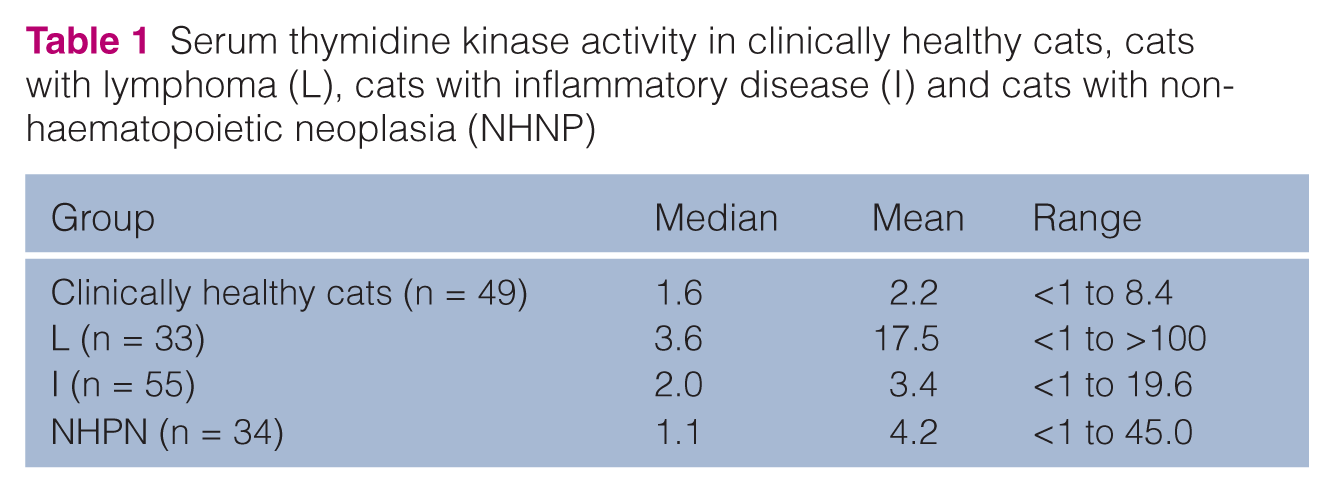
The sTK data was non-normally distributed for all four groups. The median, mean and range sTK activity values for all groups are shown in Table 1 and illustrated in Figure 2.
Serum thymidine kinase activity in clinically healthy cats, cats with lymphoma (L), cats with inflammatory disease (I) and cats with non-haematopoietic neoplasia (NHNP)
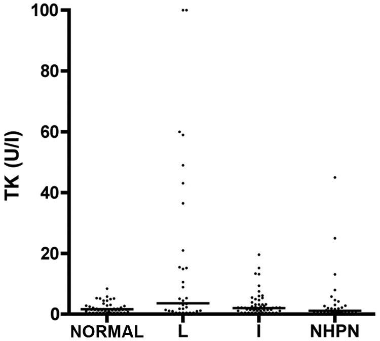
A scatter plot illustrating the serum thymidine kinase 1 (sTK) activity results for the clinically healthy cats (NORMAL), lymphoma group (L), inflammatory group (I) and non-haematopoietic neoplasia group (NHNP). Horizontal lines represent median values and outliers above 100 have been reduced to 100
Clinically healthy cats
Mean sTK activity and SD for this group was 2.2 U/l and 1.7 U/l, respectively, creating a reference interval of <5.5 U/l.
In this group only two cats had sTK activity above the reference interval at 5.8 and 8.4 U/l.
Clinical cases
Lymphoma group
Cats with lymphoma generated a wide variation in sTK activity (mean 17.5 U/l, range <1.0 to >100.0 U/l, SD 27.42), including 20 cats (60.6%) with sTK activity within the reference interval. Of cats with extranodal lymphoma only one cat (10%) had sTK activity above the reference interval (15.2 U/l in a cat with renal lymphoma), but all three cats with multicentric lymphoma generated high values (100, 100 and 43.1 U/l).
Inflammatory disease group
Mean sTK activity in this group was 3.4 U/l (range 1.0–19.6, SD 3.88). In this group, 85.5% of the cats generated results within the reference interval and no cats had sTK activity above 20 U/l. Interestingly, all five cats with pancreatitis had sTK activity above the reference interval (5.5, 7.5, 9.4, 13.4, 19.6 U/l).
NHPN group
Mean sTK activity in this group was 4.3 U/l (range 1.0–45.0, SD 8.55). Five cats (14.7%) with NHNP generated results above the reference interval, including one cat with metastatic haemangiosarcoma with a value of 45 U/l (the highest result in a cat without lymphoma in all groups), a cat with a dermal haemangiosarcoma (25 U/l), a cat with thyroid carcinoma (13.1 U/l), a cat with hepatic carcinoma (8.0 U/l) and a cat with a carcinoma in cervical region (5.8 U/l).
Comparisons between groups
Cats in the lymphoma group had significantly higher sTK activity than clinically healthy cats or cats with inflammatory disease (P <0.0001) and cats with NHPN (P = 0.0002).
There was no significant difference between the sTK activity for the NHPN group and the clinically normal cats, or the inflammatory group and clinically normal cats.
A ROC curve of sTK distinguishing between cats with lymphoma (n = 33) and cats without lymphoma (normal, inflammatory and NHNP group) (n = 138) was produced (Figure 3). The optimal cut point (operating point) for separating cats with lymphoma from cats without lymphoma was 8.9 U/l. The area under the curve (AUC) for sTK activity was 0.66. At the optimal cut point, the true positive rate was 40%, whereas the false-positive rate was 5.8%.
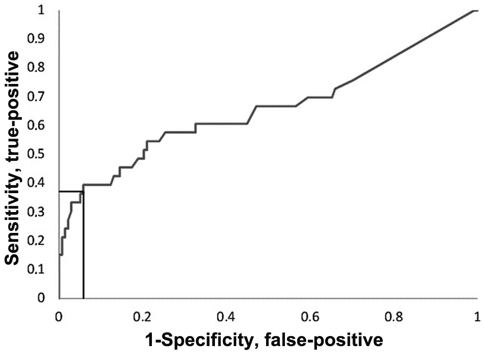
Receiver–operator curve for the diagnosis of lymphoma using serum thymidine kinase 1 (sTK) activity. Lines to axes indicate optimal cut-off point. Area under curve 0.66
Discussion
In human medicine the use of tumour biomarkers is considered standard practice, and provides clinicians with diagnostic and prognostic information. 10 Various markers have been examined in dogs, with some showing clinical utility — measurement of sTK activity being one of them. In dogs, sTK activity has been shown to be significantly higher in patients with lymphoma and higher sTK activity is associated with shorter survival in this species. 11 To our knowledge this is the first study assessing sTK activity in cats and aims to provide a starting point for further research on sTK activity in feline lymphoma.
The present study shows that there is a significantly higher sTK activity in cats with lymphoma than in clinically healthy cats, cats with inflammatory disease or cats with NHPN, suggesting that sTK activity is a biomarker for feline lymphoma. However, the ROC analysis reveals a low sensitivity for the test. This is owing to many lymphoma cases generating values within the reference interval. However, sTK activity values above the reference interval are highly suggestive of lymphoma.
For tumour markers the priority is to identify true positive cases. This is partly achieved in this study by having a fairly high bar for the reference value. Although this will result in a certain subset of lymphomas being missed, a cancer diagnosis should not be decided upon a biomarker only. The present study illustrates that biomarkers should be used as just one tool to confirm the diagnosis. Correctly used, tumour markers can also be used to prompt further investigation to confirm/reject a cancer diagnosis and, once confirmed, fluctuation in the tumour marker in the individual patient can thereafter be used as a surrogate marker for monitoring response.
The lymphoma group showed a wide variability in the sTK activity, as evidenced by the range of results obtained (<1 to >100 U/l). Lymphoma anatomical location is known to affect survival in cats,2,4,5 as does lymphoma type within some locations (small cell alimentary lymphoma is associated with a longer survival than large cell lymphoma, for example) 14 and it is this different biological behaviour, immunophenotypes and tumour burden that may affect sTK activity, but numbers within this study were insufficient for comparison.
Eight cats in the inflammatory group had sTK activity above the reference interval, which, interestingly, included all the cats with pancreatitis. It is known that inflammatory disease can result in brief transient elevations in sTK activity in humans and dogs; the aetiology is unknown, but suggested to be due to rapid proliferation and/or lysis of leukocytes or lysis of host cells at the site of inflammation.15,16
Interestingly, two cats in the present study with haemangiosarcoma had high sTK activity (one the highest in a non-lymphoma cat). Elevated sTK activity has also been reported in dogs with splenic haemangiosarcoma and suggested to be due to the high proliferation rate and intimate association with the vasculature of this tumour. 17
Limitations of the current study include the variable storage conditions of samples. However, it has been shown that sTK stability is maintained despite different durations and temperatures of storage, so this was not thought to have influenced the results. 11 Small-scale studies have been performed by one of the authors (H. von Euler, unpublished observations), demonstrating feline sTK stability despite storage and repeated freezing, and thawing.
In the current study a diagnosis of lymphoma by cytology met the inclusion criteria, but it is accepted that histopathological diagnosis is preferable; moreover, review of the samples by one pathologist, as well as immunohistochemistry, would have reduced the potential for incorrect diagnosis, but was not possible. Immunohistochemistry would also have allowed assessment of correlation between sTK activity and lymphoma subtype.
However, as lymphoma in cats can sometimes be difficult to diagnose because of the frequent occurrence of unspecific clinical signs (eg, inappetence, depression, gastrointestinal signs and weight loss) with no apparent enlargement of peripheral lymph-nodes, documenting a high sTK activity value can be a valuable tool to support further investigations to confirm a diagnosis of lymphoma.
Future studies should include an assessment of survival following a standardised treatment protocol to assess the utility of sTK activity as a prognostic marker in feline lymphoma as it is in humans and dogs.11,18 Such research should be guided towards investigating if cats with lymphoma and sTK values within the reference interval are more likely to achieve complete remission and longer survival than cats with lymphoma and high sTK activity. Furthermore, with increased case numbers it may be possible to compare sTK activity values from cats with lymphoma in different locations and of different phenotypes using immunohistochemistry.
Conclusions
This is the first study investigating the serum biomarker sTK activity in cats with and without lymphoma. It has been shown that the lymphoma group sTK activity was significantly higher than the sTK activity from the groups of cats investigated. It also shows that a high sTK activity level (above the ROC cut-off point of 8.9 U/l) is highly suggestive of a lymphoma diagnosis, while there is a low predictability for cats with low TK values.
Footnotes
Acknowledgements
The authors would like to thank Dr SE Shaw and Professor AR Fooks from the University of Bristol, Bristol, UK, and The Veterinary Laboratories Agency, Weybridge, UK, respectively, for providing samples for the normal group. The authors would also like to acknowledge the AGRIA pet insurance research foundation, Asa-Mari Pettersson for her help in sample collection and Gemma Hoskins for her help searching case records
Funding
Grants to allow completion of this work were kindly provided by the Faculty of Veterinary and Animal Sciences at SLU, the AGRIA pet insurance company research foundation and The School of Veterinary Science, University of Bristol.
Conflict of interest
At the time of data collection SST held a position sponsored by Pfizer Animal Health, and was previously funded by the Feline Advisory Bureau.