
Abstract
Bacterial paronychia or something more sinister?
Nail and nail bed disorders are commonly seen in practice, particularly in older animals. The modern-day practitioner will be all too familiar with the presentation of an ageing or elderly cat with lameness associated with a swollen toe and a layer of purulent discharge around the nail and nail bed. This often occurs because there is less natural wearing of nails in older cats, leading to a thickened nail and secondary infection. Historically, bacterial paronychia has been recognised as the most common disorder affecting feline claws. 1 However, recent studies show that 1 in 8 cases of nail and nail bed disorders are due to neoplasia, and that the common presence of secondary infection leads to many an initial misdiagnosis. 2
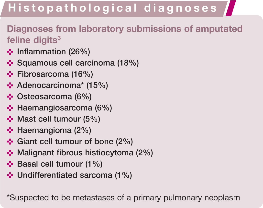
Digits suffering from recurrent infections, or additional gross pathology, are frequently amputated. Significantly, however, a 2007 clinical study of 85 feline digits submitted for histopathology found that 63 showed evidence of neoplasia, with malignant disease affecting 60 (95%) of these cases. 3 Of the tumour types identified, squamous cell carcinoma and fibrosarcoma accounted for 24% and 22% of cases, respectively. However, 21% of the tumours identified were adenocarcinoma, presumed to represent metastatic spread from a primary pulmonary neoplasm: so-called feline ‘lung–digit syndrome’ (Figures 1–3). This syndrome describes a specific clinical presentation of feline primary lung neoplasia, particularly bronchogenic adenocarcinoma, which has metastasised to one or more digits.4,5 This suggests that roughly 1 in 6 laboratory submissions of an amputated feline digit contain a metastatic lesion, where the primary disease may not have been identified.

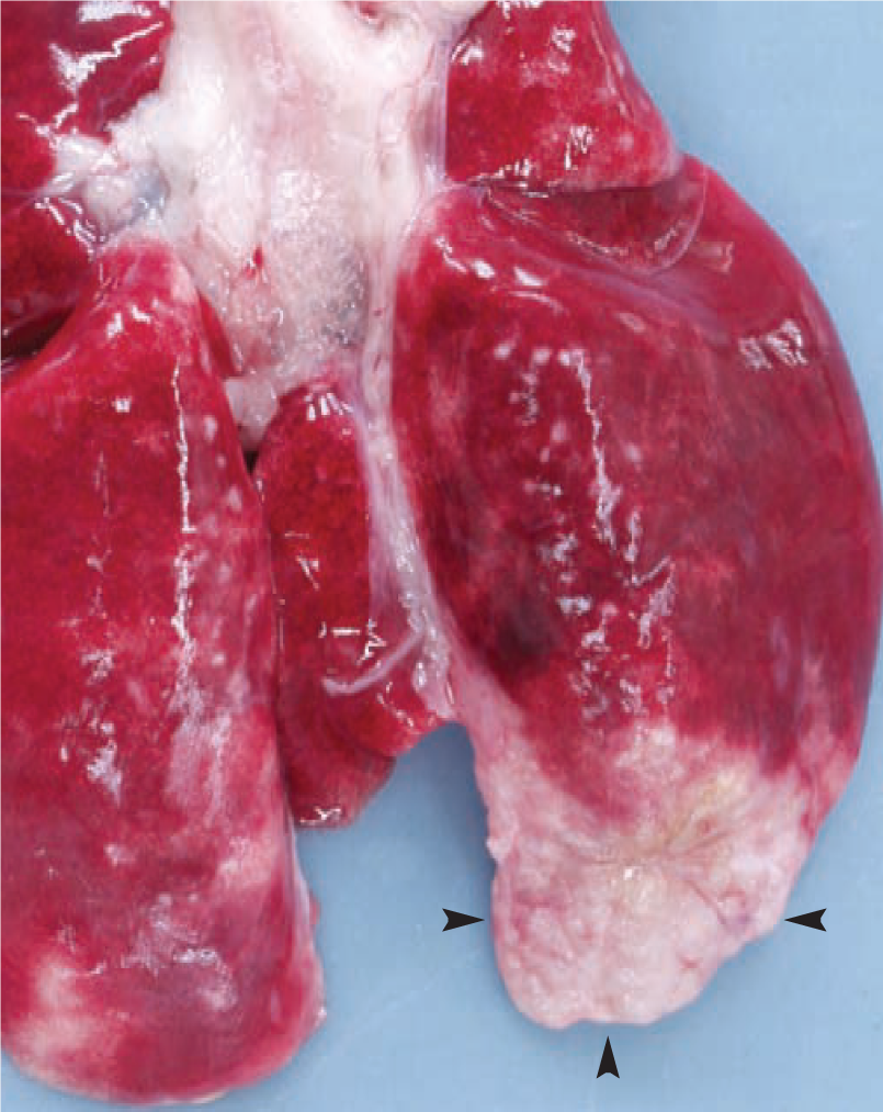
(a) Metastatic lesion from a primary pulmonary adenocarcinoma in the digit of an elderly cat. The cat presented with lameness and clinically evident deviation of the nail, local soft tissue inflammation and a serosanguineous discharge from the nail bed. (b) Radiography revealed osteolysis of the associated phalanges (arrow)

A primary pulmonary neoplasm is evident within the caudal lung fields of the same cat as in Figure 1 on thoracic radiography (lateral view)

Clinical presentation
Metastatic tumours of the digit(s) can cause a variety of different local signs, although the affected animal commonly presents for lameness or painful paws. However, some cases show only minimal signs such as deviation or fixed exsheathment of the nail. 10 Multiple digits are commonly involved, and often on multiple paws. 4 In recent case reports and case series, the most commonly recognised gross clinical signs on presentation included swelling of the distal extremity of the digit, ulceration of the digital skin or nail bed, purulent discharge, and fixed exsheathment, deviation or loss of the nail.4,5,9,11 All digits may be involved, with the exception of the dew claws, in which this condition has not yet been recognised. 11 However, retrospective studies suggest that the weightbearing digits are most commonly affected. 11 Non-specific systemic clinical signs, such as malaise, inappetence, weight loss and pyrexia, are infrequently recognised in these cases. 10

Feline lung–digit syndrome appears to be more common in the elderly cat, with an average age at presentation of 12 years, although the condition has also been seen in much younger animals (range 4–20 years).4,9,11 There is no apparent sex or breed predisposition, and the syndrome has been recognised in many different purebred lines as well as in the typical moggy.9,11
Preliminary investigations
Given the potential differential diagnoses for cats with clinical signs on multiple digits such as described (see box on page 205 and Figure 4), what can help to identify lung–digit syndrome in practice?

Metastases from primary lung tumours are not the only lesions that can affect the digit, or multiple digits, of cats. Infectious agents living in the environment (eg, Nocardia species, mycobacteria, fungi) and fastidious agents inoculated by biting rodents (rats, mice, voles) can also produce digital infections that tend to be granulomatous or pyogranulomatous in nature. Of course, these infections usually do not have associated lesions evident in chest radiographs, and cytology of aspirates or histology of excisional biopsies almost always reveals the aetiological agent. It is interesting that some infectious agents that spread haematogenously (like cryptococci) can also involve digits and other tissues that tend to be involved in the ‘lung–digit syndrome’. These images show (a and b) feline leprosy due to Mycobacterium lepraemurium; note the different severity in different toes of the same cat; (c and d) Nocardia species infections in two cats; (e) Cladosporium species infection; note that the lesion has a bluish hue imparted by the presence of this pigmented fungus in the cat’s tissues. Courtesy of Richard Malik (a–d), Joanna White (c and d) and Eamonn Lim (e)
Haematology and serum biochemistry
Complete blood counts and serum biochemistry profiles of affected cats are generally unremarkable.4,11 In some cases, non-specific changes such as anaemia (often non-regenerative) or leukocytosis (often neutrophilic) have been identified, 4 as well as biochemical changes such as azotaemia, which may be associated with further tumour metastases at different sites. 8 The vast majority of cases have tested negative for feline immunodeficiency virus and feline leukaemia virus. 4
Paraneoplastic disease was not thought to be involved with this condition, 11 although two recent case reports have suggested an association between hypercalcaemia and malignancy of feline bronchogenic carcinoma.12,13 Endogenous lipid (cholesterol) pneumonia has also been associated with feline bronchogenic carcinoma. 14

Radiography of the digits
Radiography of the digits reveals a classic picture of osteolysis of the third phalanx, with potential invasion of the intra-articular space (P2–P3) and also osteolysis of the second phalanx. 11 This is in marked contrast to the clinical picture in humans where phalangeal metastases do not show spread to neighbouring phalanges or intra-articular invasion. 17 In some feline cases, periosteal proliferation has been seen on all phalanges of the affected foot. 11
Thoracic radiography
Thoracic radiography will, in many cases, identify or suggest an underlying pulmonary neoplasm, and should always be considered prior to surgery or digital amputation.9,11 The classic radiographic appearance of feline primary pulmonary neoplasia is a solitary circumscribed mass in the caudal lung lobes.4,8 However, cases of primary feline neoplasia may present in a number of ways in any, or multiple, lung lobes: a single circumscribed mass, multiple circumscribed masses, lobar consolidation or in a diffuse pattern. Associated pleural effusion may be seen, often causing respiratory compromise.6,8,11,18 Enlargement of the tracheobronchial lymph nodes may also be evident.4,8,18
In some cases, however, the primary neoplastic lesion may not be visible radiographically. 6


Confirmation of diagnosis
Clinical signs and radiography can provide a high index of suspicion for lung–digit syndrome, and, given the associated poor prognosis, many cats are euthanased at this stage. But, in those difficult clinical situations, what can you do to confirm this condition?
True proof of metastatic spread of a pulmonary neoplasm to the digits relies on histopathological confirmation of the same tumour type in both sites, but you need to ensure that you collect appropriate material from these sites, which can often prove difficult to sample.
Digital biopsy samples
To sample the digit, there are essentially four options:
Needle aspirate of the digit (easy to perform but extremely unlikely to provide enough material to allow a good histopathological diagnosis);
Punch or incisional biopsies of affected (eg, ulcerated) tissue;
Avulsed nail;
Full digit amputation.
Any of the first three options may allow disease confirmation, but it is important to note that in a high number of samples neoplastic material may not be identified.1,11 The gold standard for histopathological diagnosis is full digit amputation,3,5 particularly if longitudinal sections are requested; 4 albeit there are obvious drawbacks associated with surgery in a case that may ultimately have a very poor prognosis.
Bacterial (aerobic and anaerobic) and fungal culture from the biopsy sample may also be relevant, and sometimes aspirate cytology can identify saprophytic pathogens (eg, Nocardia species and mycobacteria).4,19–21
Thoracic biopsy samples
Confirmation of pulmonary neoplasia can often be made based on thoracocentesis of pleural effusion, cytology of tracheal washes or bronchoscopic samples, or fine-needle aspiration of a lung mass. However, as with digital samples, biopsies that include the original tissue architecture are generally required to give a complete diagnosis, including site of origin; this is only achieved with lobar resection or on post-mortem examination.5,8,18,22
Pathophysiology
Histopathology of these tumours typically demonstrates highly cellular material with clumps, strands or cords of large mononuclear cells with epithelial morphology. Ciliated epithelial cells, as well as goblet cells, are commonly seen.8–11 Frequent cytoplasmic vacuoles are suggestive of secretory tumours (adenocarcinoma). 8 Signs of inflammation, including degenerate neutrophils, are often found, suggesting inflammation secondary to necrosis. 23 Metastatic lesions are often associated with extensive fibrosis.9,11 In digital lesions, neoplastic cells are most frequently found in the dermis, the dorsal aspect of the digit, or ventral to the footpad.4,11 Demonstration of pulmonary cellular features (ciliated epithelia, goblet cells, periodic acid Schiff-positive secretory material) and some cellular markers (positive staining for CAM 5.2 antibody against keratin) can be used to identify digital tumours as metastatic.4,9
The apparent increased frequency of metastatic spread of feline pulmonary neoplasms to the digits, in comparison with other sites and other species, is due to the angioinvasive properties of these lesions, and subsequent haematological spread. Tumour cells have been frequently identified within the pulmonary and digital arteries in histopathological samples.5,9 It has also previously been established that cats have a high digital blood flow to facilitate heat loss, and it is hypothesised that this explains the increased metastatic rate of tumours to these sites. 24 However, additional factors, such as cell markers or chemical secretions, may also play a role in the pathophysiology of this syndrome.4,25

Treatment and prognosis
The prognosis for a cat with lung–digit syndrome is grave.6,11 One study revealed a median survival time of 67 days after presentation (mean 58 days, range 12–122 days); the majority of cats were euthanased due to persistent lameness, lethargy or anorexia. 11
No effective treatment has yet been demonstrated for feline metastatic digital carcinoma. Amputation has not been shown to be palliative, as further metastases rapidly develop. 11 However, where there is no evidence of metastatic disease, solitary lung neoplasms may be surgically resected. Previous studies have demonstrated a median post-surgery survival time of 698 days for cats with well-differentiated tumours; survival time following resection of poorly differentiated lung tumours had a median of only 75 days. 18 The degree of differentiation of the pulmonary neoplasm has, therefore, been suggested to be a prognostic indicator following surgery. 18 Chemotherapy with mitoxantrone following lobar resection has been documented in some case studies. 22
Other sites of metastases from pulmonary neoplasms
Many other sites of metastases from feline primary pulmonary neoplasms have been identified, including the skin, eyes (fundus), skeletal muscles and bone, as well as more ‘common’ sites of metastases, such as the liver, spleen, kidneys, intestines, lungs and brain.5,8–10 Clinical features in these cases are associated with the site of metastasis: cutaneous lesions are frequently found on the dorsum (and have been loosely termed feline ‘lung–back syndrome’),28,29 ocular lesions tend initially to present as ischaemic areas on the fundus and may lead to blindness,30–33 and musculoskeletal and bone lesions commonly cause lameness and limb pain.34–36 In all cases, a thorough clinical examination and thoracic radiography should suggest the primary aetiology, and this may be confirmed through histopathology.
Footnotes
Key points
Funding
The authors received no specific grant from any funding agency in the public, commercial or not-for-profit sectors for the preparation of this review article.
Conflict of interest
The authors declare that there is no conflict of interest.