



Abstract
L-3-Aminoisobutyric acid (L-BAIBA) is an endogenous compound in human metabolism when thymine and valine undergo catabolism. L-BAIBA represents one of the two isomers of BAIBA in biological systems. BAIBA has been shown to reduce body fat percentage via an increase in fatty acid oxidation and a decrease in hepatic lipogenesis. However, no toxicological effects of L-BAIBA in animals or humans have been established. The present study was designed to evaluate the safety and toxic potentials of this compound, where L-BAIBA was administered orally to Sprague Dawley rats at 100, 300, and 900 mg/kg/day for 90 days. No treatment-related adverse effects were observed in any of the treatment groups. Based on the results, the No-Observed-Adverse-Effect Level (NOAEL) of L-BAIBA was 900 mg/kg/day.
Introduction
Beta-aminoisobutyric acid (also known as 3-aminoisobutyric acid, BAIBA) is a substance that was first discovered in 1951 as a new amino acid in human urine. 1 BAIBA is an endogenous non-protein amino acid (an amino acid apart from the 20 amino acids that make up proteins) in mammals, including humans, and is formed metabolically in the body when the amino acids thymine and valine undergo catabolism.1-3 There are two enantiomers of BAIBA in biological systems, which are the L-isomer (also referred to as the S-isomer, or L-BAIBA), and the D-isomer (also referred to as the R-isomer, or D-BAIBA).1,3
Both enantiomers of BAIBA are endogenously formed via distinctive pathways and studies have revealed the distribution of BAIBA in plasma, urine, and tissues at varying levels.1,4 BAIBA has been shown to reduce body fat percentage through an increase in fatty acid oxidation and a decrease in hepatic lipogenesis in animals.5,6 These observations could potentially benefit humans as well.
The majority of the supplementation studies in animals were done using the total BAIBA. 1 The total BAIBA level, including the L- BAIBA isomer, increases in human plasma and urine after exercise and maybe inversely related to cardiovascular disease.4,7 In physiological investigations after exercise, BAIBA is considered a myokine because it is released by myocytes during muscle contraction and helps to regulate genes in brown adipocytes.1,7 Both isomers of BAIBA are involved in various forms of biological activities related to exercise. A few studies have reported the presence of both isomers in human serum, with the D-isomer being present at a higher level after acute exercise and in women with osteoporosis.4,8 The L-isomer of BAIBA is the predominant form related to skeletal muscle tissue. 7 Kitase et al. 9 observed a greater potency of the L-isomer compared to the D-isomer on osteocyte viability. A cell death assay in the osteocyte cell line MLO-Y4 showed a much greater productive effect of L-BAIBA on MLO-Y4 cells when subjected to oxidative stress.
BAIBA was reported to induce hepatic fatty acid beta-oxidation, browning of white adipose tissue, and contribute to exercise-induced protection from metabolic diseases, by enhancing the expression of peroxisome proliferator-activated receptor-gamma co-activator-1α (PGC-1α). 7 The characterization of brown adipose tissue in the study was conducted through quantification of the uncoupling protein-1 (UCP1) and PGC-1α expressions. 7 Furthermore, BAIBA mitigates insulin resistance (caused by incomplete fatty acid oxidation), inhibits inflammation, and promotes fatty acid oxidation in skeletal muscle tissue by activating the AMP-activated protein kinase (AMPK)-peroxisome proliferator-activated receptor (PPAR)-delta. 10 Increased fatty acid oxidation in skeletal muscle tissue is associated with many beneficial effects such as reduced body fat percentage, reduced body weight, and increased skeletal muscle metabolic rate. 10
BAIBA has also been widely studied in animal models for various outcomes relating to health, notably (but not limited to) the relationship to physical activity and metabolism. For example, more recent studies have shown that BAIBA elicits a protective effect from diet-induced obesity in animal models.5,6,10 The reason for this phenomenon is that L-BAIBA could induce change of white adipose tissue into a beige/brown phenotype, thereby leading to fatty acid oxidation and increased insulin sensitivity. 1 The overall profile of this endogenously produced compound suggests it is innocuous at the dosage tested in animal models.
D-BAIBA and L-BAIBA appear to be two disparate yet highly biologically active compounds. Since the majority of the studies do not distinguish between the two in terms of the metabolic and downstream effects, it is critical to evaluate each isoform separately. L-BAIBA, the main enantiomer in plasma, 11 was chosen for this study.
Given the beneficial effects observed on animal models, L-BAIBA could also stimulate similar effects in humans. However, the safety and risk profile of L-BAIBA has not been established in humans, nor has the recommended daily intake of L-BAIBA in humans been assessed. Therefore, to determine the recommended dosage and safe duration of use of L-BAIBA (as a food ingredient in multiple food categories) in humans, the safety and potential toxicity as well as information pertaining to the No Observed Adverse Effect Level (NOAEL) needs to be established. Once a NOAEL is determined, the value can then be used to calculate an acceptable daily intake (ADI, ADI is 1/100 of NOAEL).
This study was the first study to assess the safety profile of L-BAIBA in an in vivo repeated dose subchronic toxicity study by oral (gavage) route in Sprague Dawley rats.
Materials and Methods
Study Compliance
The subchronic toxicity study of L-BAIBA was conducted in accordance with Organisation for Economic Co-operation and Development (OECD) Principles on Good Laboratory Practice (GLP) [C(97)186/Final] 12 ; OECD Guideline for Testing Chemicals; Health effects; Test Guideline No. 408, “Repeated Dose 90-Day Oral Toxicity Study in Rodents” (adopted on June 25, 2018) 13 ; Standard Operating Procedures (SOPs) at Vedic Lifesciences Pvt. Ltd., Mumbai, India, and a mutually agreed/signed Study Plan. The study was approved by the Institutional Animal Ethics Committee (IAEC) and Committee for the Purpose of Control and Supervision of Experiments on Animals (CPCSEA). The test facility was AAALAC (Association for Assessment and Accreditation of Laboratory Animal Care) accredited. All procedures were conducted in compliance with CPCSEA guidelines.
Test Item
The test item, L-BAIBA (CAS #4249-19-8, MW 103.12 g/mol) (Figure 1), used in this study was manufactured as per cGMP by Nanjing Nutrabuilding Bio-tech Co., Ltd., Nanjing, China. Purity was confirmed using titration assay. The presence of heavy metals was analyzed using Inductively Coupled Plasma Mass Spectrometry (ICP-MS) and microbiological examination was determined using United States Pharmacopeia USP <61> and USP <62>. Structure of L-BAIBA (C4H9NO2). CAS# 4249-19-8 with a molecular weight of 103.12 g/mol.
The test item, L-BAIBA (Batch No. MB20200704), is a white to off-white crystalline powder with an assay purity (titration) of 99.77% and was stored at ambient room temperature (21 -29°C) in a tightly sealed container, away from moisture and direct sunlight. The test item has a shelf life of 2 years.
Animals
Sprague Dawley rats used in this study were procured from a CPCSEA approved vendor, Vivo Bio Tech Ltd. All animals were physically examined and those without visual signs of illness were selected for this study. Before the initiation of the study, nulliparous and non-pregnant female and male rats were acclimatized in the experimental room for 6 and 5 days, respectively. The experimental room was a monitored control environment with adequate air supply (19 air changes per hour), room temperature of 20.2 to 22.8°C, relative humidity of 40 to 69%, and a light/dark cycle of approximately every 12 hours. The experimental room floor and worktops were sanitized with disinfectant (0.5% sodium hypochlorite and/or 0.3% lizol) twice a day on weekdays and at least once a day on holidays.
Up to three animals were housed in clean, sterilized polycarbonate (size: L 34 × 23.5 × H17.5 cm)/polypropylene (size: L 41× B29 ×H17.5 cm) cages and stainless-steel mesh grid top with facilities for holding a water bottle and pellet feed. Autoclaved clean corn cob procured from Ritusri Life Sciences Pvt. Ltd. was used as bedding material. The bedding material was subjected to microbial and contaminant testing and results were recorded as part of the study raw data. Cage rotation was performed at least once per week throughout the study.
One hundred (100) rats were divided into six (6) study groups: four main groups with 10 animals/sex and two recovery groups with 5 animals/sex. The average age at the start of acclimatization was 6 to 7 weeks old with a body weight ranging from 128.11-154.60 g for males and 123.98-144.28 g for females. Weight variation of animals on the day of randomization was within ± 20% of the mean body weight for each sex. All animals were fed ad libitum with autoclaved pellet feed (product code RM3) from SDS (Special Diets Services) except during the scheduled fasting period. Drinking water was processed by reverse osmosis and autoclaved prior to being bottled and was provided ad libitum throughout the study.
Treatment
Animal grouping was conducted according to body weight stratification and randomization, which allocated 10 animals per sex per group to main groups 1 to 4 (G1 to G4) and 5 animals per sex per group to recovery groups G5 and G6. Upon completion of randomization, three sentinel male and female rats were selected from the leftover animals and were subjected to sentinel health monitoring on days 43, 86, and 119.
Animals in groups G2, G3, G4, and G6 were administered orally (gavage) with test item L-BAIBA once daily for 90 consecutive days at a dose volume of 10 mL/kg body weight at the following dose level: 100 mg/kg body weight for G2 (low dose), 300 mg/kg body weight for G3 (mid dose), and 900 mg/kg body weight for G4 (high dose) and G6 (high dose recovery). Animals in groups G1 (vehicle control) and G5 (vehicle control recovery) were administered orally (gavage) once daily with vehicle (autoclaved reverse osmosis water) for 90 consecutive days. Dose formulation solutions were freshly prepared at 0, 10, 30, and 90 mg/mL and were administered within 2 hours of preparation, at a volume of 10 mL/kg, to animals using a 3 mL disposable syringe with either a 14, 16, or 18 sized oral gavage needle. Homogeneity of the test item was maintained by continuous stirring with a magnetic stirrer at ambient room temperature. The homogeneity and stability of the solution were not analyzed since the dose formulation was well dissolved in the vehicle and used immediately after preparation.
Observations
(1) Clinical Signs. All animals were observed for cage side clinical signs once daily throughout the experiment, and for mortality and morbidity twice daily during weekdays and once daily during weekends and public holidays. Detailed clinical signs were performed outside the cage once a week and care was taken to minimize variations in the observation conditions. Observations included changes in the skin, fur, eyes, mucous membranes, the occurrence of secretions and excretions and autonomic activity, changes in gait, posture and response to handling, the presence of clonic or tonic movements, and stereotypy. (2) Functional Observational Battery. Functional observation battery was performed as per in-house Standard Operating Procedure (SOP) in the last treatment week for animals in the main groups and in the last week for the recovery groups. Observations included home cage observations, handling observations, open field observations, measurement of sensory reactivity, and foot splay. Gait and rearing observed during FOBs were used to assess motor activity. (3) Body Weight and Feed Consumption. Individual body weights were measured on the day of receipt, before randomization, prior to first dosing, and weekly thereafter for all groups and additionally on Days 97, 104, 111, 118 for recovery groups (G5 and G6). Terminal fasting body weights were measured on Day 91 for main groups and Day 119 for recovery groups. Feed consumption was recorded weekly for all groups and additionally on Days 90-97, 97-104, 104-111, 111-118 for recovery groups G5 and G6. (4) Ophthalmological Examination. Ophthalmological examination was performed on all animals using a Welch Allyn direct ophthalmoscope prior to the administration of the test item, and in the last treatment week to high dose and control groups. Mydriasis was induced by instilling one to two drops of 1% Tropicamide five minutes before the examination.
Clinical Pathology
Animals were fasted overnight for 13–18 hours prior to blood collection. Water was provided ad libitum during the fasting period. Blood samples (approximately 4.5 mL) were collected before terminal sacrifice on Day 91 for main groups and Day 119 for recovery groups. Blood samples were withdrawn through retro-orbital sinus under isoflurane anesthesia for hematology and clinical chemistry analyses. (1) Hematology. Blood samples (approximately 0.5 mL) were collected in pre-labeled EDTA vials and evaluated for the following haematology parameters using Siemens ADVIA 2120i Haematology System/Analyzer: white blood cells (WBC), red blood cells (RBC), haemoglobin concentration (HGB), haematocrit (HCT), mean corpuscular volume (MCV), mean corpuscular haemoglobin (MCH), mean corpuscular haemoglobin concentration (MCHC), red cell distribution width (RDW), platelet count (PLT), mean platelet volume (MPV), neutrophils (Neut), lymphocytes (Lymph), monocytes (Mono), Eosinophils (Eos), Basophils (Baso), and reticulocyte count (Retic). Blood smears were utilized to evaluate morphological abnormalities and immature cells. Bone marrow smears were fixed in methanol and stained with Giemsa to evaluate for cellularity and morphology. Coagulation analysis of prothrombin time (PT) and activated partial thromboplastin time (APTT) was analyzed using blood (approximately 1.0 mL) collected in sodium citrate vials on the Sysmex CA-50 coagulation analyzer. All clinical pathology data were evaluated by a board-certified veterinary clinical pathologist. (2) Clinical Chemistry. Blood samples (approximately 2.0 mL) were collected in pre-labeled heparinized vials and centrifuged at 4000 rpm for 10 minutes at 4°C to harvest plasma. The following clinical chemistry parameters were evaluated using Siemens Dimension RxL Max Clinical chemistry analyzer: albumin (XALB), Albumin Globulin Ratio (A:G Ratio, calculated), Alkaline Phosphatase (ALPI), Alanine aminotransferase (ALTI), Aspartate aminotransferase (AST), Blood Urea Nitrogen (BUN), Calcium (CA), Total cholesterol (CHOL), Creatinine (CRE2), Glucose (GLUC), Phosphorus (PHOS), Total bilirubin (TBI), Total Protein (TP), Globulin (GLOB, calculated), Triglyceride (TGL), Low-Density Lipoprotein Cholesterol (ALDL), High-density Lipoprotein Cholesterol (HDL), and Urea (calculated from BUN). Sodium (Na+), potassium (K+), and chloride (Cl-) were evaluated using the Easy Lyte plus Na+/K+/Cl- analyzer. All clinical pathology data were evaluated by a board-certified veterinary clinical pathologist. (3) Thyroid Parameter Measurements. Blood samples (approximately 1.0 mL) were collected through retro-orbital sinus under isoflurane anesthesia into pre-labeled clot activator vials on Day 91 for main groups and Day 119 for recovery groups and were centrifuged at 4000 rpm for 10 minutes at 4°C to harvest serum. Blood collection was conducted in a randomized manner in the morning (approximately between 9:30 am and 1 pm). T3, T4, and TSH parameters were evaluated using ELISA (Enzyme-Linked Immunosorbent Assay, commercially available kit). (4) Urinalysis. Urine samples were collected one day before terminal sacrifice and were centrifuged at 1500 rpm for 3 minutes. Parameters including appearance, clarity, color, and microscopic examination of urine sediment were determined manually. Biochemical parameters such as pH, specific gravity, blood, bilirubin, urobilinogen, ketones, protein, nitrite, glucose, and leucocytes were analyzed using Siemens Cliniteck status Urine analyzer with Multistix® 10 SG urine analysis strips.
Reagent strips were dipped into urine and removed immediately to avoid the dissolving of reagents. Wetted strips were then held horizontally and placed in the urine analyzer for analysis. (5) Estrus Cycle. The estrus cycle of all female rats was evaluated by taking vaginal smears on the day of necropsy.
Pathology
(1) Euthanasia and Gross Pathology (2) Organ Collection The following organs and tissues were collected: adrenal, aorta, bone (sternum) with bone marrow, bone marrow smear (femur), brain (cerebrum, cerebellum, midbrain), caecum, colon, duodenum, epididymides, esophagus, eyes, heart, ileum with peyer’s patches, jejunum, kidneys, liver, lungs, mesenteric and auxiliary lymph nodes, peripheral nerve (sciatic nerve), ovaries and oviducts, pancreas, prostate plus seminal vesicles with coagulation glands, rectum, skeletal muscle (bicep femoris), skin with mammary glands, spinal cord (cervical, thoracic, lumbar), spleen, stomach, testes, thymus, thyroid and parathyroid, trachea, urinary bladder, uterus with cervix, vagina, pituitary, salivary glands, and all gross lesions (if any). All organs collected (except for eyes, testes, and epididymides) were preserved in a 10% neutral buffered formalin solution. Eyes, testes, and epididymides were preserved in modified Davidson’s fluid. Bone marrow smears were prepared from the femur for all animals. (3) Organ Weights. Liver, kidneys, adrenals, testes, epididymides, uterus, ovaries, thymus, spleen, lung, brain, pituitary, prostate plus seminal vesicles with coagulation glands and heart were weighed right after collection and prior to fixation. Thyroid and prostate were weighed after fixation. Paired organs were weighed together. Relative organ weights were calculated by dividing the respective organ weight by the body weight of the animal x 100. (4) Histopathology. Fixed tissues were processed for Hematoxylin and Eosin (H&E) staining. The complete histopathology of preserved organs was conducted on control and high dose groups only. Gross lesions were observed in the uterus of animals No. 61, 65, and 95, thus microscopic evaluation was performed on these animals.
The severity of findings was analyzed using a 4-point scoring system: 0 indicated observations within normal limits; a score of 1 indicated change that slightly exceeds the normal limit; a score of 2 indicated that the lesion can be easily identified but was of limited severity; a score of 3 indicated that the lesion is prominent with a significant potential for increased severity; a score of 4 indicated that the degree of change is severe and involved the majority of the organ.
The findings plus organ weights and gross lesions were reviewed and evaluated by a veterinary pathologist certified with the Diplomate Indian Board of Toxicologic Pathology. Organ weights were compared with the historical range whenever needed (historical ranges were not included in the report).
Statistical Analysis
All raw data (body weight, body weight gain, feed consumption, hematology, clinical chemistry, functional observation battery (foot splay, rearing, grip strength), urine analysis (volume, pH, specific gravity), absolute and relative organ weights) were subjected to statistical analyses using the Systat Version No. 13 Software.
Basic statistics, homogeneity of variance by Bartlett’s Test, ANOVA, Dunnett’s two-sided test for equal variance, and Dunnett T3 for unequal variance were conducted for groups G2, G3, and G4 in comparison to group G1. T-test was conducted for group G5 in comparison to group G6. All test parameters were analyzed at p<0.05 level of significance.
Results
Clinical Signs and Mortality
No mortality or adverse clinical signs of toxicity and mortality/morbidity were observed in all treatment groups in comparison to the control groups. No adverse clinical signs were observed during a detailed clinical examination.
Functional Observational Battery, Body Weight, and Feed Consumption
No treatment-related changes in FOB observations were observed in either sex.
Body Weight and Feed Consumption
No treatment-related changes in body weights were observed (Figure 2 and Figure 3) and no treatment-related changes in body weight gain were observed (Table 1). Effect of L-BAIBA on body weights in male rats. Mean body weights are shown for male rats during a 90-day oral (gavage) toxicity study. The values are presented as means ± standard deviation (10 rats/sex/group for main study group and 5 rats/sex/group for recovery group). Effect of L-BAIBA on body weights in female rats. Mean body weights are shown for female rats during a 90-day oral (gavage) toxicity study. The values are presented as means ± standard deviation (10 rats/sex/group for main study group and 5 rats/sex/group for recovery group). Effect of L-BAIBA on Mean Body Weight Gain. *Statistically significant changes at p < 0.05. Not Applicable (necropsy performed on day 91 for groups G1 to G4).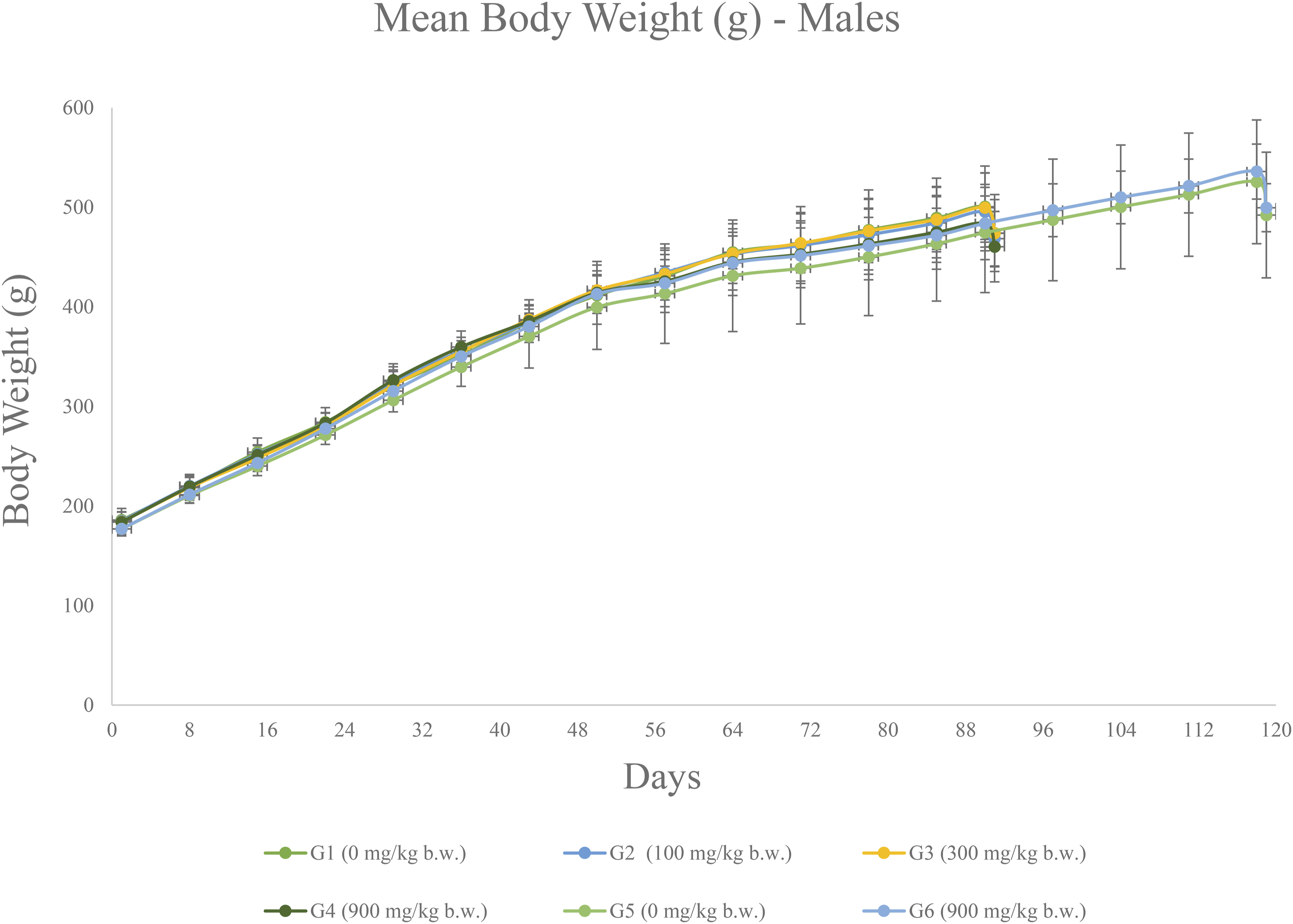


The food consumption in both sexes showed no treatment-related adverse effects and the findings between the treatment groups were comparable to that of the control groups.
Ophthalmological Examination
No abnormality was detected in any of the animals prior to the administration of the test item. Ophthalmologic examination performed in the last treatment week for high dose and control groups revealed no abnormality. Hence, the ophthalmologic examination was not extended to the lower dose and reversal groups.
Clinical Pathology
(1) Hematology. No treatment-related changes in hematology parameters were observed in any treatment groups (Table 2). No morphological abnormality was detected in the blood smears for all treatment groups. No cellular or morphological abnormality were detected in the bone marrow smears in any treatment group. (2) Clinical Chemistry. There were no treatment-related changes in clinical chemistry parameters (Table 3). Even though creatine kinases is a quick indicator of skeletal muscle damage, it was not analyzed because there were no macroscopic findings observed. (3) Thyroid Hormone Evaluation. In males, there were statistically significant increases of T4 in groups G3 (300 mg/kg) and G4 (900 mg/kg), a statistically significant decrease of T4 in group G6 (recovery, 900 mg/kg), and a statistically significant increase of T3 in group G6 (recovery, 900 mg/kg) (Table 4). In females, there was a statistically significant increase of TSH in group G4 (900 mg/kg) (Table 4). Effect of L-BAIBA on Hematological Parameters. Effect of L-BAIBA on Clinical Parameters. *Statistically significant changes at p < 0.05. Summary of Thyroid Measurements. *Statistically significant changes at p < 0.05.


No persistent findings were observed for both the main group and recovery group. T3 and T4 values are inversely proportional to TSH and interlinked. The statistically significant differences observed in T4 were considered incidental since the changes were only observed in Groups 3 and 4, and were not observed in the recovery group.
No prominent histological changes were observed in the thyroid for any treatment group. The observed changes were considered to be incidental and not treatment-related since no correlative histopathology findings were observed. (4) Urinalysis. No significant treatment adverse effects and changes were observed in any treatment group and the results were comparable to that of the control groups (Table 5). The statistically significant decrease in urine volume in group G2 (100 mg/kg) male, the statistically significant decreases in urine volume and pH in group G3 (300 mg/kg) and G4 (900 mg/kg) male, and the statistically significant decrease in urine volume in group G3 (300 mg/kg) female were not dose-dependent and no correlative findings were determined in the clinical pathology. Thus, the changes were considered normal biological variations and not having toxicological significance. Summary of Urinalysis. *Statistically significant changes at p < 0.05. Absence of findings.

Nitrite (as an indication of the presence of nitrite-producing bacteria) and leukocytes are screening tests used to determine the necessity to examine urine sediment and/or culture urine for humans, but are not valuable to animal specimens, especially for urine collected overnight. Thus, the changes in nitrite and leukocytes were not treatment-related. (5) Estrus Cycle. The estrus cycle of all females was determined by taking vaginal smears on the day of necropsy and information was recorded (Table 6). Summary of estrus cycle. Absence of findings.

All clinical pathological changes, statistically significant or not, were determined to represent individual animal variation because they were observed in only one gender, were not dose-dependent, were noted in control animals, or were small.
Pathology
(1) Gross Pathology. No treatment-related abnormality in gross pathology was observed in any treatment group on day 91 or recovery groups on day 119 (Table 7). (2) Organ Weights. There were no treatment-related changes in either the absolute or relative organ weights, and the results were comparable to those of the control groups. (Table 8 and Table 9). No correlative histopathology findings were observed. (3) Histopathology. No test item-related changes were observed in any animals administered with L-BAIBA at the highest dose of 900 mg/kg b.w./day (Table 10 and Table 11). Summary of gross necropsy findings. Absence of findings. Summary of Absolute Organ Weights (g). *Statistically significant changes at p < 0.05. Summary of Relative Organ Weights (%). *Statistically significant changes at p < 0.05. Summary of histopathology findings (male). Absence of findings or not applicable. Summary of histopathology findings (female). Absence of findings or not applicable. In groups G2 and G5, gross lesions of animal no. 61, 65, and 91 were microscopically examined; mild to moderate luminal dilation of the uterus was observed.


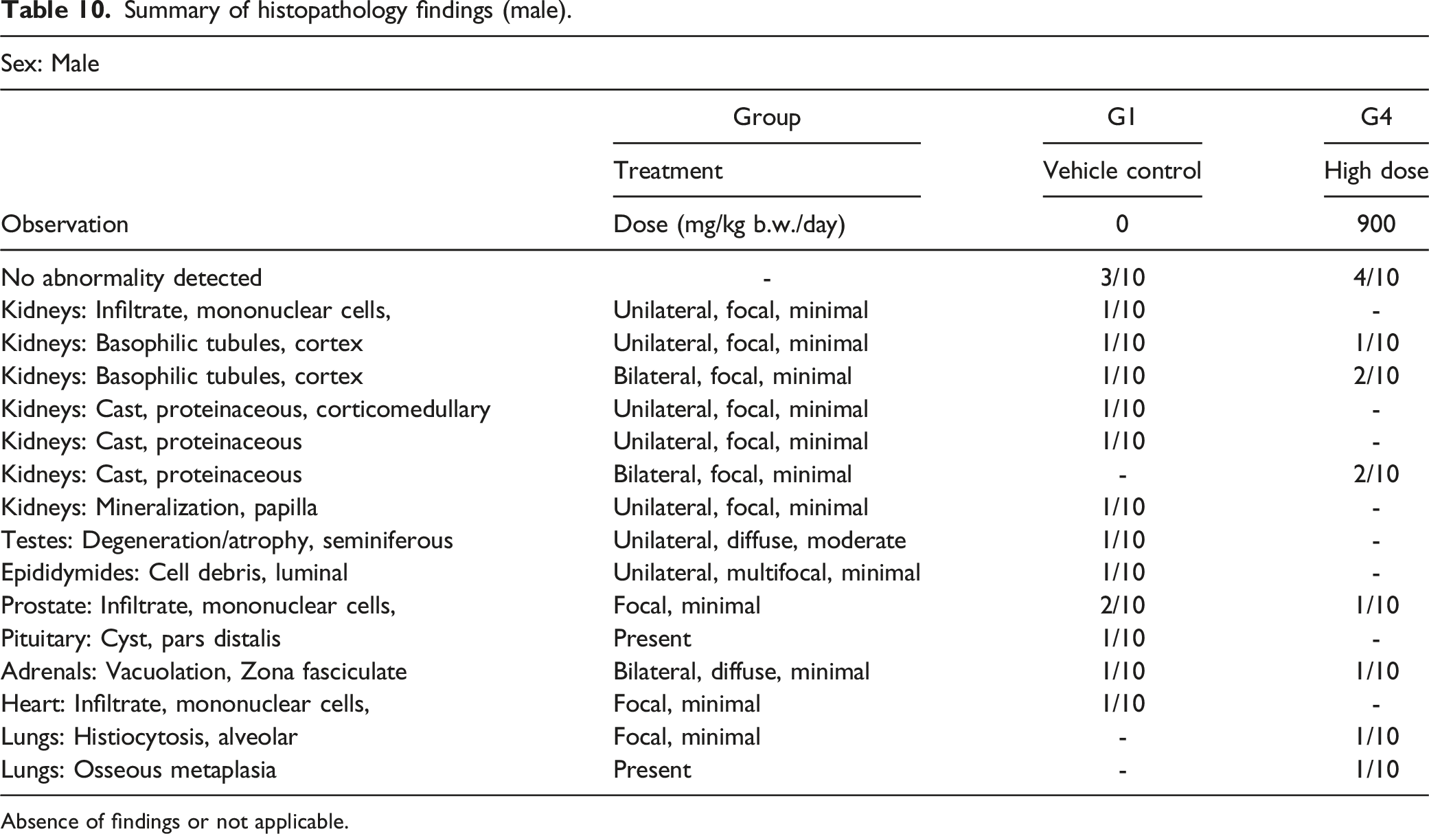
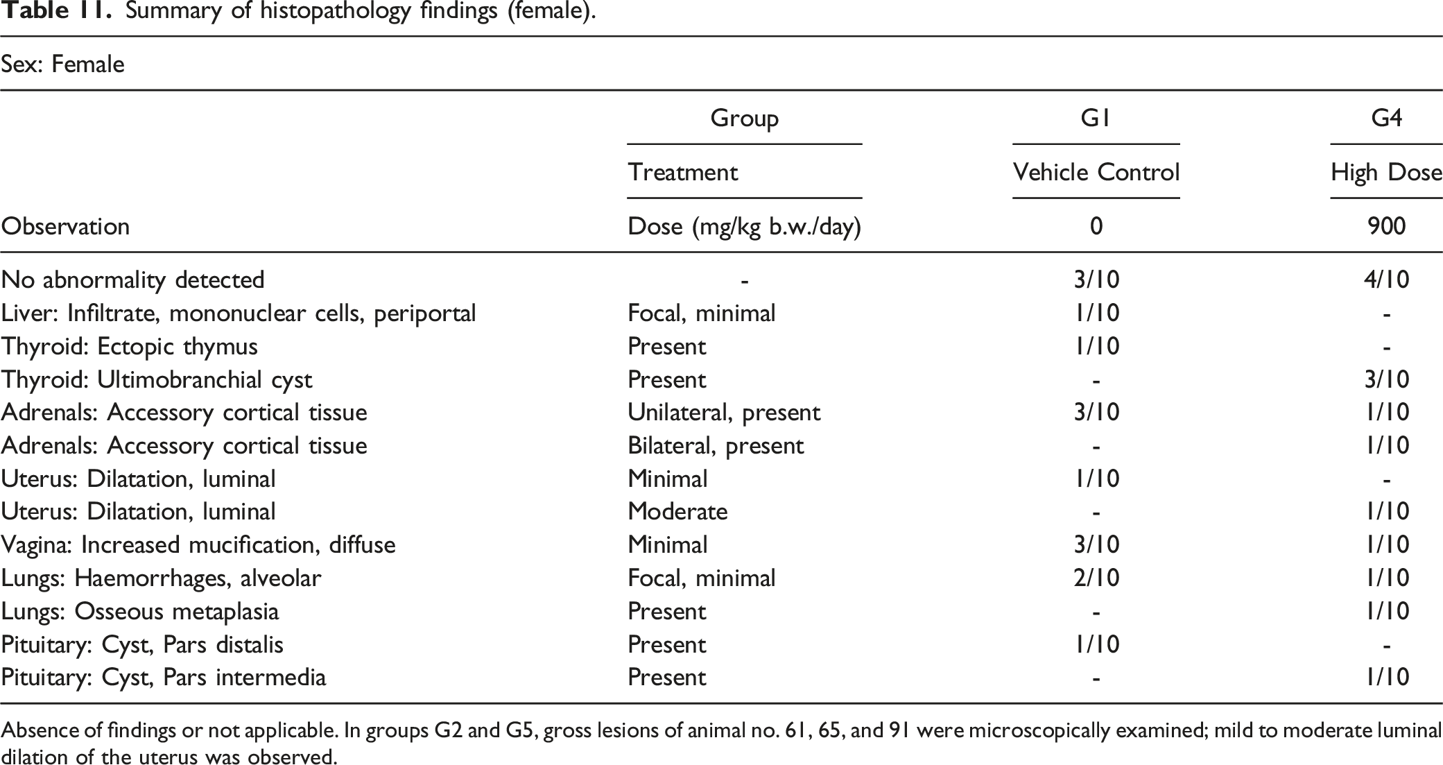
All other microscopic findings were determined to represent individual animal variation because they were observed in only one gender, were not dose-dependent, were noted in control animals, or were small, and were, therefore, considered not test article-related.
Discussion
Subchronic repeated dose toxicity study of L-BAIBA given once daily for 90 days consecutively by oral gavage in Sprague Dawley rats showed no adverse clinical signs of toxicity and mortality/morbidity. There were no treatment-related adverse effects on body weight, body weight gain, feed consumption, functional observational battery, ophthalmological examination, hematology parameters, clinical chemistry parameters, thyroid hormone parameters, and urinalysis parameters analyzed.
Statistically significant changes in the absolute and/or relative brain, adrenal, spleen, and thymus weights were considered incidental because there were no correlative histopathology findings observed. Hence, the changes were not regarded as treatment-related. The small-sized testis and distended uterus observed during gross pathology were also incidental because the change in the testis was seen in only one male from the vehicle control group, and the change observed in the uterus was the normal physiological change and hence, the changes were not treatment-related.
The microscopic changes (such as mononuclear cell infiltrates, mineralization in kidneys, vacuolation in adrenal, cyst in pituitary and degeneration of testis, cell debris in epididymides, etc.) observed in vehicle control and vehicle control recovery groups, low dose, high dose, and high dose recovery groups were not treatment-related because these were either incidental or spontaneous background changes commonly seen in this species and strain.
As noted by the National Toxicology Program, focal liver inflammation is a spontaneous occurrence in rodents, as it is observed in many pre-chronic studies. 14 Mononuclear cell infiltrates seen in the kidney and prostate of rodents are common microscopic observations and represent a background finding in rats. 15 Casts observed in the kidneys of rats were associated with chronic nephropathies and are commonly seen in both rats and mice. 16 Mineralization in kidneys is frequently accompanied by spontaneous and minimal baseline findings due to basophilic deposits in rats and mice. 17 Even though male and female rodents react slightly differently in terms of microvascular vacuolation, it is a common observation in the adrenal cortex of this species. 18 Focal infiltrates of inflammatory cells observed in the hearts of rats are a common feature seen in rodents, while the foci of alveolar histiocytosis are more frequently observed in older and aged rats.18,19 Hemorrhages observed in lungs resulted from the carbon dioxide used for euthanasia, and the osseous metaplasia observed was a background lung lesion. 20 Lung lesions in rodents could arise during the aging process 21 ; no other evidence of irritation was observed with the administration of L-BAIBA. Aging-related lesions were observed in normal rats at as early as 5 weeks of age. 21 These lesions, in rodents euthanized by carbon dioxide asphyxiation, include alveolar histiocytosis, peribronchial lymphocytic aggregates, mesothelial hypertrophy and septal calcification in the lung, capsular fibrosis, cyst(s) in the liver; deep mucosal fibrosis in the duodenum; fatty infiltration in the pancreas; fatty infiltration in the musculoskeletal system; atrophy in the seminal vesicle. 21 In this study, rats were euthanized at 20 to 24 weeks. Therefore, the lesions observed were considered to be incidental and spontaneous lesions in older rats.
Ectopic thymus observed in rats is a developmental abnormality that could occur from time to time.19,22 Cysts observed in the thyroid and adjacent parathyroid of rats are an indication of embryonal rests. Low incidence of tubular degeneration and/or atrophy found in testes is a common observation in rats and mice. 23 Pituitary cysts identified in the par distalis are observed occasionally in rats. 24 The sloughed germ cells and cell debris found in the epididymis were due to testes' degenerations. Uterine dilatation found in rats is a common physiological change and is of no significance. The elevated mucification level is commonly seen even in rats and represents no significant changes. 25 As such, overall, the effects described in the toxicity study were not deemed to be treatment-related.
Several studies have investigated the oral ingestion of BAIBA, which have generally been efficacy-related. The systematic absorption of L-BAIBA is not fully understood. It is hypothesized that L-BAIBA enters the bloodstream after ingestion and travels to the destined organs to induce metabolic effects. Further studies are required in this area.
Kitase et al. 9 observed additional effects of L-BAIBA on osteocytes in mice when compared to the D-isomer. In the study, mice were administered a dose of 100 mg/kg/day to evaluate health outcomes relating to bone health. No deleterious effects were observed. This finding aligns with the absence of toxicity as reported in this study. Kitase et al. 9 demonstrated the innocuous nature of this compound over 2 weeks of supplementation in mice at 100 mg/kg/day of L-BAIBA.
Begriche et al. 5 conducted an oral study in mice with full or partial leptin deficiency to investigate the metabolic effects of BAIBA. The study mice were dosed daily at either 100 or 500 mg/kg/day continuously for 4 months. Quantitative PCR (polymerase chain reaction) results of the elevated expression of LCTP1 (liver carnitine palmitoyltransferase-1) and the elevated plasma level of β-hydroxybutyrate suggested an increase in fatty acid oxidation. In partially leptin-deficient mice, the administration of BAIBA limited the gain of body fat, steatosis and necroinflammation, glucose intolerance, and hypertriglyceridemia. 5 No deleterious effects were seen in the study animals and this finding aligns with the findings in this study.
Roberts et al. 7 conducted several studies in mice for investigations related to exercise and the induction of brown adipocyte specific genes. In a short-term BAIBA study utilizing 6-week old C57BL6/J mice, mice were treated with either 100 or 170 mg/kg/day BAIBA in their drinking water for 2 weeks. Results showed that BAIBA activates the thermogenic program in white adipocytes via PPARα. 7 In another study, 129S4/SvJae-Pparatm1Gonz/J mice were treated with 100 mg/kg/day BAIBA in their drinking water for 2 weeks. For the long-term BAIBA treatment cohort, 6-week-old C57BL6/J mice were treated with 100 mg/kg/day BAIBA in their drinking water for 14 weeks. It was determined that BAIBA decreased weight gain and improved glucose tolerance in mice after 14 weeks. 7 The association of plasma BAIBA levels with metabolic traits was examined in a large human cohort study, in which 2067 random subjects were enrolled in the longitudinal, community-based Framingham Heart Study. BAIBA plasma concentrations in humans were examined before and after an exercise training intervention for 20 weeks. The circulating BAIBA levels in humans increased with exercise, which is in agreement with the findings in mice. 7 No deleterious effects were seen in mice examined in this study for metabolic-related health outcomes of this substance.
Shi et al. 6 conducted a 4-week study of orally administered BAIBA (150 mg/kg/day) in mice to investigate glucose/lipid metabolic changes and effects. Deleterious effects were not observed in this oral study and favorable health outcomes of L-BAIBA related to metabolic disease were observed at a dose of 150 mg/kg/day.
Jung et al. 11 conducted two studies to investigate the effects of BAIBA on insulin resistance and inflammation in mice. The studies demonstrated that BAIBA treatment attenuates insulin resistance, suppresses inflammation, and induces fatty acid oxidation via the AMPK– PPARδ pathway in skeletal muscle. Deleterious effects were not observed in this study and favorable health outcomes relating to insulin resistance and inflammation of L-BAIBA were observed at a dose of 150 mg/kg per day.
Maisonneuve et al. 26 evaluated the effects of zidovudine (d4T), stavudine (AZT), and BAIBA (100 mg/kg per day) on lipid homeostasis in mice for 6 weeks. Results indicated that BAIBA, d4T, and AZT all increased plasma beta-hydroxybutyrate in lean mice, indicating an increased hepatic fatty acid oxidation and ketogenesis. Deleterious effects were not observed in this oral study and favorable effects on lipid homeostasis by BAIBA were observed at a dose of 100 mg/kg per day.
Currently, there are only investigational and metabolic studies about BAIBA in the public search domain. We have conducted the first toxicity study of L-BAIBA and established the first safety profile through an in vivo subchronic study in Sprague Dawley rats. The results demonstrated that repeated oral administration of L-BAIBA at doses up to 900 mg/kg body weight/day are safe and would not result in toxic effects or treatment-related changes. The NOAEL of L-BAIBA was determined to be 900 mg/kg body weight/day when administered to Sprague Dawley rats orally for 90 days followed by a 28-day recovery period. Therefore, the equivalent ADI of L-BAIBA is calculated to be 9 mg/kg body weight.
Rats were chosen for this study because they are the standard and recommended rodent species to evaluate the toxicity of the test item. However, given the differences in endogenous metabolism of amino acids across species, further studies might be needed to evaluate the toxicological effects in mice and other species.
Footnotes
Author Contributions
Shanmugasundaram, D. contributed to conception and design and contributed to acquisition; Fan, Q. contributed to conception and design and contributed to analysis and interpretation; Wang, M. contributed to conception and design and contributed to analysis and interpretation; Yi, R. contributed to conception and design and contributed to analysis and interpretation; Wang, O. contributed to conception and design and contributed to analysis and interpretation. All authors drafted manuscript, critically revised manuscript, gave final approval, and agree to be accountable for all aspects of work ensuring integrity and accuracy.
Declaration of Conflicting Interests
The author(s) declared no potential conflicts of interest with respect to the research, authorship, and/or publication of this article.
Funding
The author(s) disclosed receipt of the following financial support for the research, authorship, and/or publication of this article: This work was supported by Nanjing Nutrabuilding Bio-tech Co., Ltd (NNB NUTRITION).