
Abstract
Antidepressants are the most commonly prescribed drugs for psychiatric treatment, and venlafaxine (VEN) is one of the most popular options. Venlafaxine is a nontricyclic dual-acting serotonin–norepinephrine reuptake inhibitor. Although an increased incidence of acute toxicity and addiction has been reported, controlled studies examining its toxic effects on different organs are still lacking. This study investigated the possible toxic effects of VEN on the liver, kidney, and gastric tissues. Three groups of rats were administered saline, a single LD50 dose (350 mg/kg), or 100 mg/kg VEN daily, followed by increases in the dose of 50 mg/kg every 10 days for 30 days (about 10 times the therapeutic doses). The following parameters of liver and kidney injury were then assayed: alanine aminotransferase, aspartate aminotransferase, γ-glutamyl transferase, prothrombin time, partial thromboplastin time, blood urea nitrogen, and serum creatinine. A histopathological examination was then conducted. Both acute and subchronic administration of VEN produced multiple clinical manifestations in the experimental animals, including seizures, coma, and even death. Moreover, the liver and renal function tests indicated injury in these tissues. Furthermore, the histopathological examination showed signs of organ toxicity after both acute and chronic VEN exposure. This study has shown that VEN has harmful effects on the liver, kidney, and stomach in either a single high dose (LD50) or repeated exposure to 10 times the therapeutic doses. As a result, strategies to increase awareness of these effects among physicians and the public are needed because this drug may be addictive.
Keywords
Introduction
Venlafaxine (VEN) is a nontricyclic phenylethylamine derivative antidepressant. The drug was first marketed in the United States and the United Kingdom in 1994 for the management of major depressive disorder. 1 Venlafaxine is now one of the most frequently prescribed antidepressants worldwide. 2 It is also indicated for neuropathic pain, 3 anxiety, 4 fibromyalgia, 5 among other disorders.
The main pathway of VEN metabolism is its hepatic biotransformation to O-desmethylvenlafaxine, N,O-desmethylvenlafaxine, and to a lesser extent, N-desmethylvenlafaxine. O-desmethylvenlafaxine is the most important active metabolite. 6 Venlafaxine and its metabolites are potent inhibitors of various neuronal reuptake channels in the postsynaptic membrane. Venlafaxine principally acts on the neurotransmitters: serotonin, norepinephrine, and dopamine. In humans, serotonin is affected by low-dose VEN (<150 mg/d). However, increasing VEN doses affects both serotonin and norepinephrine (>150 mg/d). Dopamine is altered by higher drug doses (>300 mg/d). 2
In 2014, VEN was described as a new psychoactive substance by the European Monitoring Center for Drugs and Drug Addiction. 7 Several reports noted the ability of VEN to cause abuse, with amphetamine-like manifestations. A previous study 8 reported a case report of a patient who used more than 2 g/d for over 2 years. Another case of chronic ingestion of VEN reported the use of about 2,250 mg/d. 9
In an acute toxicity study, a survey was conducted between 2001 and 2011 and found that a dose of 64 mg/kg produced moderate to severe toxic manifestations. Manifestations included fever, tachycardia, tachypnea, hypertension, abdominal pain, and seizures. 10
A review of the literature reveals that many case reports describing VEN toxicity are available. 6,11 -13 The reported toxicity included neurological effects, 14 cardiovascular effects, 15 and rhabdomyolysis. 16 Other toxic effects include interstitial pneumonitis 17 and prostatism. 18
However, only a small number of controlled studies have detailed poisoning with this drug. 19 Although VEN exerts well-known toxic effects on the nervous and cardiovascular systems, the potential toxic effects of this drug on other major organ systems must be investigated. Therefore, the aim of this study is to investigate acute and subchronic toxicity of VEN in liver, kidney, and gastric tissues.
Materials and Methods
All experimental procedures were conducted in the Forensic Medicine and Clinical Toxicology Department, Faculty of Medicine, Minia University. Adult female Westar rats were obtained from the Faculty of Veterinary Medicine, Minia University. Ethical approval for the study was granted based on the adherence to the guide on the use and care of laboratory animals accepted by the Committee of Minia University. Female rats were chosen as their hepatocytes were more sensitive to hepatotoxicants than that of males. 20 Animals were housed in stainless steel cages under standard laboratory conditions. Animals were fed standard rat pellets and water was provided ad libitum. The animals were acclimated in the laboratory for 2 weeks before initiating the experiment. Venlafaxine (Effegad ER, 75 mg; Hikma Pharma, Cairo, Egypt) was dissolved in saline immediately prior to oral administration.
This study used 3 groups of rats weighing 200 (7) g (each group consisted of 20 rats). The control group (group I) received normal saline via gavage. The acute toxicity group (group II) was administered a single dose of 350 mg/kg VEN. 21 The third group (group III) was treated with 100 mg/kg/d and the dose was increased by 50 mg/kg every 10 days for 30 days. Francesconi et al documented that patients addicted to VEN may use up to 10 to 15 times the therapeutic doses. 2 We selected doses that were 10 times the uppermost human therapeutic dose (225 mg/d; the maximum therapeutic dose recommended for major depression 22 ) as the maximum dose that will be given to the animal. The dose conversion from human to animals was performed using a previously developed formula depending on the body surface area. 23 The following formula was used 24 :

where Km is a correlation factor that is calculated by dividing the average body weight of the animal (kg) by its surface area (m2): human Km = 37 and rat Km = 6. 24
The body weight of the rats was recorded at the start of the experiment and then every 10 days thereafter. Rats were observed twice daily for the occurrence of convulsions, coma, or death throughout the study. A modified Glasgow Coma Scale was used to assess coma in rats. 25 Rats that died of acute or chronic toxicity exposure to VEN were excluded from biochemical or histological analysis.
Biochemical Analysis
Rats were euthanized by decapitation 24 hours after the administration of the last drug dose. Blood was collected (0.5 mL) via cardiac puncture. Blood samples were centrifuged for 10 minutes at 5,000 rpm. The separated serum samples were used to assess kidney and liver function. The levels of the following enzymes were measured to evaluate liver injury: aspartate aminotransferase (AST), alanine aminotransferase (ALT), alkaline phosphatase (ALP), and γ-glutamyl transferase (GGT). Renal function was assessed by measuring serum creatinine (SC) and blood urea nitrogen (BUN) levels. The assay used in the present study was an enzyme-linked immunosorbent assay manufactured by Ranbaxy Diagnostics Ltd (Hayes, United Kingdom). The prothrombin time (PT) and partial thromboplastin time (PTT) were measured in seconds. The PT, PTT, and serum albumin levels were assessed using the available commercial kits (Quimica Clinica Aplicada s.a., Tarragona, Spain) following the manufacturer’s notes.
Histological Studies
The right lobe of the liver, right kidney, and stomach were excised and then fixed with a buffered 10% formalin solution for 24 hours. Tissues were then embedded in paraffin, sectioned and stained with hematoxylin and eosin for histological examination under a light microscope. 26 A simple scoring system was used to evaluate histological changes of the examined tissues. Changes were classified as follow: (—) none, (+) slight change, (++) moderate change, and (+++) severe change.
Statistical Analysis
All data are presented as the means (standard deviation) and were compared using 1-way analysis of variance. As the data of clinical and histopathological examination did not show normal distribution, these changes were analyzed by Mann-Whitney U test with Bonferroni correction. All P values ≤0.05 were considered statistically significant. All statistical procedures were conducted using SPSS 22 software on a personal computer.
Results
Clinical Findings
Table 1 summarizes the clinical findings in rats with acute and subchronic VEN exposure. It is noteworthy that seizures developed after 30 minutes of VEN administration in rats of the acute toxicity group. Two rats developed convulsive twitches. Myoclonic jerks were observed in another 2 rats. Three rats developed myoclonic jerks that progressed to generalized tonic–colonic seizers. Generalized tonic–clonic convulsions were reported in 5 rats from the start. Seven rats that developed generalized convulsions died. In the chronic toxicity group, only 4 rats developed mild convulsive states. Symptoms ranged from ear and facial twitches to convulsive twitches. Twitches repeated 3 to 4 times in the past 10 days of the study. In acute toxicity study group, death occurred in 9 rats (7 after generalized convulsions and 2 without preceding convulsions). Death occurred within 2 hours after VEN administration. A significant reduction in body weight was observed in rats chronically treated with VEN (Table 2).
Clinical Manifestations of Rats Exposed to Acute and Subchronic VEN Toxicity.a
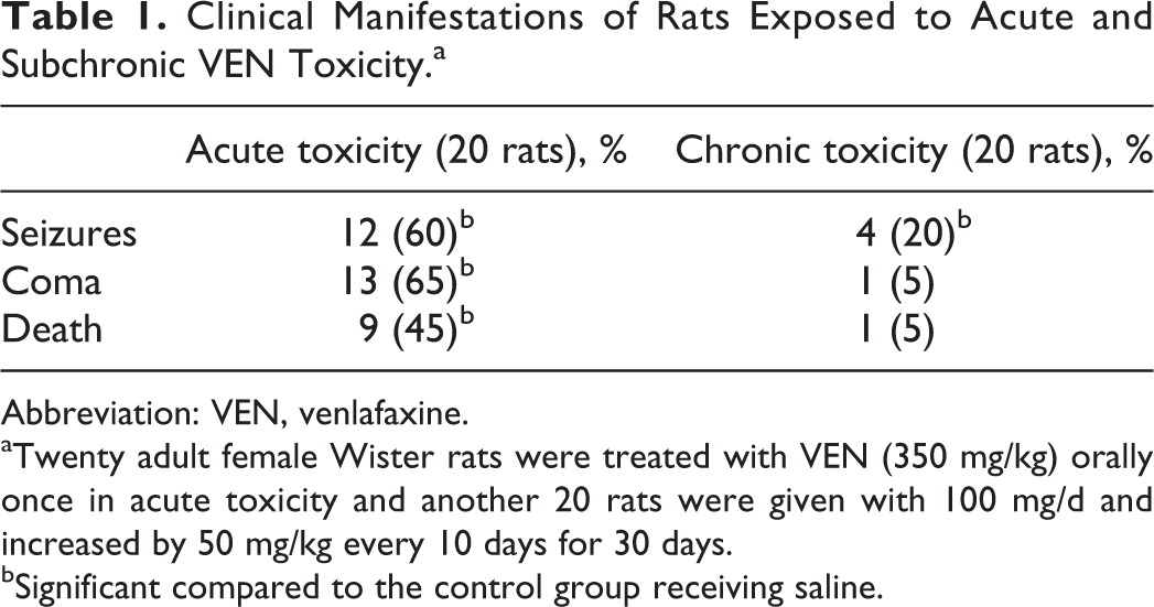
Abbreviation: VEN, venlafaxine.
aTwenty adult female Wister rats were treated with VEN (350 mg/kg) orally once in acute toxicity and another 20 rats were given with 100 mg/d and increased by 50 mg/kg every 10 days for 30 days.
bSignificant compared to the control group receiving saline.
Changes in the Mean Body Weight (in g) of Rats Treated With 100 mg/d VEN Orally and Increased by 50 mg/kg Every 10 days for 30 Days.
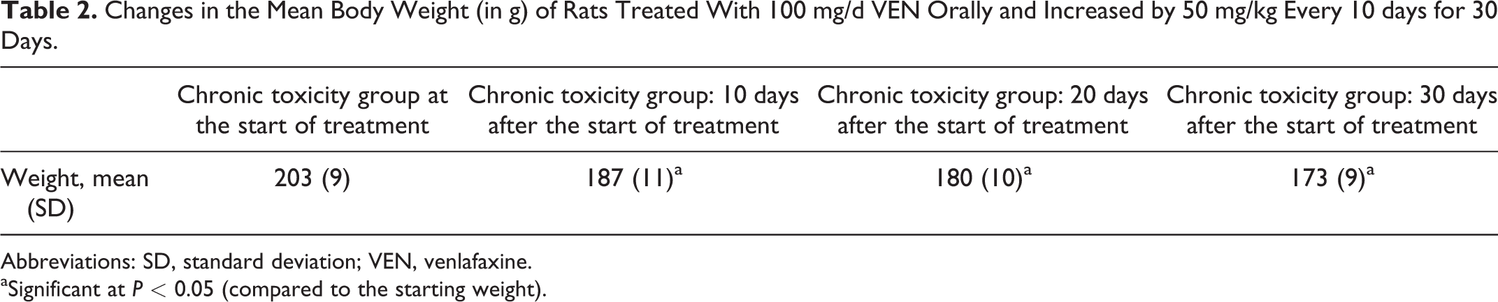
Abbreviations: SD, standard deviation; VEN, venlafaxine.
aSignificant at P < 0.05 (compared to the starting weight).
Biochemical Results
A single treatment with a high dose of VEN produced liver injury in rats. Table 3 shows significant increases in AST, ALT, ALP, and GGT levels in rats that were acutely and chronically treated with VEN. The PT was significantly diminished in both treatment groups. The liver function tests in the subchronic group differed from the control group but did not show the same extent of changes as the acute exposure group.
Effects of VEN Administration on Liver and Kidney Functions in Adult Female Rats.a
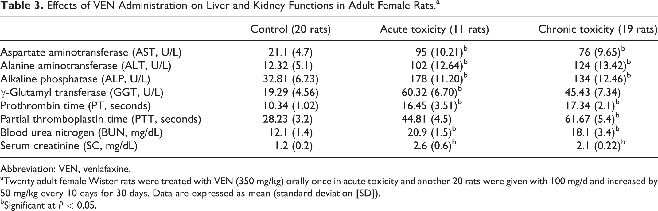
Abbreviation: VEN, venlafaxine.
aTwenty adult female Wister rats were treated with VEN (350 mg/kg) orally once in acute toxicity and another 20 rats were given with 100 mg/d and increased by 50 mg/kg every 10 days for 30 days. Data are expressed as mean (standard deviation [SD]).
bSignificant at P < 0.05.
Regarding the renal functions, the exposure to acute dose of VEN impaired kidney function. Significant elevations in the BUN and SC levels were observed. Subchronic treatment with VEN also altered the kidney function (Table 3).
Histological Results
The control group showed the normal architecture of the gastric tissue (Figure 1A and B). In acute VEN toxicity, there were limited changes in the gastric mucosa in the form of edema of the gastric mucosa that widely separated the gastric glands (Figure 1C and D). The subchronic administration of VEN for 30 days induced numerous superficial mucosal ulcers, degenerated and vacuolated cells of the gastric glands, and submucosal inflammatory cell infiltration.
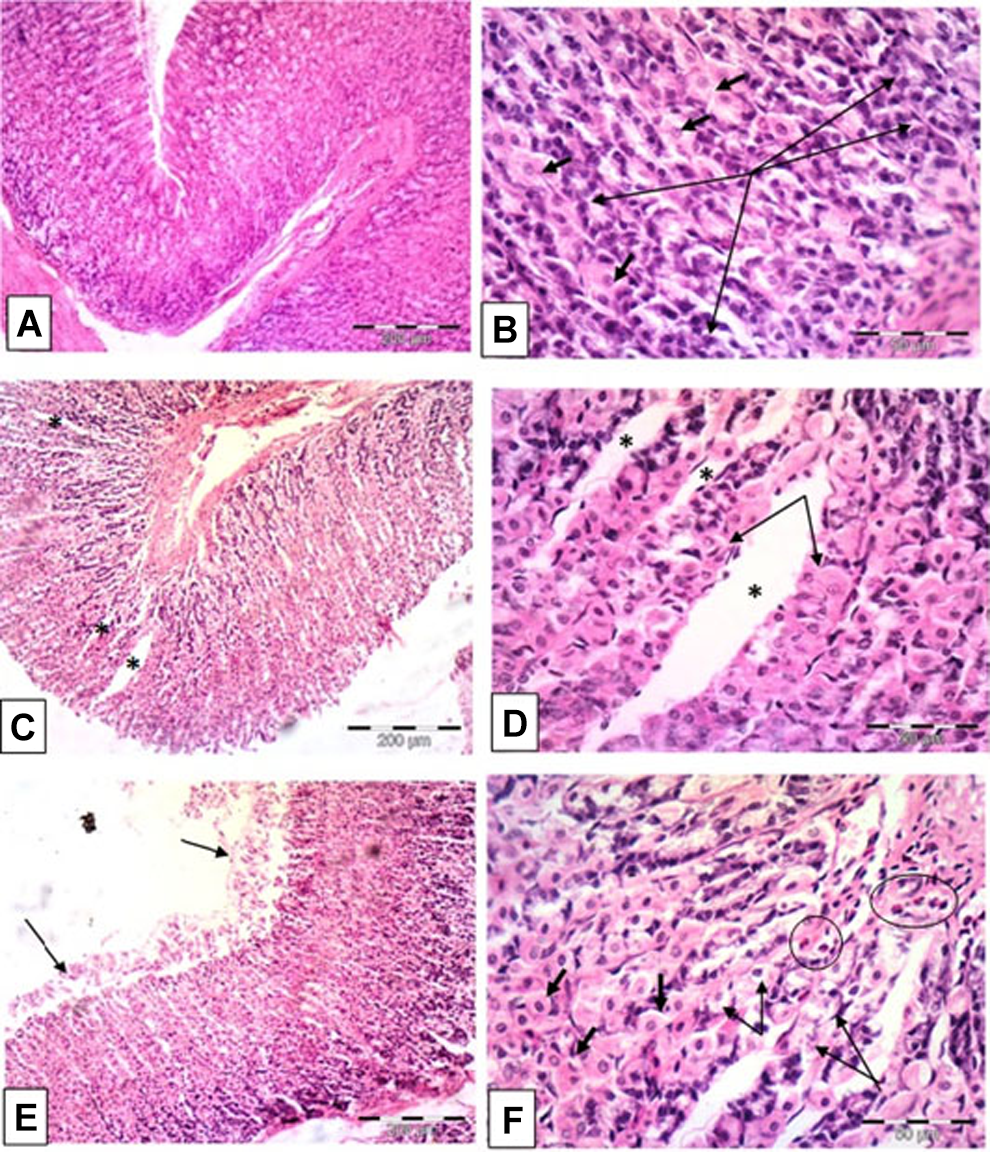
Photomicrographs of the gastric tissue. A and B, The control group (group I) showing the normal architecture of the gastric tissue. Notice the basophilic chief cells (long arrows) and the acidophilic parietal cells (short arrows). C and D, Group II (venlafaxine [VEN] acute toxicity) showing edema (*) of the gastric mucosa widely separating the gastric glands (arrows). E, Group III (VEN subchronic toxicity) showing numerous superficial mucosal ulcers (arrows). F, Group III (VEN subchronic toxicity) showing degenerated and vacuolated cells (long thin arrows) of the gastric glands with predominate parietal cells (short thick arrows) and submucosal inflammatory cell infiltration (circles). Hematoxylin and eosin (H&E): A, C, and E ×100; B, D, and F ×400.
The histological examination of hepatic tissues showed the normal hepatic architecture of the control group (Figure 2A). Rats receiving a single LD50 dose of VEN showed a loss of lobular architecture with markedly degenerated hepatic tissue and inflammatory cell infiltration. Additionally, some degenerated areas showed interstitial hemorrhage; apoptotic hepatocytes with densely stained cytoplasm and pyknotic or fragmented nuclei were also observed (Figure 2B and C). The subchronic toxicity group showed less impairment of liver function and pathology than the acute exposure group. Liver sections from rats treated with VEN for 30 days showed distortion of the architecture, dilated blood sinusoids, and degenerated vacuolated cells surrounding the central vein and portal tract (Figure 2D and E). Histological examination of the renal sections showed the normal cortical architecture of the kidney in the control group formed of renal corpuscles and proximal and distal convoluted tubules (Figure 3A). Acute toxicity group showed marked congestion of the glomeruli with interstitial hemorrhage (Figure 3B). The subchronic treatment with VEN for 30 days produced distortion of the cortical architecture, tubular degeneration, hyperplasia of some tubules; the tubular epithelium was formed of multiple layers instead of the normal single layer, and glomerular hyperplasia (Figure 3C). Table 4 summarizes the histological changes in the liver, kidney, and stomach using the scoring system.
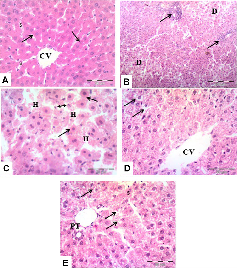
Photomicrographs of the liver tissues. A and B, The control group showing the normal lobular architecture. Notice the hepatocytes with an acidophilic cytoplasm and rounded vesicular nuclei arranged in plates (arrows) surrounding the central veins (CV) and separated by blood sinusoids (S). C, Group II (venlafaxine [VEN] acute toxicity) showing a loss of the lobular architecture with markedly degenerated hepatic tissue (D) and inflammatory cell infiltration (arrows). D, Group II (VEN acute toxicity) demonstrating a degenerating area that shows interstitial hemorrhage (H) and apoptotic hepatocytes with densely stained cytoplasm and pyknotic (arrows) or fragmented nuclei (double head arrow). E, Group III (VEN subchronic toxicity) showing dilated blood sinusoids (s), scattered degenerated vacuolated cells (arrows) surrounding the central vein (CV) or portal tract (PT). Hematoxylin and eosin (H&E): A, C, D, and E ×400; B ×100.
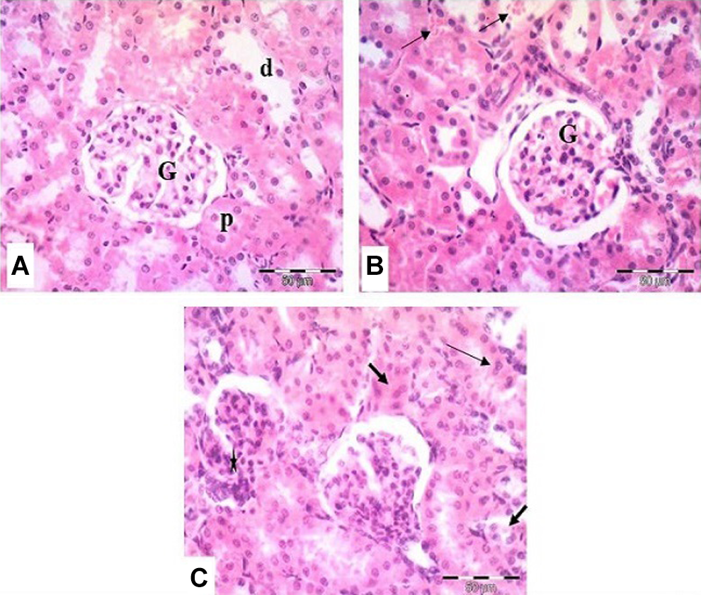
Photomicrographs of the renal cortex. A, The control group showing the normal architecture of the renal cortical tissue. Notice the renal corpuscles (G), proximal convoluted tubules (p), and distal convoluted tubules (d). B, Group II (venlafaxine [VEN] acute toxicity) showing a markedly congested glomerulus (G) and interstitial hemorrhages (arrows). C, Group III (VEN subchronic toxicity) showing tubular distortion (thick arrows), hyperplasia of some tubules, multiple epithelial layers (thin arrows), and glomerular hyperplasia (star). Hematoxylin and eosin (H&E): A-C ×400.
Semiquantitative Scoring System for Histopathological Changes of the Rats’ Livers, Kidneys, and Stomach (Mean [SD]).a,b
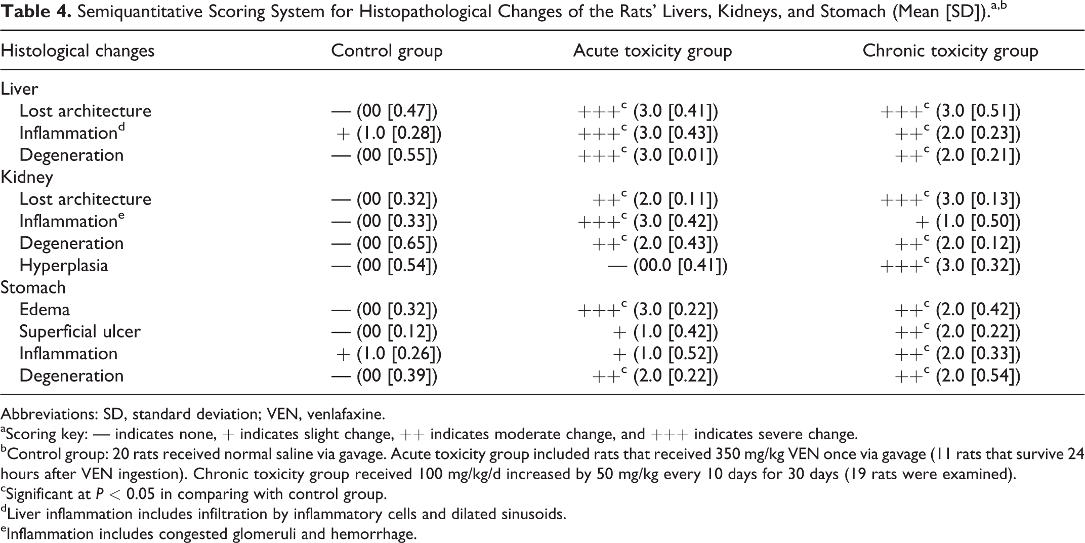
Abbreviations: SD, standard deviation; VEN, venlafaxine.
aScoring key: — indicates none, + indicates slight change, ++ indicates moderate change, and +++ indicates severe change.
bControl group: 20 rats received normal saline via gavage. Acute toxicity group included rats that received 350 mg/kg VEN once via gavage (11 rats that survive 24 hours after VEN ingestion). Chronic toxicity group received 100 mg/kg/d increased by 50 mg/kg every 10 days for 30 days (19 rats were examined).
cSignificant at P < 0.05 in comparing with control group.
dLiver inflammation includes infiltration by inflammatory cells and dilated sinusoids.
eInflammation includes congested glomeruli and hemorrhage.
Discussion
Antidepressants are the most commonly prescribed drugs for psychiatric treatment, and VEN is one of the most popular options. Kumar et al 27 reported a sharp increase in antidepressant toxicity.
Acute VEN treatment in rats produces a high incidence of seizures in 60% of exposed rats. Seizures occur at a lower rate after subchronic treatment (20%). As reported in previous studies, 27,28 seizures are the most common severe complication of VEN overdose, and the probability of seizures increases as the dose increases. 29 The convulsant effect of VEN is attributed to its action on dopamine. At high doses, VEN inhibits dopamine reuptake and increases its presynaptic levels, which induces convulsions. 30
A single LD50 dose of VEN (350 mg/kg) treatment induced fatality in 45% of rats. Conversely, chronic exposure to high doses induces death in 6% of exposed rats. Several reports discussed VEN-associated mortality. 31,32 In most circumstances, the cause of death is related to cardiovascular complications. 10,33
Subchronic administration of high doses of VEN significantly decreased the body weight of the treated rats throughout the experimental period. These results are consistent with findings from previous studies using therapeutic 34,35 and toxic doses. 8 According to another study, VEN has no effect on body weight, in contrast to other antidepressants that led to weight gain. The authors attributed weight gain to the improved mood and appetite caused by antidepressants. 36
Rats exposed to a single highly toxic dose of VEN (350 mg/kg) or subchronically exposed to an increased dose from 100 to 200 mg/kg daily for 30 days showed significant biochemical and pathological changes in the liver. To our knowledge, our study is the first controlled study of the toxic effect of VEN on the hepatic tissue in laboratory animals. The literature review identified several published case reports of VEN-induced hepatotoxicity in humans. 37 -40 Most of the reported cases were exposed to therapeutic doses. In clinical trials, VEN elevated liver enzymes in 0.4% of the 3,000 patients receiving therapeutic doses. 41 The liver damage was attributed to idiosyncratic reactions (dose independent and erratic), which may only occur following the administration of lower therapeutic doses. In the present study, toxic doses of VEN exerted cytotoxic effects on liver cells. One research group 42 reported a case study of a patient exposed to a toxic dose of VEN. Both the biochemical and pathological changes were similar to our findings. In a study on isolated hepatocytes, VEN produced a cytotoxic effect on liver cells. Moreover, VEN induces oxidative stress in liver tissues. The mitochondria and lysosomes are the main sites of VEN-induced cellular injury. 43
Few reports have examined the effects of antidepressants on renal function. A study 44 reported acute renal failure following the ingestion of 15 g of VEN. The authors did not determine whether the renal failure was due to the direct effect of VEN on the kidney or if it was secondary to circulatory failure. Acute exposure to a toxic dose of VEN impairs kidney function, as evidenced by the elevated BUN and SC levels. These changes were accompanied by pathological changes in the renal cortex in the form of congested glomeruli with interstitial hemorrhage and a disturbed architecture of the renal tubules. Subchronic exposure to toxic doses produces glomerular degeneration and hyperplasia. The BUN and SC levels were higher than normal levels but lower than the levels in the acute exposure group. Recent data have confirmed VEN-induced increases in reactive oxygen species–mediated apoptosis and cellular degeneration of nervous tissues after exposure to therapeutic doses. 45,46 Exposure to high, toxic doses of VEN may extend this oxidative stress to other tissues such as the liver, kidney, and stomach. Another report confirmed that antidepressants, including VEN, exert cytotoxic effects on cancer cells. 47
Several studies described the association of gastrointestinal bleeding with antidepressants. 48 -51 In the study by Opatrny et al, 48 VEN had the highest risk of inducing gastrointestinal bleeding among antidepressants. The authors attributed the increased gastrointestinal bleeding risk to the trend toward increased bleeding observed following the use of antidepressants, 52 without any reference to the local effect of VEN on the gastrointestinal mucosa. Based on the results of the present study, VEN exerted a local detrimental effect on the gastric mucosa. The effect of the subchronic treatment was more evident than the effect of the acute treatment. This result may help explain the cause of the increased gastrointestinal bleeding in VEN users.
One limitation of this study is the highly variable toxic VEN dose. A literature review reported an acute toxic dose of 1,500 to 7,500 mg (10 mg/kg in humans). 10 Other studies 19 used intravenous injections of acute toxic doses of 40 to 50 mg/kg in pigs. This dose is similar to the LD50 of female rats (350 mg/kg). Several reports evaluating chronic VEN intake found that variable doses were ingested by abusers. For example, some subjects reported taking 2,100 mg/d, 53 2,100 to 3,750 mg/d , 54 1,950 to 2,100 mg/d, 8 3,600 to 4,050 mg/d, 55 1,500 mg/d, 56 and 2,250 to 3,375 mg/d for variable peroids. 9 Francesconi et al summarized that VEN abusers might use 3 to 5 and up to 10 to 15 times the recommended therapeutic doses. 2
Although VEN is widely used as an antidepressant and a growing incidence of acute toxicity and addiction has been reported, controlled studies examining its effects on different organs are still lacking. To our knowledge, the present report describes the first animal study examining the effects of acute and chronic VEN exposure to toxic doses. Venlafaxine induces harmful effects on the liver, kidney, and stomach. Thus, strategies that increase awareness of these effects among physicians and the public are needed, particularly due to the possibility of abuse of this drug. Further work may address the reversibility of these toxic effects after VEN exposure stops.
Footnotes
Author Contribution
Melad G. Paulis contributed to conception and design, contributed to acquisition and analysis, drafted the manuscript, and critically revised the manuscript. Essam Hafez contributed to conception and design and critically revised the manuscript. Nashwa F. El-Tahawy contributed to conception, contributed to interpretation, drafted the manuscript, and critically revised the manuscript. Mohmed K Aly contributed to design, contributed to acquisition and analysis, drafted the manuscript, and critically revised the manuscript. All authors gave final approval and agree to be accountable for all aspects of work ensuring integrity and accuracy.
Declaration of Conflicting Interests
The author(s) declared no potential conflicts of interest with respect to the research, authorship, and/or publication of this article.
Funding
The author(s) received no financial support for the research, authorship, and/or publication of this article.