
Abstract
The toxicological effects of nanoparticles (NPs) on humans, animals, and environment are largely unknown. Assessment of NPs cytotoxicity depends on the choice of the test system. Due to NPs optical activity and absorption values, they can influence the classical cytotoxicity assay. Eight NPs were spiked in the 3-(4,5-dimethylthiazol-2-yl)-2,5-diphenyltetrazolium bromide (MTT) and crystal violet assays and tested with HaCaT human skin cells. The MTT assay standard curve optical density (OD) measurements were altered by the presence of trisilanol phenyl and trisilanol isooctyl polyhedral oligomeric silsesquioxane particles. The crystal violet standard curve OD measurements were significantly shifted by gold NPs, but they did not affect the MTT assay. Carbon black decreased ODs in the MTT and crystal violet assays and was localized in the cell cytoplasm. These findings strongly indicate that a careful choice of in vitro viability systems is required to avoid flawed measurement of NPs toxicity.
Keywords
Introduction
The wide use of nanomaterials in many areas, including chemistry, medicine, and biology, and incomplete characterization of the toxicity of many nanoparticles (NPs) heighten the demand for occupational health and safety guidelines. 1,2 The creation of these guidelines relies on experimental investigation to characterize the framework for hazards and identification of the risk of exposure to newly synthesized NPs. 3 –6 Industrial workers are in daily risk from occupational exposure. 7 –10 Therefore, it is essential to identify the cytotoxicity of these newly developed NPs. 11 –15 Research has identified the interference of some NPs with some assays, including Alamar Blue, neutral red (NR), 3-(4,5-dimethylthiazol-2-yl)-2,5-diphenyltetrazolium bromide (MTT), and 2-(4-iodophenyl)-3-(4-nitrophenyl)-5-(2,4-disulfophenyl)-2H-tetrazolium (WST-1). 16 –23
Tetrazolium salts are extensively used in the measurements of metabolic activity of cells due to the ease of carrying out the assays. 24 –27 An MTT assay is a versatile and popular assay to assess mitochondrial activity. 28,29 Metabolically active cells reduce water-soluble MTT salts to the form of MTT formazan crystals. 30 This leads to an insoluble MTT formazan, which is found in the cytoplasm, mitochondria, and in some parts of the plasma membranes. 31 The decrease in MTT activity in cells is considered an indicator of cell redox activity. 32 –34
In the current study, we examined the interference of 8 NPs with 2 classical cytotoxicity assays. We selected commonly used cytotoxicity assays with 2 different cytotoxic end points (metabolic activity [MTT] and cell death [crystal violet]). The NPs studied were 4 novel polyhedral oligomeric silsesquioxane (POSS) particles, carbon black, cadmium sulfide (CdS) quantum dots (QDs), silicon dioxide (SiO2), and gold nanoparticles (AuNPs).
Materials and Methods
Particles Used for Testing
Table 1 illustrates the physical and chemical characteristics of POSS particles, SiO2 and carbon black. Polyhedral oligomeric silsesquioxane, CdS QDs, and AuNPs were provided by the Department of Chemical and Physical Sciences, Flinders University in South Australia. Silicon dioxide and carbon black were purchased from Sigma-Aldrich, Australia.
Physical and Chemical Characteristics of Nanoparticles Tested.a
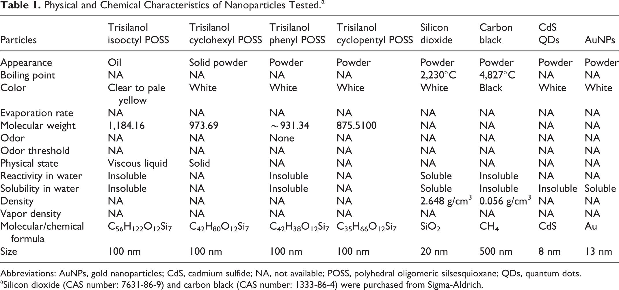
Abbreviations: AuNPs, gold nanoparticles; CdS, cadmium sulfide; NA, not available; POSS, polyhedral oligomeric silsesquioxane; QDs, quantum dots.
aSilicon dioxide (CAS number: 7631-86-9) and carbon black (CAS number: 1333-86-4) were purchased from Sigma-Aldrich.
Cell Culture
HaCaT cell line originated from American Type Culture Collection. Cells were grown in Roswell Park Memorial Institute (RPMI) medium with 10% fetal bovine serum and incubated at 37ºC, 5% CO2 in a humidified incubator. Cells were established at 2 × 10⁶ cells/mL and subcultured when confluence reached 60% to 70% every 2 to 3 days. 35,36
Cell Exposure to Carbon Black for Transmission Electron Microscope
HaCaT cells were seeded, and a cell suspension of 2 × 10⁶ cells/mL was exposed for 24 hours to 10 mg/mL carbon black. The cells were fixed in 1% glutaraldehyde, 4% formaldehyde, and in 0.1 mol phosphate buffer (PB; pH 7.4) by mixing equal volume of fixative and cell suspension. 37,38 Cells were transferred to a centrifuge tube and spun for 10 minutes at 1,200 rounds per minute and then the fixative was removed. Fresh fixative was added for approximately 2 hours and then it was also removed. Three repetitions for 15 minutes of 8% of (0.2 mol) sucrose in 0.1 mol PB at 4°C were done, followed by 1-hour postfix with 1% osmium tetroxide (OsO4) in 0.1 mol PB. Lastly, OsO4 was aspirated and the pellet was rinsed in 0.1 mol PB 3 times for 10 minutes.
Dehydration of cell suspension was carried out once by 50%, 70%, and 95% of ethanol and twice by 100% for 15 minutes, then twice by 100% propylene oxide for 15 minutes. The cell suspension was embedded in beam capsules and baked in 60°C oven for 48 hours. Finally, the cell suspension was sectioned (0.5-2 μm) and stained with uranyl acetate and lead citrate.
3-(4,5-Dimethylthiazol-2-yl)-2,5-Diphenyltetrazolium Bromide Assay Standard Curve Procedure
3-(4,5-Dimethylthiazol-2-yl)-2,5-diphenyltetrazolium bromide assay standard curves were carried out in a 96-well flat bottom plate. 39 The standard curves were prepared by halving serial dilutions of cells in 4 technical replicate wells, starting from 40,000 cells/well to 78 cells/well. 40 The final volume of cell suspension in each well was 100 μL. The plates were incubated at 37°C, 5% CO2 in a humidified incubator for 18 hours. Media was then removed and 0.5 mg/mL of MTT solution was added to each well (200 μL/well). The plates were incubated for 4 hours at 37°C, 5% CO2 in a humidified incubator. Sodium dodecyl sulfate (SDS; 80 μL of 20% solution) was added, and the plates were incubated in the dark at room temperature overnight. Absorbance was read at a primary wavelength of 570 nm and a reference wavelength of 630 nm. In addition, the absorbance of the dilutions of specific NPs in RPMI was also determined at the primary and reference wavelength.
3-(4,5-Dimethylthiazol-2-yl)-2,5-Diphenyltetrazolium Bromide Assay Particle Interference Experimental Procedure
Four replicate standard curve plates were prepared for each interference assay. Replicate 1 was the media control plate. Replicate 2 was the solvent control plate, if required, when the particle was suspended in other than media. Replicate 3 had NPs added at the same time as the MTT. Replicate 4 had NPs added with the SDS-solubilizing solution. Doses tested for each NP are shown in Table 2.
Particles Toxicity and Interference Doses Tested.a

Abbreviations: CdS, cadmium sulfide; DMSO, dimethyl sulfoxide; POSS, polyhedral oligomeric silsesquioxane; QDs, quantum dots; SPB, sodium phosphate buffer.
aThey are based on the doses predicted to be approximately 1 in a 1,000 dilution of the maximum solubility for each nanoparticle. 41 This dose was chosen because it was predicted to be the maximum residual dose after performing a treatment experiment and then washing it 3 times.
Crystal Violet Assay Standard Curve Procedure
Crystal violet standard curve plates were set up identically to the MTT standard curve plates, except that 6 technique replicate wells were used per concentration of cells/well. After overnight incubation, media was aspirated and replaced by 50 μL of crystal violet stain and then incubated at room temperature for 15 minutes. The stain was washed off with demineralized water and plates left to dry overnight. A 33% (vol/vol) acetic acid solution was then added and the optical densities (ODs) read using a spectrophotometer at 570 nm. 42
Crystal Violet Assay Experimental Procedure Particle Interference
Three standard curve replicate plates were used. Replicate 1 was the media control; replicate 2 was the solvent control (solvent added with the stain); and replicate 3 had the test particle added with the stain.
Statistical Analysis
The data were analyzed as mean ± standard error of the mean of at least 3 independent experiments using 1-way analysis of variance and Tukey-Kramer multiple comparisons test using SPSS software to compare exposure groups. All comparisons were considered with the significant level of P < 0.05.
Results
For the analysis of NPs interference with popular in vitro toxicity test systems, we tested 8 NPs on HaCat skin cells for 2 classical bioassays. Polyhedral oligomeric silsesquioxane particles and carbon black with significant interference were also tested with RPMI media only in a dose response to verify any OD properties of those particles in the absence of cells (Table 3).
ODs for Dilutions in Media of Nanoparticles That Showed Significant Interference With MTT Standard Curves (Figure 1).a
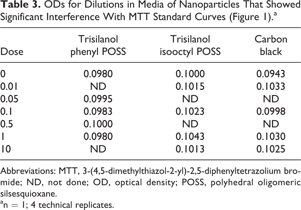
Abbreviations: MTT, 3-(4,5-dimethylthiazol-2-yl)-2,5-diphenyltetrazolium bromide; ND, not done; OD, optical density; POSS, polyhedral oligomeric silsesquioxane.
an = 1; 4 technical replicates.
Particle Interference—MTT Assay
Of the 8 NPs examined for potential interference with the components of the MTT assay, the presence of trisilanol phenyl POSS and trisilanol isooctyl POSS resulted in a significant alteration in MTT standard curve (Figure 1). There were decrease in ODs for ≥10,000 cells/well when the particles were added with either the MTT or the SDS. These changes were significant (P ≤ 0.05). As it can be seen from Figure 1, there were smaller nonsignificant changes for ethanol and trisilanol cyclopentyl POSS when added with SDS in MTT.
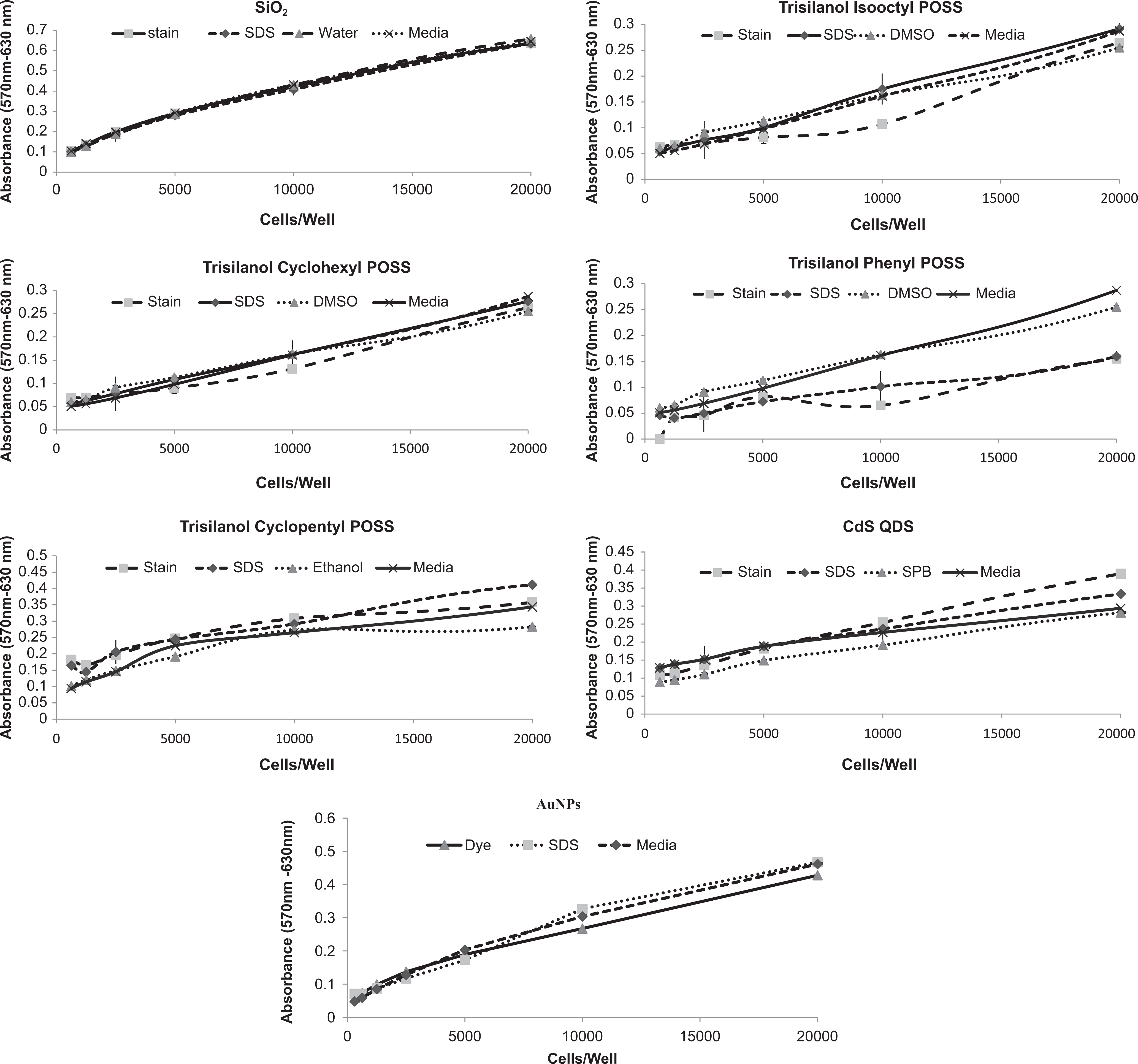
The MTT standard curves generated using HaCaT cells in the absence and presence of nanoparticles, and the relevant solvents indicated. Media indicates untreated standard curve. MTT indicates NP added concurrently with MTT. SDS indicates NP added concurrently with SDS. Water, DMSO, ethanol, and SPB indicate solvent controls with no particles added. DMSO indicates dimethyl sulfoxide; MTT, 3-(4,5-dimethylthiazol-2-yl)-2,5-diphenyltetrazolium bromide; NP, nanoparticle; SDS, sodium dodecyl sulfate; SPB, sodium phosphate buffer.
Particle Interference—Crystal Violet Assay
Altered OD values in the crystal violet assay standard curves were observed when NPs and solvents were added to the assay plates. Standard curves for the solvent controls of SiO2, trisilanol isooctyl, cyclohexy, and phenyl showed similar OD readings to the NPs (Figure 2). In all the particles, the differences between control standard curve media OD values and the OD values with NPs increased, as the cell densities (cells/well) increased. Gold NPs significantly shifted the crystal violet standard ODs. In contrast, for SiO2 and trisilanol cyclopentyl POSS, there was no significant change in the shape of the crystal violet standard curve line when the particles of the negative solvent was present (P ≤ 0.05).
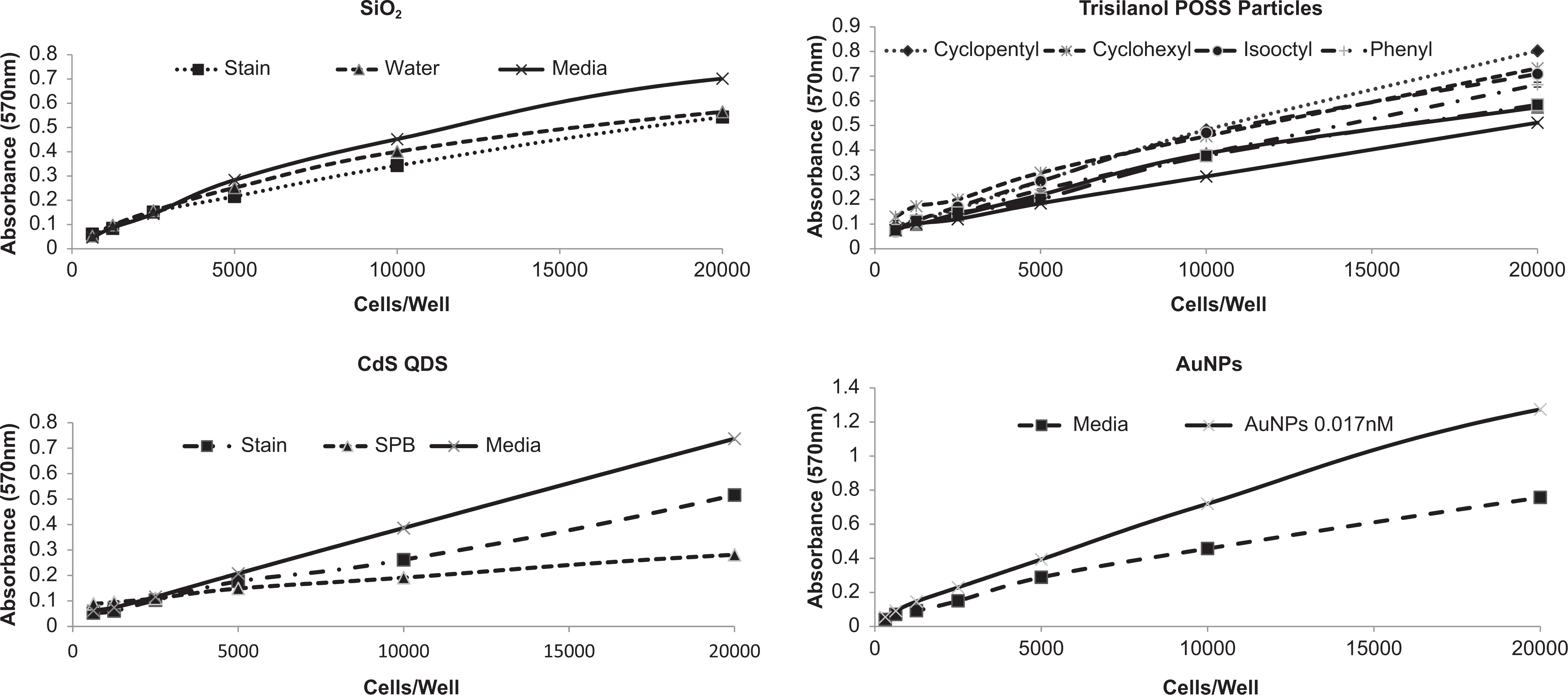
Crystal violet standard curves generated using HaCaT cells in the absence and presence of nanoparticles, and the relevant solvents indicated. Media indicates untreated standard curve. Stain indicates NP added concurrently with crystal violet. SDS indicates NP added concurrently with SDS. Water, DMSO, ethanol, and SPB indicate solvent controls with no particles added. DMSO indicates dimethyl sulfoxide; NP, nanoparticle; SDS, sodium dodecyl sulfate.
Carbon Black Interference Assay (Positive Control)
In the current study, 10 mg/mL reduced the absorption values of the MTT and crystal violet assay standard curves (Figure 3). In the MTT assay, for the solvent control dimethyl sulfoxide (DMSO), there was no effect, and when carbon black was added with the MTT or SDS, there was a small nonsignificant decrease in the ODs when carbon black was present between 203 and 103 cells/well. The effect of the solvent control, in contrast, in the crystal violet assay significantly decreased the ODs for all values of ≥5,000 cells/well. The standard curve in the presence of carbon black was the same as the DMSO. After 1-hour exposure to carbon black, the particle was not seen in the cytoplasm of HaCaT cells; however, it was seen after 24-hour exposure (Figure 4).
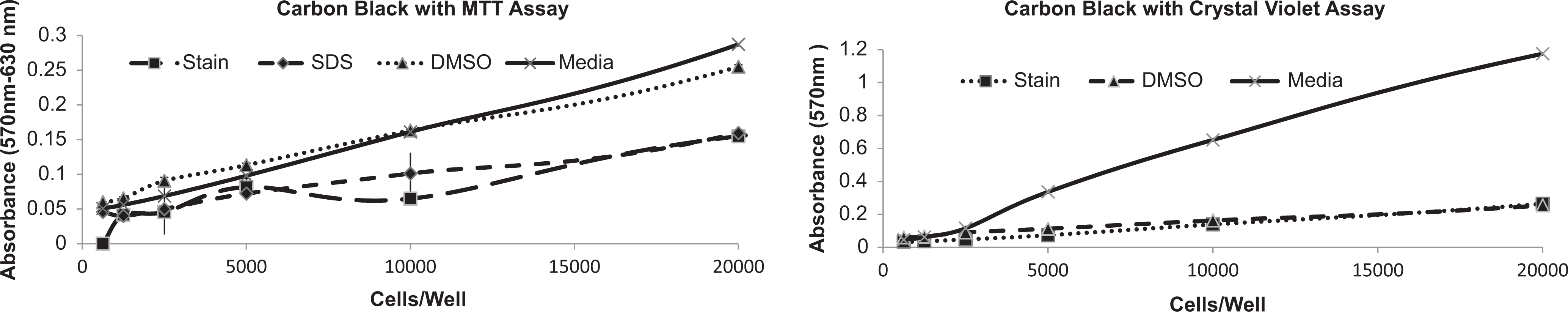
Absorbance (OD) versus cells/well obtained from the MTT assay and crystal violet assay using HaCaT cells. Media indicates no changes to standard curve. DMSO indicates DMSO added with MTT or crystal violet stain. Dye and stain indicate carbon black at 10 mg/mL added with MTT and crystal violet. SDS added with media and carbon black. DMSO indicates dimethyl sulfoxide; MTT, 3-(4,5-dimethylthiazol-2-yl)-2,5-diphenyltetrazolium bromide; OD, optical density; SDS, sodium dodecyl sulfate.
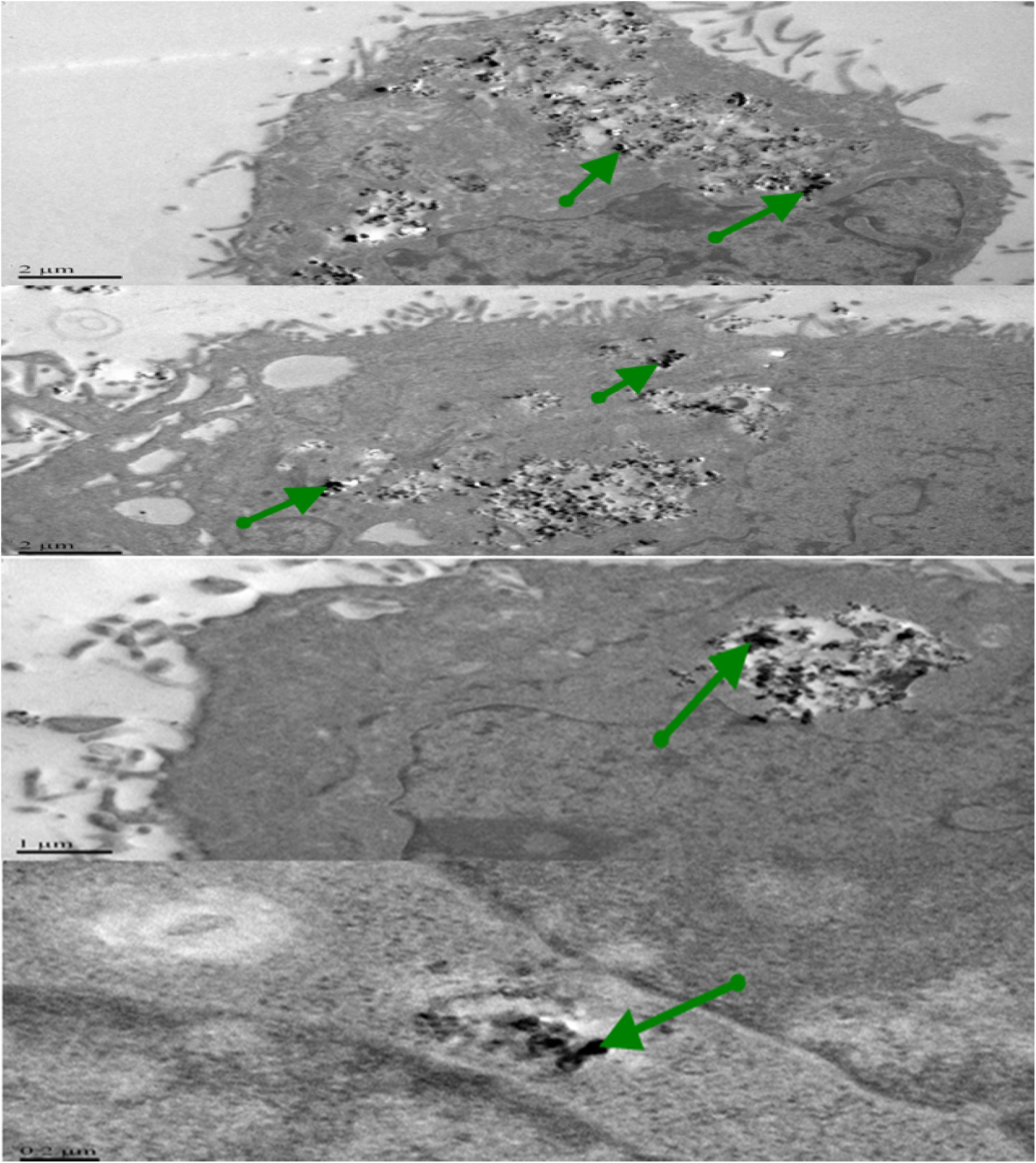
Carbon black can be seen primarily in the cytoplasm of the cells at 24 hours using TEM. Green arrows indicate particle accumulation. Bar = 2, 1, 0.2 μm.
Discussion
Single-walled carbon nanotubes (SWCNTs) and a variety of carbon-based nanomaterials interact in common cytotoxicity assays, such as MTT and crystal violet assays, and could interfere with fluorescence/absorption data used to evaluate the cytotoxicity level. 43 There is only a small database on the effects of NPs on in vitro assays. 44,45 Thus, in this study, we related the detection of NPs interference principally to previous research on SWCNTs and metal oxide NPs (see Introduction).
Wörle-Knirsch et al 43 reported that when SWCNTs were exposed to A549, there was a false strong cytotoxic effect within the MTT assay after 24 hours that reached approximately 50%; however, the same treatment with SWCNTs, but with the cell proliferation reagent WST-1, did not reveal cytotoxicity. Thus, SWCNTs appear to interfere with some tetrazolium salts such as MTT but not with others such as WST-1. The effect of these NPs does not seem to be on the enzymatic reaction but appears to be with the insoluble nature of MTT formazan.
Despite the vast research efforts, the cellular responses to nanomaterials are often variable and even contradictory. 45-46 Additionally, correlations between the nanomaterial properties and responding cell types are not well understood. 46 These effects were studied in an in vitro study using 3 cell lines of different origins (RAW 264.7 macrophages, human telomerase reverse transcriptase, and 3T3 fibroblasts) and comparing nanomaterials of various ranges of compositions (multiwall carbon nanotubes [MWCNTs], SiO2 and TiO2) and size (MWCNTs of diameters <8 , 50, and 20-30 nm, although the same length of 0.5-2 μm) to detect the influence of size and composition on toxicity assessment. 46 Nanomaterial composition and size showed a distinct role in the cellular response. Additionally, the response varied between cell types and was more likely to be attributed to the physiological function of the type of cell origin. 46 Moreover, depending on the exposed cell type, the same materials can cause different intracellular responses and potential mechanism of toxicity. These findings both emphasize the priority of analyzing the effects of NPs in the most related exposure model and uphold the idea that NP engineering methods should be focused on the cell types that are likely to be exposed to the particles.
Applications in vitro and inhalation studies in vivo have been known to yield divergent results. The aim was to find why some particles yield strong positive cytotoxicity results with some in vitro assays but not in other assays. Such conflicting data are what we are investigating here by studying the presence of particles for potential interference with the MTT and crystal violet assays. Four trisilanol POSS particles with similar compositions, shape, and diameter were synthesized to compare with CdS QDs, SiO2,, AuNPs, and the control carbon black at different composition size, shape, and diameters. Interaction of trisilanol cyclopentyl POSS, trisilanol cyclohexyl POSS, SiO2, and CdS QDs particles was clearly observed not to interfere with standard curve via the MTT dye as compared with carbon black (Figures 1 and 3). However, trisilanol phenyl POSS, trisilanol isooctyl POSS, and AuNPs indicate similar results with carbon black (Figures 1, 2, and 3).
In a recent study, the influnce of structure, surface, polymer compatibility on composite morphology, and bulk properties of POSS particles was examined. Nylon 6-clay hybrid was chosen as the matrix polymer. 46 Two POSS strcutrues, one is a full condense nonpolar octansubstituted octaisobutyl-POSS cage and another with an open cage polar trisilanolphenyl-POSS, were scattered in the nylon matrix at different concentrations. 46 A variation was observed in the dispersion, surface charactristics, bulk (thermomechanical) properties, and solubility in the polymer. The variation of POSS particle interference may be attributed to the alteration of POSS particles after been dispersed in suitable matrices; therefore, the results of POSS with MTT assay highlight the potential to clearly design POSS nanocomposites with particular surface enhancement capabilities by the control of POSS chemical structure.
Carrying out the crystal violet assay method with the crystal violet stain showed contradicting results compared to the MTT assay with the exception of carbon black, which interfered with both assays (Figures 2 and 3). The differences possibly occurred from either particles interaction with stains or with HaCaT cells. Previously, Hela cells were used as a bioindicator of cell viability in an in vitro assay method that combined dyes WST-1, NR, and crystal violet. 47 The joined cell viability assay using WST-1, NR, and crystal violet presented an absorbance that correlated linearly with the number of cells over the range of 1,000 to 50,000 cells/well. 48 This supports that human cell lines interact linearly with crystal violet and that HaCat cells are not causing the reduction in OD values with crystal violet stain.
Due to their small size and surface area, insoluble NPs are approximately not affected by the gravitation force; therefore, cells may not be exposed to the majority of NPs in suspension. 49,50 On the other hand, particles may aggregate at the bottom of a culture vessel preventing staining cells by crystal violet. 51 Carbonaceous material and several metal oxide particles interrupt light through spectral regions, which could affect the outcome of the assay. 52 Mass concentration and composition are the main causes of spectrum absorbance. 53 Carbon black and single-walled nanotubes decrease the tetrazolium compound in the XTT (2,3-bis-(2-methoxy-4-nitro-5-sulfophenyl)-2H-tetrazolium-5-carboxanilide) and MTT assays even in the absence of cells. Furthermore, silver NPs interfere with lactate dehydrogenase enyzme leading to false vaiablity assessment. 54,55
Monteiro-Riviere and Inman examined carbon black with different sizes on human epidermal keratinocytes, adult using the NR 47 and MTT assays. 11 Four types of carbon black particles were examined. Conflicted results were observed across the cytotoxicity end points at 0.1 to 0.4 mg/mL, suggesting that adsorbing properties of carbon were interfering with the components of the assays.
This could be an influence of particle dispersion or agglomeration. 56 Variants in particle dispersion have recently shown an important role in nanomaterial toxicity. 56 Sohaebuddin et al 46 assessed the size distribution of nanomaterials using dynamic light scattering. Apparent particles increased in both media and phosphate-buffered saline (PBS). For example, MWCNT > 50 nm at 10 μg/mL increased to 100 μg/mL after dispersed in both media and PBS. Compared with similar particle composition, SiO2 did not show a reduction in formazan and solubilizing solution, whereas CdS QDs informed overlap with MTT and SDS. Binding of particles with crystal violet stain was observed to reduce the OD readings for most of the particles tested except with AuNPS. This could attribute to the particle composition, size, and surface area. In this study, we tested the particle interruption with cells using transmission electron microscope. The 24-hour exposure of carbon black at 10 mg/mL shows aggregation in the cytoplasm of HaCaT cell line but not in the nucleus (Figure 4).
In conclusion, the interference of particles does not seem to affect the enzymatic reaction of HaCaT cells but interrupt the insoluble nature of MTT formazan and crystal violet. Our findings strongly suggest verifying cytotoxicity data with independent test systems for this new class of materials (nanomaterials). Transmission electron microscope was chosen to verify disruption of cells by particles. Basically, a clear reference material was needed to evaluate the interference with fluorescence/absorption within in vitro assays; therefore, carbon black was chosen. Nanomaterials cytotoxicity could pose artifacts, if it was not combined with the screening of interruption of the insoluble formazan and crystal violet dye. This study demonstrates that interference assays need to be included as an important part of any screening of novel NPs for potential cytotoxic hazard.
Footnotes
Acknowledgments
The authors would like to thank the Ministry of Higher Education, Saudi Arabia, for partial support of this project.
Author Contributions
A. Almutary and B. J. S. Sanderson are equal contributors. A. Almutary carried out the experiment work, contributed to acquisition, analysis, or interpretation of data, drafted the manuscript, critically revised the manuscript for important intellectual content, gave final approval, and agrees to be accountable for all aspects of the work in ensuring that questions relating to the accuracy or integrity of any part of the work are appropriately investigated and resolved. Barbara Sanderson contributed to conception and design, contributed to acquisition, drafted the manuscript, critically revised the manuscript, gave final approval, and agrees to be accountable for all aspects of work ensuring integrity and accuracy.
Declaration of Conflicting Interests
The author(s) declared no potential conflicts of interest with respect to the research, authorship, and/or publication of this article.
Funding
The author(s) received no financial support for the research, authorship, and/or publication of this article.