
Abstract
Although reverse transfection cell microarray (RTCM) is a powerful tool for mammalian cell studies, the technique is not appropriate for cells that are difficult to transfect. The lentivirus-infected cell microarray (LICM) technique was designed to improve overall efficiency. However, LICM presents new challenges because individual lentiviral particles can spread through the cell population, leading to cross-contamination. Therefore, we designed a cell-defined lentivirus microarray (CDLM) technique using cell-friendly biomaterials that are controlled by cell attachment timing. We selected poly-
Introduction
Following the development of a lipid-mediated DNA transfection method, the reverse transfection cell microarray (RTCM) approach was adopted to perform functional genetics in a high-throughput screening format. 1 The clear advantage of RTCM is that it allows rapid visualization and analysis of cellular phenotypes in live cells; the technique has revealed novel functions of genes in many pathways, including apoptosis,2,3 mitogen-activated protein kinase (MAPK) and c-Jun N-terminal kinase (JNK) signaling, 4 and cAMP-dependent signaling.5,6 However, RTCM is limited by the amenability of target cell lines to transfection using lipid-based reagents. Therefore, research groups have implemented lentivirus-infected cell microarrays (LICMs) because the lentivirus has high transduction efficiency in a wide range of cell types.6–8 However, LICM-based methods are susceptible to the phenomenon of “spreading,” and improvements are needed.
Some investigators have suggested applying the cell-defined method for small interfering RNA (siRNA) reverse transfection microarrays, which facilitates control of attachment timing by allowing cells to attach only to the spot region; cell adhesion is further reinforced by growth factor–reduced Matrigel.
9
Here, we report the development of a cell-defined lentivirus microarray (CDLM) method and demonstrate its application. In brief, a lentivirus solution was prepared from siGLO Red dye, Matrigel, and poly-
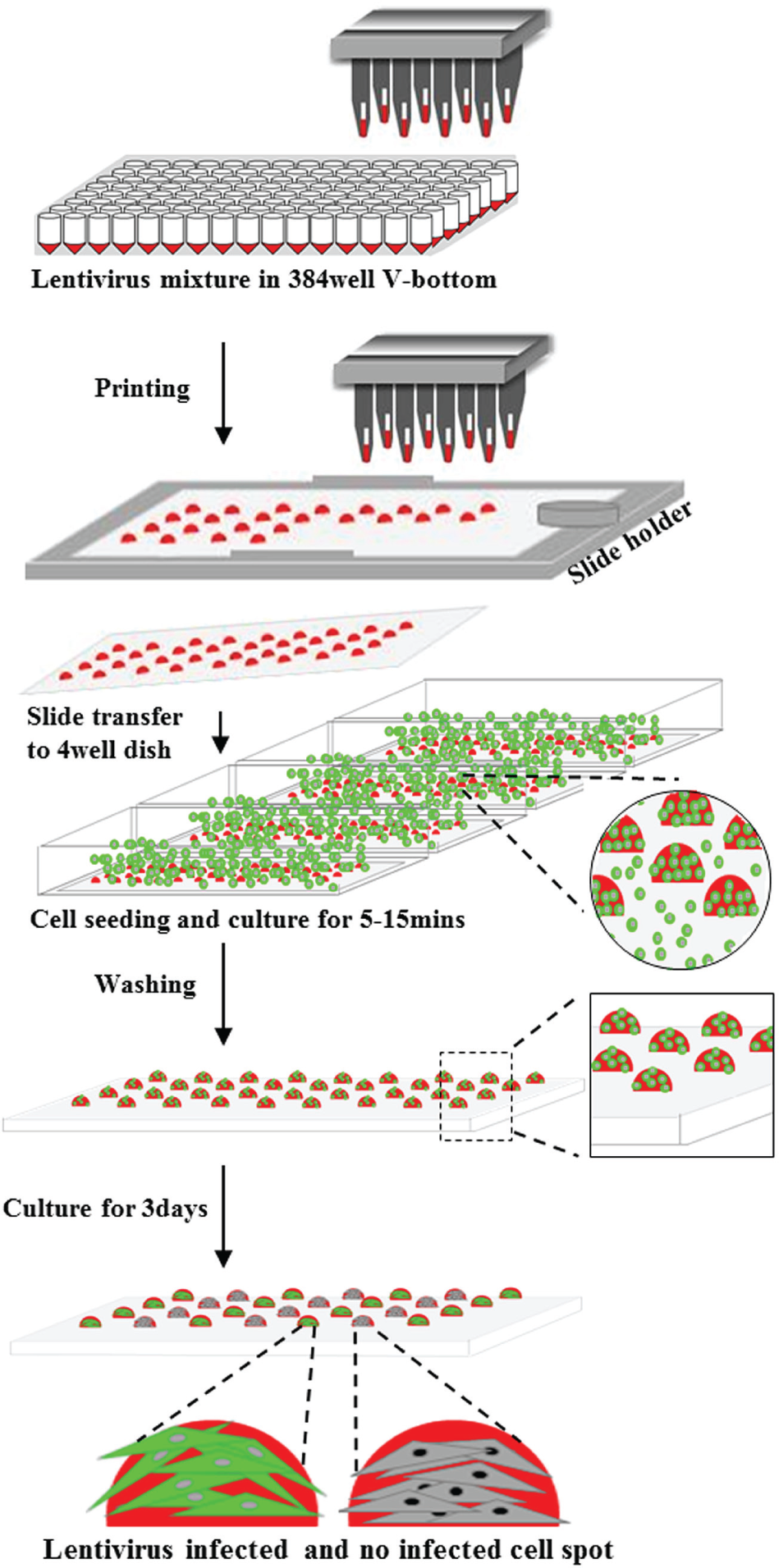
Schematic diagram of cell-defined lentivirus microarray. The lentivirus mixture, prepared in a 384-well V-bottom plate, was printed on glass coverslip by a stealth pin-based microarray printer. The cells were seeded and cultured for 5 to 15 min to induce cell adhesion on the spot region in a 4-well dish. The unattached cells were removed by washing three or four times with fresh media, and attached cells were cultured for 3 days to induce for lentivirus reverse infection.
Materials and Methods
Cell Culture
The cell lines used for this experiment (U2OS, HeLa, EKVX, PC9, A549, and A375) were maintained in Dulbecco’s modified Eagle’s medium (DMEM) supplemented with 10% (v/v) fetal bovine serum (FBS) and 1% (v/v) penicillin streptomycin (Gibco, Invitrogen, CA) in an incubator at 37 °C and 5% CO2. Individual patient-derived primary lung cancer cells were established (Asan Hospital, Seoul, South Korea) and cultured in bronchiolar epithelial basal medium (BEGM; Lonza, Walkersville, MD) comprising bovine pituitary extract (BPE), human epidermal growth factor (hEGF), GA-1000, insulin, and a triiodothyronine-SingleQuots Kit (Lonza, Walkersville, MD). 10
Lentivirus Production
The protocol of lentivirus production was based on the method reported by Bailey et al. 7 In brief, HEK293T cells were cultured for 24 h and cotransfected with 9 µg pCDH-EF1-MCS-T2A-copGFP lentiviral plasmid (System Biosciences, Palo Alto, CA), 6 µg Delta VPR 8.9, and 3 µg VSV-G, using FuGENE 6 (Roche Applied Science, Indianapolis, IN). The virus-containing supernatant was collected after 48 h, filtered through a 0.45-µm cellulose acetate filter (Millipore, Darmstadt, Germany), and concentrated by ultracentrifugation at 21,000 rpm for 2 h. The viral pellets were resuspended in microarray printing solution using the protocol reported in the literature. 7 The working lentivirus concentration for printing is roughly 1 × 10⁸ infectious units (IFU)/mL. Solutions of this stock were stored at −80 °C until required for printing.
Printing the CDLM
The prepared lentivirus solution was printed on uncoated glass coverslips (24 × 60 mm; Marienfeld, Lauda-Königshofen, Germany), using SMP9 stealth pins (Telechem, Atlanta, GA) and a high-throughput microarray printer (Genomic Solutions, Ann Arbor, MI) at 22 to 25 °C and 55% to 65% relative humidity. The printed spots were 200 to 300 µm in diameter, and the distance between spots was 1000 µm. The lentivirus microarrays were stored at −80 °C in a deep freezer until required.
Reverse Infection of the CDLM
Cell lines were cultured to 70% to 80% confluence in 25T or 75T flasks, detached by incubation with 0.05% trypsin–ethylenediaminetetraacetic acid (Gibco, Invitrogen, Carlsbad, CA), and seeded (1 × 10⁶/4 mL/slide) with assay media (Opti-MEM–GlutaMAX (Gibco, Invitrogen, Carlsbad, CA), 5% [v/v] fetal bovine serum, 1% [v/v] penicillin streptomycin) onto the CDLMs, which were transferred to a 4-well rectangular dish (Thermo Fisher Scientific, Waltham, MA). Cell attachment was induced for 5 to 15 min, and unattached cells were removed by washing three to five times with fresh medium. 8 The cells attached to the spot regions were cultured for 3 days in an incubator at 37 °C and 5% CO2 to induce reverse infection. The primary lung cancer cells were subjected to the same process, using a primary lung cancer–specific culture medium.
Imaging Acquisition and Analysis
For imagining acquisition, the cells were washed once with phosphate-buffered saline (PBS) and fixed with 4% (w/v) paraformaldehyde in PBS for 10 min. The cells were then washed again with PBS and incubated with 5 mM DRAQ5 (1:2000 in PBS; BioStatus, Shepshed, UK) for nuclear staining. Confocal images were acquired using an ImageXpress Ultra point scanning confocal microscope (Molecular Devices, Sunnyvale, CA) equipped with four solid-state lasers for simultaneous excitation at 405, 488, 561, and 635 nm; a galvanometer for X scanning; and a stage for Y scanning. Each cell spot was scanned at 10× or 20× magnification using a Nikon (Tokyo, Japan) air immersion objective lens with specific filter sets for Alexa 488, Alexa 568, and Alexa 635. The image quantification was performed using cell scoring and the nuclei count parameter with MetaXpress software (Molecular Devices) to evaluate the proportion of green fluorescent protein (GFP) fluorescent-positive cells by intensity, nuclei numbers, and size (
Results and Discussion
Establishment of Cell-Defined Culture on the Spots by Biomaterials
Cell-friendly biomaterials such as sucrose, gelatin, Matrigel, and PLL facilitate the attachment and growth of cells.9,11,12 We determined the optimal combination of biomaterials for defined culture on the spots before developing the CDLM. The combinations of 0.1% (0.0125%) PLL, 0.01% (0.00125%) PLL, 0.3% (0.0375% and 0.01875%) sucrose, and 0.2% (0.025% and 0.0125%) gelatin were tested with printing solution (50%–75%), siGLO Red dye (12.5%), and Matrigel (12.5%) (
To determine optimal PLL concentration, we titrated PLL with 10 µL (62.5%) printing solution, 2 µL (12.5%) siGLO Red dye, and 2 µL (12.5%) Matrigel in HeLa and EKVX cells (
Fig. 2
,
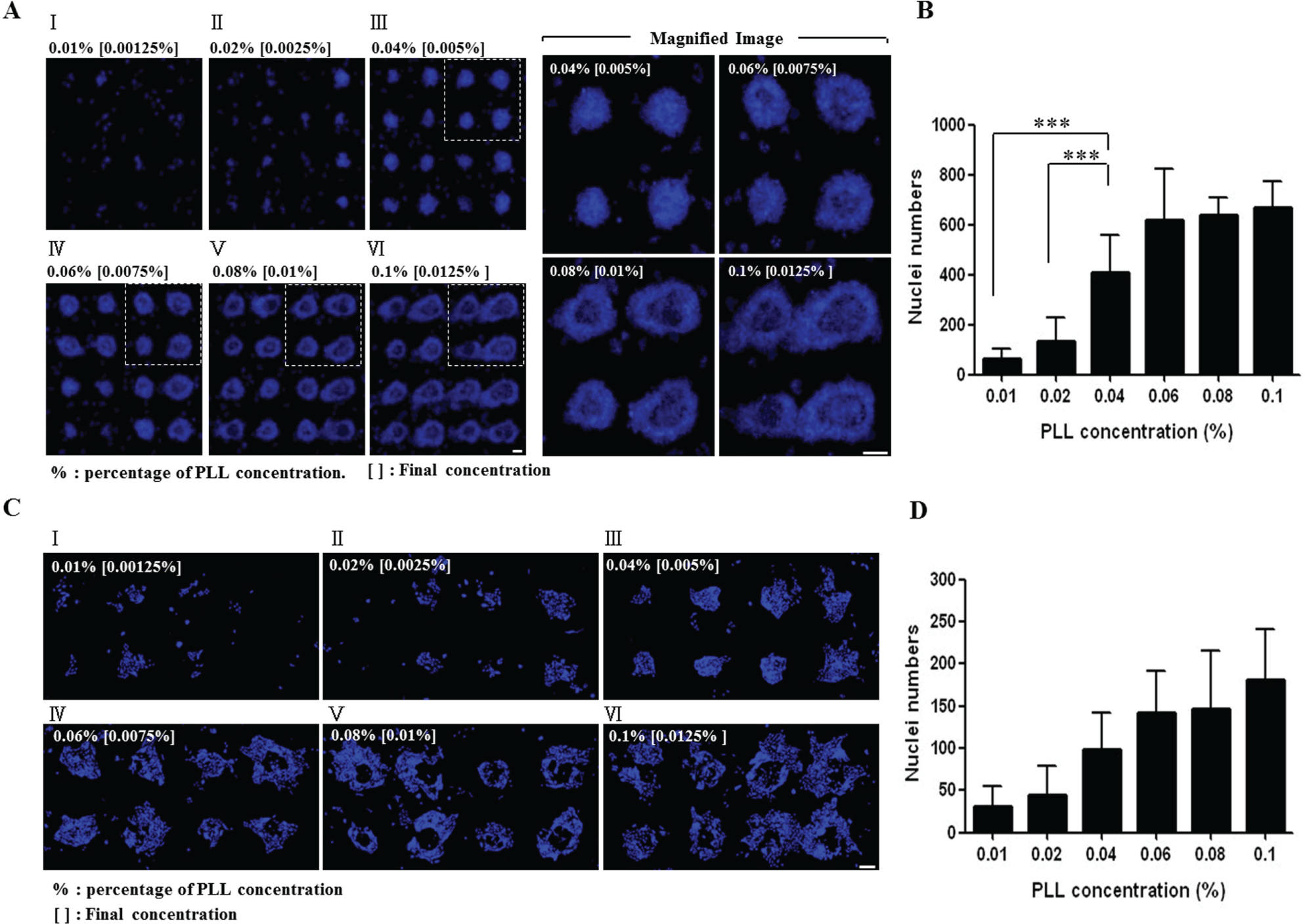
Quantification of poly-
Optimization of Cell-Defined Lentivirus Microarray
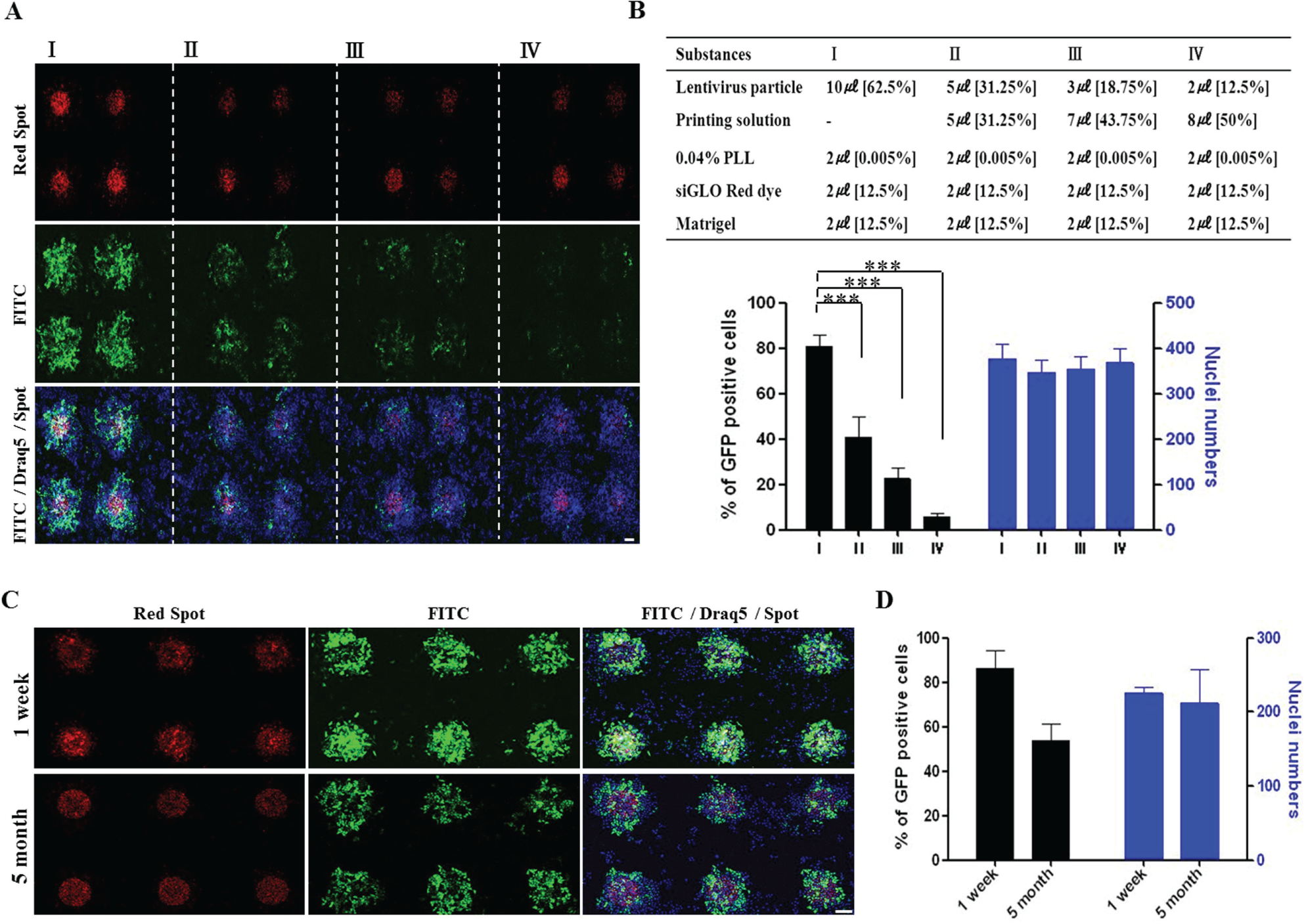
We used the optimal combination of biomaterials described above to perform the titration of lentivirus particles for validation in U2OS cells ( Fig. 3A , B ). First, 2 to 10 µL of lentivirus particles was transferred to the 384-well V-bottom plate, 2 µL (12.5%) each of siGLO Red dye and Matrigel was added, and the combination was gently mixed by pipetting. Next, 2 µL (0.005%) of 0.04% PLL was added and the mixture was centrifuged (1200 rpm, 30 s). The prepared lentivirus solution was printed using a microarrayer. The U2OS cells were seeded and cultured for 5 min on the lentivirus spot array, then washed to remove unattached cells. The attached cells on the spots were incubated for 3 days to induce reverse infection. Significant infection efficiency was observed with 10 µL (62.5%) of lentivirus particles and decreased sharply at half that volume. According to these results, we chose a mixture comprising 10 µL (62.5%) lentivirus particles, 2 µL (12.5%) each of siGLO Red dye and Matrigel, and 2 µL (0.005%) of 0.04% PLL ( Fig. 3B , upper).
The optimization of the cell-defined lentivirus microarray in U2OS cells. (
Conservation of Lentivirus Activity on CDLM
We confirmed the storage stability of the lentivirus by determining the activity of the lentivirus spots, which had been printed using the optimal mixture. The efficiency of lentivirus infection was 86% after short-term storage (1 week) and 52% after long-term storage (5 months) ( Fig. 3C , D ). Based on these results, we observed that lentivirus spot activity decreased (by 32%) after long-term storage. Therefore, activity is linked to the length of storage of the spot; however, specificity using the microarray was not considered.
Confirmation of Reverse Infection Effect on CDLM in Various Cell Lines
To validate CDLM, using the optimal lentivirus mixture, we examined reverse infection efficiency in various cell lines (HeLa, EKVX, A549, A375, PC9, and patient-derived primary lung cancer) ( Fig. 4 ). Lentivirus spots and negative control spots were printed at intervals of 1 mm, with a space of 4 mm between the two groups. In the HeLa cells, each group of 16 lentivirus spots showed high reverse infection efficiency (88%–98%) without spot-to-spot variation or contamination of the negative control ( Fig. 4A , B ). Five other cell lines were tested to confirm the effect of reverse infection, and high efficiency (83%–100%) was verified in all ( Fig. 4C , D ).
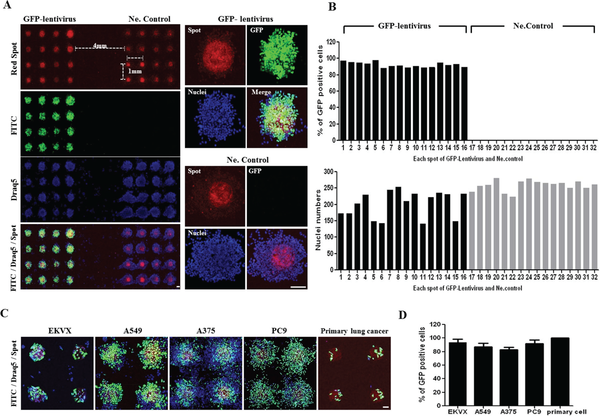
The confirmation of green fluorescent protein (GFP)–tagged lentivirus reverse infection efficiency in mammalian cells. (
Some researchers have investigated cell-defined microarrays using a combination of siRNA and a liposome transfection reagent.9,13 Although the “cell-defined” concept is effective for the control of spreading, liposome reverse transfection is limited to gene delivery for various cell types in microarray systems, despite the development of numerous transfection reagents. Researchers have suggested the use of a lentivirus microarray to overcome the transfection limitations of liposomes, but it is unclear whether the use of lentivirus resolves the spreading issue.6–8 Therefore, we propose a CDLM technique that is more effective for gene delivery and the control of spreading.
The CDLM technique was developed by optimization of a lentivirus mixture, examination of lentivirus spot activity conservation, and application to various cell lines. The technique has two advantages: it facilitates easy gene transfer for various cell types using lentivirus and allows miniaturization for HTS by microarray. However, cell-defined culture on an uncoated slide by only controlling attachment timing is not possible in all cell lines; human bone marrow stromal cells (hBMSCs) attached and grew on an uncoated glass coverslip, despite a weak environment for the cells. Previously, we used slides coated with specific substances, such as a hydrogel that restricted cell attachment, for the development of a cell-defined siRNA microarray (CDSM) for hBMSCs, and the slide allowed greater clear cell spot formation. 13 Therefore, if hydrogel-coated slides are used, we expect to improve the quality of CDLM.
In conclusion, we developed an optimized protocol for lentivirus microarray, which is a powerful tool for gene functional studies, and demonstrated its application to various cell lines. This result offers a basis for the development of more effective cell-based microarray systems.
Footnotes
Declaration of Conflicting Interests
The authors declared no potential conflicts of interest with respect to the research, authorship, and/or publication of this article.
Funding
The authors disclosed receipt of the following financial support for the research, authorship, and/or publication of this article: This work was supported by the National Research Foundation of Korea (NRF) grant funded by the Korea Government (MSIP) (NRF-2014K1A4A7A01074642), Gyeonggi-do, and by a grant from the Bio-industry Technology Development Program (no. 312062-05) and by a grant (2010-491) from the Asan Institute for Life Sciences, Asan Medical Center, Seoul, Korea.
References
Supplementary Material
Please find the following supplemental material available below.
For Open Access articles published under a Creative Commons License, all supplemental material carries the same license as the article it is associated with.
For non-Open Access articles published, all supplemental material carries a non-exclusive license, and permission requests for re-use of supplemental material or any part of supplemental material shall be sent directly to the copyright owner as specified in the copyright notice associated with the article.