
Abstract
We describe a polyacrylamide gel casting cassette that overcomes limitations of commercially available gel electrophoresis equipment. This apparatus molds a single polyacrylamide gel that can evaluate more than 200 samples in parallel, is loaded with a multichannel pipettor, and is flexible with respect to composition of the separating matrix. We demonstrate its use to characterize inhibitors of enzymes that modify protein and nucleic acid substrates. Throughputs of greater than 1000 samples per day were achieved when this system was paired with a quantitative laser-based imaging system, yielding data of remarkable quality.
Keywords
Introduction
High-throughput screening (HTS) of chemical libraries is employed in the academic and pharmaceutical sectors to discover chemical scaffolds that pharmacologically modulate the target under study.1-3 It is not unusual for an HTS assay to be sensitive to both genuine modulators as well as effectors that act through nonspecific or technology-dependent mechanisms. 2 As such, it is an advisable and generally accepted practice to confirm the activity of hit matter in a method whose detection format is orthogonal to the discovery assay.
For some targets, a limited number of methods/detection technologies can be employed to gather the several hundred to thousands of data points needed to triage a primary HTS hit list. 3 For enzymes that modify the structure of nucleic acids or proteins (e.g., those that catalyze group-transfer reactions, DNA helicases, nucleases and isomerases, and proteases), their action imparts a change in mass or charge density such that the substrate and product species can be separated electrophoretically. Thus, the reactions are amenable to the development of electrophoretic mobility shift assays (EMSAs).4-6
Many aspects make EMSAs a desirable format for an HTS follow-up assay. EMSAs allow for the direct observation of enzyme and binding reactions, a feature that is highly desired, as it eliminates the opportunity for compounds to interfere with detection reagents or complicated detection cascades. In addition, the visual nature of data arising from EMSAs provides a greater depth of information about reaction under study: it is our experience that visual inspection of EMSA gel images can provide insight to spurious behaviors of false-positive hits (e.g., intrinsic compound fluorescence, absorptivity, and colloid formation). In addition, the technique’s ubiquitous use in practically all molecular and cell biology laboratories provides the benefit that results are easily communicable between scientists of diverse backgrounds. Finally, the relatively low cost of electrophoresis equipment makes it an attractive format to groups whose access to advanced instrumentation may be limited. Taken together, these aspects provided the impetus for us to explore the application of EMSAs for high-throughput hit confirmation purposes.
Typically, electrophoretic approaches are considered cumbersome and time-consuming; it has been our experience that even the most skilled of researchers can collect only 100 to 200 data points per day with traditional vertical electrophoresis equipment. As a result, these techniques are incorporated into the discovery workflow late in the process and at a limited capacity. Over the course of one discovery campaign,1,2 we encountered the situation that no alternative assay format presented as suitable for hit triaging, and we sought to employ a gel-based EMSA immediately following HTS. Through this work, we found ourselves limited by commercial equipment due to low sample densities and a general incompatibility of well spacing with multichannel liquid handling equipment.
Thus, we pursued the design of custom equipment to mold gels with increased sample densities, and these efforts produced a multitiered polyacrylamide gel electrophoresis (PAGE) gel casting form. We have combined this cassette with the commercially available Multiphor-II (GE Healthcare, Piscataway, NJ) electrophoresis unit to assemble a high sample-capacity polyacrylamide electrophoresis platform that we have dubbed Elph. Compared with traditional vertical electrophoresis systems, this Elph platform possesses significant advantages that simplify the execution of EMSA protocols. In several instances, the use of this system has enabled a single scientist to achieve EMSA throughputs of greater than 1000 data points per day. Herein, we detail the physical parameters of this gel cassette and demonstrate its use in three separate EMSA-based experiments where it has enabled the acceleration of the respective discovery programs.7-10
Materials and Methods
Chemicals
Separation matrix was prepared from a Protogel 40% (w/v) acrylamide stock solution (0.8% bis-acrylamide, EC-850; National Diagnostics, Atlanta, GA) and supplemented with Rhinohide gel strengthener (Thermo Scientific, Waltham, MA). Ammonium persulfate and N,N,N′,N′-tetramethyl-1,2-diaminoethane were from Bio-Rad (Hercules, CA). PAG-bond was from GE Healthcare (Piscataway, NJ). All other chemicals were reagent grade or better from Sigma Aldrich (Milwaukee, WI).
Electrophoretic Apparatus
The horizontal PAGE apparatus was assembled as described in Westmeier 11 and included the Pharmacia Multiphor-II flatbed electrophoresis unit (GE Healthcare), PowerPac High Voltage power supply (Bio-Rad), and a Neslab RTE 7 Recirculating chiller (Thermo Scientific).
Elph Gel Casting Cassette
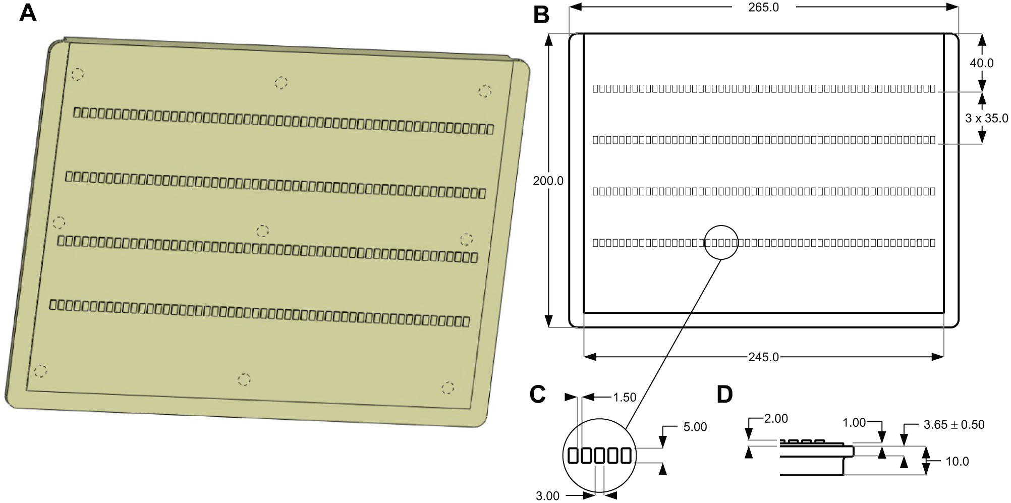
The Elph gel casting mold (ELPH001B) cassette was designed using SolidWorks CAD software (Dassault Systèmes Solidworks Corp., Waltham, MA) and CNC-machined from a 0.5-inch-thick polycarbonate sheet. Gaskets were hand-cut from a 0.125-inch-thick (~3 mm) silicone sheet (Durometer 50A, cat. 1460N34; McMaster-Carr, Robbinsville, NJ). The cassette backing plate (ELPH002A) was cut from a 0.25-inch acrylic sheet. The casting cassette was assembled with office-grade 1-inch binder clips. Schematic diagrams for ELPH001B and ELPH002A are presented in
Figure 1
and
Design of the Elph gel casting mold. (
Gel Casting
The Elph cassette parts were cleaned with 95% ethanol and allowed to air dry prior to use. In instances where the gel did not easily release from the mold, the cassette was pretreated with 0.5% (v/v) silicone oil in hexane, the solvent evaporated in a fume hood, and excess oil removed with Kim-wipes. A sheet of PAG-bond film was positioned on the cassette backing plate, and the cassette was assembled (
Data Analysis
Gels were imaged with a Typhoon FLA-9500 (GE Healthcare) and band intensities quantified using the ImageQuant 8.0 suite (GE Healthcare). For dose-response analysis, nonlinear regression was performed with GraphPad Prism v5.00 (GraphPad Software, La Jolla, CA) using the four-parameter Hill equation and default initiation parameters.
Compound Handling and Assay-Ready Plate Preparation
Compound stock solutions were prepared and maintained as previously described. 12 Compound source plates were prepared as 3-fold serial dilutions from a top concentration that was suitable for the range of potencies under investigation for each assay. Assay-ready plates were freshly spotted on the morning of experimentation. For the Sfp assay, compound spots (0.500 µL) were transferred manually with a multichannel pipettor. For apurinic endonuclease 1 (APE1) and Bloom (BLM) assays, compound spots (100 nL) were transferred into wells of the microplate using a Mosquito X1 outfitted with accurate nanoliter transfer tips (TTP Labtech, Cambridge, MA).
Fluorescence EMSA for Sfp 4′-Phosphopantetheinyl Transferase
The preparation of reagents for this method has been previously described.7,8 To begin the assay, a 1.33× enzyme solution (15 µL, containing 26.6 nM Sfp, 66 mM HEPES·Na, 13.3 mM MgCl2, 0.0133% NP-40, and 0.133% bovine serum albumin, pH 7.6) was added to the assay-ready plate. After a 10-min room temperature incubation, the enzymatic reaction was initiated by the addition of 4× substrate solution (4 µL, containing 50 µM rhodamine CoA and 50 µM apo-actinorhodin-ACP). The reaction was allowed to proceed for 30 min at room temperature, after which it was terminated by the addition of 2× quench solution (20 µL, containing 4 M urea, 25 mM EDTA, and 0.004% phenol red, pH 8.0). Samples (10 µL) were separated under native conditions on a 15% polyacrylamide gel matrix that contained 6% Rhinohyde and a Tris-glycine buffer (25 and 192 mM). Following electrophoresis, the gels were imaged with the Typhoon FLA-9500 using the default TAMRA settings.
Gel-Based EMSA for BLM
The BLM helicase gel-based assay employed a fluorescently labeled, forked duplex DNA substrate. 9 This substrate was prepared by annealing the 3′-TAMRA-labeled A1 oligonucleotide (T)30CGTACCCGATGTGTTCGTTC with the unlabeled partially complementary strand, A2 GAACGAACACATCGGGTACG(T)30. Reactions were initiated by the addition of enzyme solution (15 µL, 5 nM BLM in BLM buffer [50 mM Tris-HCl pH 7.5, 50 mM NaCl, 2 mM MgCl2, 2 mM ATP, 1 mM dithiothreitol (DTT), 0.1% Tween-20]) into assay-ready plates. The reactions were initiated by the addition of 200 nM TAMRA-labeled forked duplex substrate. Following incubation at room temperature for 30 min, the reactions were terminated by the addition of loading dye (5% glycerol, 2 mM EDTA, and 2.5% bromophenol blue). Reaction products were then separated on 15% native polyacrylamide gel containing 1× TAE buffer, which was subsequently imaged with the Typhoon FLA-9500 imager using the default TAMRA settings.
Gel-Based EMSA for Apurinic Endonuclease 1
APE1 enzyme was obtained from New BioLabs (Ipswich, MA) and has been described in detail elsewhere.
10
The substrate for these experiments represents a duplexed DNA fragment of annealed oligonucleotides sourced from Biosearch Technologies (Novato, CA). TAMRA-Strand: 5′-TAMRA
Results and Discussion
In assessing the state of the art in electrophoretic equipment and techniques, we were intrigued by description of horizontal PAGE systems that used the Multiphor-II apparatus. 11 We envisioned a PAGE platform that employed this equipment to provide a polyacrylamide gel in a format that would allow for direct sample loading with a multichannel pipettor to ensure rapid multisample transfer from a microtiter plate to gel. Furthermore, given the size of the apparatus relative to modest separating distances that are required for most EMSAs, we reasoned that it could be configured in a multitiered system such that we could achieve a higher sample density by incorporating a multiplicity of rows.
The design of the Elph casting mold is diagrammed in Figure 1A . To maximize sample density, we settled on overall dimensions for Elph that use the entire cooling tray of the apparatus (190 × 245 mm; Fig. 1B ). We determined that the inclusion of samples in four tiers of 52 wells each would provide a maximum density while maintaining well-to-well barrier integrity and a modest resolution distance of 2 cm. In total, the cassette creates a polyacrylamide gel with 208 wells and in practice accommodates 48 samples in each tier—a number that is directly divisible by 8, 12, 16, and 24, thus matching whole integer values of rows or columns of both 96- and 384-well plates—as well as additional wells for reference samples. This imparts a high degree of flexibility in the hands of an experimenter who can work in either plate type and configure plate-based experiments in either columnar or row-based formats.
With regard to intersample spacing, we used a 4.5-mm center-to-center distance to match the 384-well plate formatting. In this configuration, 96-well plate spacing (9 mm center-to-center) is accommodated when samples are loaded into every other well with a multichannel pipettor. The optimized well dimensions are presented in Figure 1C and accommodate a maximum sample volume of 15 µL.
Finally, to facilitate assembly of the Elph cassette, we also incorporated a gasket alignment lip around the perimeter of the system at a depth of 1 mm ( Fig. 1D ). When combined with a seal that is cut from standard 1/8-inch silicone sheet (~3 mm), this cassette will produce a gel with a thickness of 1 mm at the bottom of the wells—a barrier that is robust against puncture during all but the most aggressive of sample loading mistakes. Production units were cut according to this design from half-inch polycarbonate sheet by a contract machining service provider.
To assess the ability of gels from this system to produce uniform data, we employed the phosphopantetheinyl transferase (PPTase) reaction,7,8 where a rhodamine-containing group is transferred from a coenzyme A analogue (rhodamine CoA) onto an acyl carrier protein acceptor substrate by action of the PPTase enzyme. This reaction is monitored as shift in electrophoretic mobility of the rhodamine label, which is visualized by fluorescence imaging of a PAGE gel after separation. We prepared a bulk reaction mixture and loaded the same sample into every well of a gel to monitor for the presence of positional effects, and an image of the gel is presented in Figure 2A . The pixel intensity of the bands was quantified and is presented as a scatterplot in Figure 2B . The analysis, revealing a mean value of 32.5% ± 1.9% product (i.e., a standard deviation of only 1.9%), clearly demonstrates that the system can reproducibly return a uniform value for the bulk sample independent of position. From an uncertainty standpoint, this equates to a 5.7% coefficient of variation over the entire Elph data set.
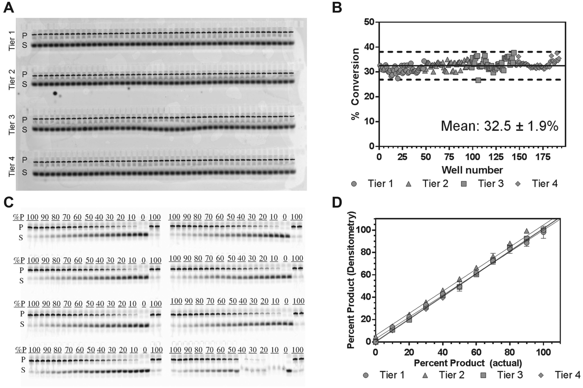
Sample and signal uniformity in Elph gel samples. (
Building on this, we sought to bolster confidence in the equipment to reliably report on samples that exhibit differing levels of activity as would be encountered when an enzyme reaction is only partially inhibited. In this experiment, we prepared solutions that contained equimolar concentrations of rhodamine label but differed by the extent to which the fluorophore was conjugated to the protein product. The resulting gel image from this experiment is presented in Figure 2C , and the normalized densitometric data are plotted against the input percentage of product value in Figure 2D . Linear regression of the data revealed slopes near unity for all four tiers of the gel and clearly demonstrated that the system possessed the capacity to distinguish between reactions of varying activity.
Subsequent to our validation studies, we built out an EMSA for Sfp PPTase on the Elph platform.7,8 With this technique, we profiled 210 structurally diverse compounds in 6-point dose response immediately following HTS. Exemplary data from this effort are shown in Figure 3 , which demonstrates the ability of this platform to characterize compounds with differing pharmacological activity. In Figure 3A , the activity of the compounds can be visually observed to track with increasing compound dose. Following digitization of the image, we were able to quantitate the pharmacological inhibition via nonlinear regression ( Fig. 3B ). The system functioned so satisfactorily that this EMSA became the preferred method to quantitate the pharmacological activity of test compounds and was performed on multiple occasions. During these production runs, throughputs of >1000 samples per day were achieved by a single experimenter equipped with two Multiphor-II units.
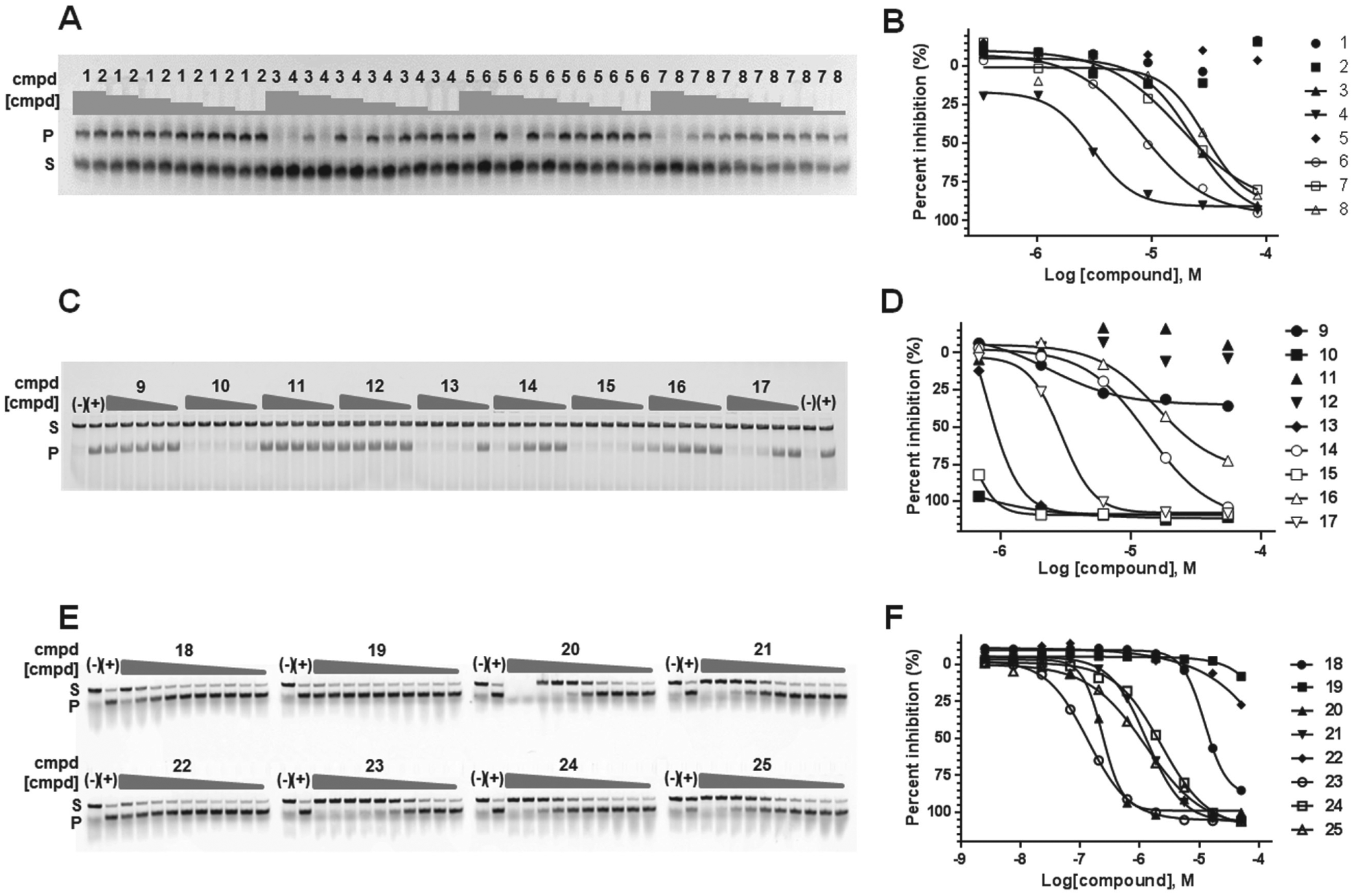
Application of Elph to pharmacological profiling. (
In addition to our work with Sfp PPTase, we explored the application of this platform to conduct EMSAs for other targets, including BLM helicase 9 and APE1 endonuclease. 10 Both of these enzymes modify nucleic acid structure and specifically accomplish the ATPase-driven strand separation of duplex DNA or digest oligo-duplex DNA at an engineered DNA cleavage site, respectively. In both of these cases, we successfully built out EMSA platforms that were used to assess potency for inhibitors in development, and exemplary results are shown in Figure 3C–F . A partial image of a gel from a BLM helicase assay is shown in Figure 3C , where compounds were tested at concentrations ranging from 0.7 to 55 µM. Densitometric analysis and curve fitting of the normalized product band intensities are shown in Figure 3D , and the resulting data were consistent with the potencies observed in the HTS method, as well as a radionuclide-based EMSA performed using a traditional vertical gel apparatus. 9 Similarly, a portion of an APE1 gel is shown in Figure 3E , where a panel of compounds that display a broad range of potencies was tested at concentrations ranging from 5 nM to 100 µM. The normalized product band intensities are plotted in Figure 3F , and the calculated IC50 confirmed those that we observed earlier in the HTS format. Thus, the direct observation of enzyme inhibition through EMSA boosted our confidence in both the respective chemical series under development as well as the multistep triaging funnels we had assembled to eliminate false-positive behaviors, such as DNA binding/intercalation found during HTS.9,10
In conclusion, we have described the Elph platform, which can significantly accelerate EMSA data collection rates and represents an approximately 10-fold improvement in capacity over vertical gel systems. We have demonstrated the ability of Elph to provide high-quality data that can be used to quantify pharmacological parameters. In addition, we have demonstrated the flexibility of our system and adapted it to a diverse panel of enzymes that modify the structure of proteins and nucleic acids. This has allowed us to apply EMSAs and make impactful contributions to multiple inhibitor development campaigns. We are hopeful that this report will bolster enthusiasm for EMSAs within the discovery community and lower the operational hurdle for the technique’s implementation when it is considered as a pharmacological profiling platform.
Footnotes
Acknowledgements
We thank Opher Gileadi for supplying BLM protein. The content of this publication does not necessarily reflect the views or policies of the Department of Health and Human Services, nor does mention of trade names, commercial products, or organizations imply endorsement by the U.S. government.
Declaration of Conflicting Interests
The authors declared no potential conflicts of interest with respect to the research, authorship, and/or publication of this article.
Funding
The authors disclosed receipt of the following financial support for the research, authorship, and/or publication of this article: This research was supported by the intramural research program of the National Center for Advancing Translational Sciences, as well as NIH award numbers 1R03MH083266 and R21AI090213 (M.D.B.).
References
Supplementary Material
Please find the following supplemental material available below.
For Open Access articles published under a Creative Commons License, all supplemental material carries the same license as the article it is associated with.
For non-Open Access articles published, all supplemental material carries a non-exclusive license, and permission requests for re-use of supplemental material or any part of supplemental material shall be sent directly to the copyright owner as specified in the copyright notice associated with the article.