
Abstract
Most of the current antiherpetics target viral DNA polymerase, but with the emergence of drug-resistant viruses, antiherpetics with different targets have become necessary. Inhibition of herpes simplex virus (HSV) replication at the early stages of infection minimizes cytotoxicity and immune suppression induced by HSV infection. In this report, quantitative reporter systems that use recombinant HSV and a stably transfected cell line were developed for the screening of agents targeting the early stages of HSV infection. The reporter genes in both systems were directed by HSV immediate-early (IE) promoters, so considerably less time was required for the quantification of HSV infection than the traditional plaque reduction assay. The results show that both reporter assays were sensitive to antiherpetic screening. Both assays were quantitative, rapid, easy to perform, and highly adaptable for automatic high-throughput screening. Exploiting the flexibility of these 2 assays, modified assays were also proposed for the detailed analysis of antiherpetic mechanisms.
Introduction
H
The general methods of antiherpetic screening include plaque reduction assay (PRA), dye uptake assay, cytopathic effect inhibition, virus yield reduction, and DNA hybridization. These assays are time-consuming or laborious, and they are ineffective for antagonists targeting the early phases of HSV infection. With the goal of developing assays for antiherpetics targeting the early stages of HSV infection, methods based on reporter systems were established in this study. Viral or cellular reporter systems have been described previously as rapid methods for monitoring viral infection and replication. These reporter-expressing recombinant viruses or cell lines are generally obtained by introducing a reporter protein under a specific viral promoter. Tanaka et al. 6 reported the construction of a recombinant HSV-1 (YK333) containing enhanced green fluorescent protein (EGFP) expression cassette driven by the Egr-1 promoter. The recombinant HSV-1 is capable of detecting antiviral activity within 48 h after infection. Several reporter cell-based assays for testing the drug susceptibility of HSV isolates have been reported, including methods based on Vero-ICP10-SEAP 7 and BHKICP6LacZ-5. 8
In this study, 2 methods based on HSV IE promoter-directed reporter systems were established. The first method, HSV-1/Blue Assay (HBA), is based on a recombinant HSV-1. HSV-1/Blue contains an HSV-1 ICP4 promoter directing a lacZ gene inserted into the HSV-1 TK gene. 9 The second method, Vero-ICP10-Luc assay (VILA), is based on a reporter cell line stably transfected with a HSV-2 ICP10 promoter-directed luciferase (Luc) expression cassette. There are 2 assay versions for both HBA and VILA: the long and short varieties. Performing these assays is easy, and antiherpetics, especially those targeting the early stages of HSV infection, can be rapidly identified. Modified reporter assays were also described, which can be used to further determine the working mechanisms of candidate entry inhibitors.
Materials and Methods
Cells
Vero cells (ATCC, Manassas, VA) were grown in Dulbecco’s modified Eagle’s medium (DMEM) supplemented with 5% fetal calf serum (FCS). The Vero-ICP10-Luc cell line was generated using Vero cells transfected with a plasmid, pGL4-ICP10- Luciferase, which contains the firefly Luc gene under the control of the HSV-2 ICP10 promoter. Neomycin-resistant clones were selected with G418 (1000 µg/mL; Merck, Whitehouse Station, NJ). A stable clone was selected and maintained in DMEM supplemented with 5% FCS and G418 (400 µg/mL).
Viruses
Drs. Gary H. Cohen and Roselyn J. Eisenberg (University of Pennsylvania) provided HSV/Blue, which has an ICP4::lacZ insertion in its TK gene. 9 Drs. Wang Yifei (Jinan University, Guangzhou, China) supplied HSV-1 (F strain). Both viruses were cultured and titrated using Vero cells.
Antiviral drugs and antibodies
Acyclovir (ACV; Sigma, St. Louis, MO), foscarnet sodium (PFA; Chia-tai Tianqing Pharmaceutical Co., Ltd., Jiangsu, China), and dextran sulfate (DS; mw 9000-2000, Sigma) were used. Drs. Gary H. Cohen and Roselyn J. Eisenberg (University of Pennsylvania) provided Anti-gD MAb DL6 and anti-gL1 MAb L4.
Long version assay for HBA and VILA
HBA
Confluent Vero cells in a 96-well tissue plate were inoculated in triplicate with 50 µL of virus suspension (HSV/Blue, at multiplicity of infection [MOI] 0.5) and 50 µL of culture medium containing testing compounds at different concentrations (ACV, PFA, or DS). Cells were lysed with 1% Nonidet P-40 in DMEM at 24 h postinfection. Lysates from each well were mixed with CPRG (chlorophenol red-β-D-galactopyranoside; Boehringer, Ingelheim, Germany), and β-galactosidase (β-Gal) activity was measured by taking absorbance readings at 570 nm every 2 min for a total of 25 readings, using an enzyme-linked immunosorbent assay (ELISA) plate reader (BioTek, Winooski, VT). The slope of the line was used to quantify β-Gal activity as milli-optical density units/min (mOD/min). 10 The 50% inhibitory concentration (IC50) was defined as the concentration of the antiviral drug that reduced the mOD/min values by 50% relative to the virus control. Inhibitory concentrations were calculated using the probit regression method.
VILA
Confluent Vero cells were inoculated in triplicate with virus suspension (HSV1-F strain, at MOI 5) and testing compounds at different concentrations (ACV, PFA, or DS). Twenty-four hours after infection, a supernatant was aspirated from each well and added to confluent Vero-ICP10-Luc cells prepared in another 96-well plate. Luciferase activity was measured 5 h later (Steady-GloR Luciferase Assay System; Promega, Madison, WI). Inhibitory concentrations were calculated.
Short version of assay for HBA and VILA
HBA
Vero cells were prepared in a 96-well plate as described above. The plate was precooled at 4°C for 10 min, and the virus mixture (HSV/Blue, at MOI 5) with serially diluted ACV or antibodies was added. The plate was incubated at 4°C for 90 min and then transferred to a 37°C incubator for incubation for 5 h. The β-Gal activity was then measured.
VILA
Confluent Vero-ICP10-Luc cells were prepared in a 96-well plate and precooled at 4°C for 10 min; the virus mixture (HSV1-F strain, at MOI 5) and serially diluted ACV or antibodies were added. The plate was incubated at 4°C for 90 min and then transferred to a 37°C incubator for incubation for 5 h. The activity of luciferase was then measured.
Modified HBA for mechanism determination
As shown in Figure 1 , Vero cell monolayers in 96-well plates are precooled at 4°C for 10 min, and then the assay can be continued using one of the following 3 slightly different procedures:
A: Cells are infected with HSV/Blue at an MOI of 5 for 1.5 h at 4°C, followed by replacement and incubation with the candidate compound for 1.5 h at 4°C. The plate is shifted to 37°C for 5 h. The assay is completed by measuring the β-Gal activity of the cell lysates.
B: The candidate compound is added to the cells for 1.5 h at 4°C, followed by replacement and inoculation with HSV/Blue at an MOI of 5 for 1.5 h at 4°C. The cells are washed with the medium and then shifted to 37°C for 5 h. The assay is completed by measuring the β-Gal activity of the cell lysates.
C: The plate is precooled to 4°C. At the same time, HSV/Blue and the candidate compound are mixed and incubated at 4°C for 1.5 h. The mixture is then added and incubated with the precooled plate at 4°C for another 1.5 h. Subsequently, the plate is washed with the medium and then shifted to 37°C for 5 h. The assay is completed by measuring the β-Gal activity of the cell lysates.
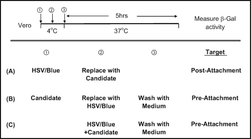
Modified HSV-1/Blue Assay (HBA) for mechanism determination. Vero cell monolayer in 96-well plates was treated with HSV-1/Blue, and the candidate compound in the sequences shown in panels
Plaque reduction assay
A modified PRA was performed, in which the Vero cell monolayers in a 24-well plate were inoculated with HSV mixed with serially diluted testing compounds. After incubation at 37°C for 1.5 h, the inoculum was replaced with the medium containing different concentrations of testing compounds and 1% methylcellulose. The plate was incubated at 37°C for 3 days until plaques appeared. Cells were fixed and stained with crystal violet. Plaques were then counted, and IC50 was calculated.
Results and Discussion
In this study, rapid screening methods were established for antiherpetics targeting the early stages of HSV infection. Two methods, HBA and VILA, were developed using the HSV IE promoter-directed reporter system. The reporter proteins in both methods were determined to be efficiently expressed within 5 h after HSV infection (data not shown).
Both HBA and VILA had 2 assay versions. In the short assay, the reporter protein expression was assayed 5 h after HSV infection. Thus, the inhibition activity displayed in the short assay was most likely due to the inhibition of the early stages of HSV infection. In the long assay, the reporter protein expression was assayed 24 h after HSV infection. Thus, the inhibition activity against any step of the HSV replication cycle is displayed in the long assay.
To determine whether the long assay for HBA or VILA was sensitive enough for antiherpetic screening, IC50 for ACV, PFA, and DS was compared with HBA, VILA, and PRA. As shown in Table 1 , there was no significant difference in the IC50 values in HBA and PRA (Mann-Whitney p > 0.05). When the correlation was analyzed with Spearman’s rank test, a significant correlation was observed (Spearman r = 1; p < 0.01). Similarly, when VILA was compared with PRA, the IC50 values had no significant difference (Mann-Whitney p ≥ 0.05) and were closely correlated (Spearman r = 1; p < 0.01). Because these compounds target different stages of the HSV life cycle (ACV and PFA inhibit HSV DNA replication, and DS inhibits HSV entry), these results indicate that the long HBA and VILA assays were sensitive to and effective for anti-HSV activities with different targets. Table 1 also shows that the IC50 for ACV in HBA and PRA using HSV-1/Blue was higher than those in VILA and PRA using HSV-1 F. This result was consistent with HSV-1/Blue as an HSV thymidine kinase (TK) gene null mutant. Thus, HSV-1/Blue was resistant to ACV. These data suggest that the long HBA and VILA assays could be used to screen for antiherpetics with other targets beyond the early stages of HSV infection and provide similar IC50 values as PRA while requiring a much shorter quantification time for HSV infection (24 h for HBA and 30 h for VILA vs. 72 h for PRA).
Comparison of Long HBA and VILA Assay with PRA
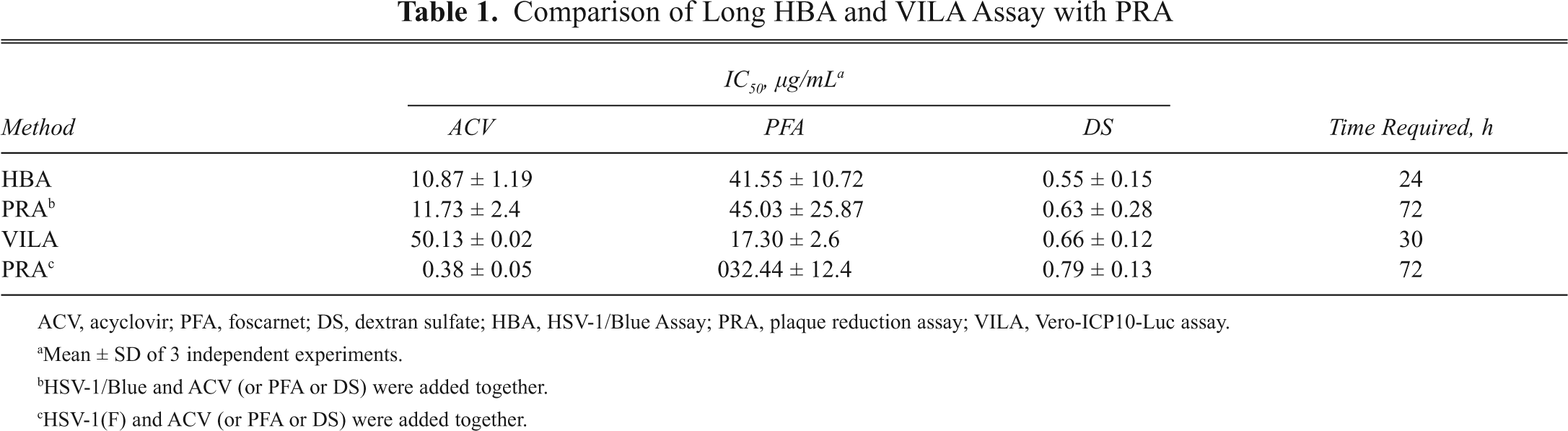
ACV, acyclovir; PFA, foscarnet; DS, dextran sulfate; HBA, HSV-1/Blue Assay; PRA, plaque reduction assay; VILA, Vero-ICP10-Luc assay.
Mean ± SD of 3 independent experiments.
HSV-1/Blue and ACV (or PFA or DS) were added together.
HSV-1(F) and ACV (or PFA or DS) were added together.
To determine whether the short HBA or VILA assay measured the inhibition of the early stages of HSV infection, ACV, HSV neutralizing antibodies (neut-Ab, a mixture of anti-gD MAb DL6 and anti-gL1 MAb L4), 11,12 and DS were assayed. Acyclovir acts on viral DNA polymerase, representing late-stage inhibitors, whereas HSV neut-Abs and DS inhibit viral entry, representing early stage inhibitors. When the detection was performed at 5 h postinfection, a dose-dependent effect was observed in the neut-Abs and DS ( Figs. 2B , C , E , F ), whereas no effect was observed with ACV ( Figs. 2A , D ). These results suggest that the short HBA and VILA assays were effective for the quantitative selection of antagonists against the early stages of HSV infection.
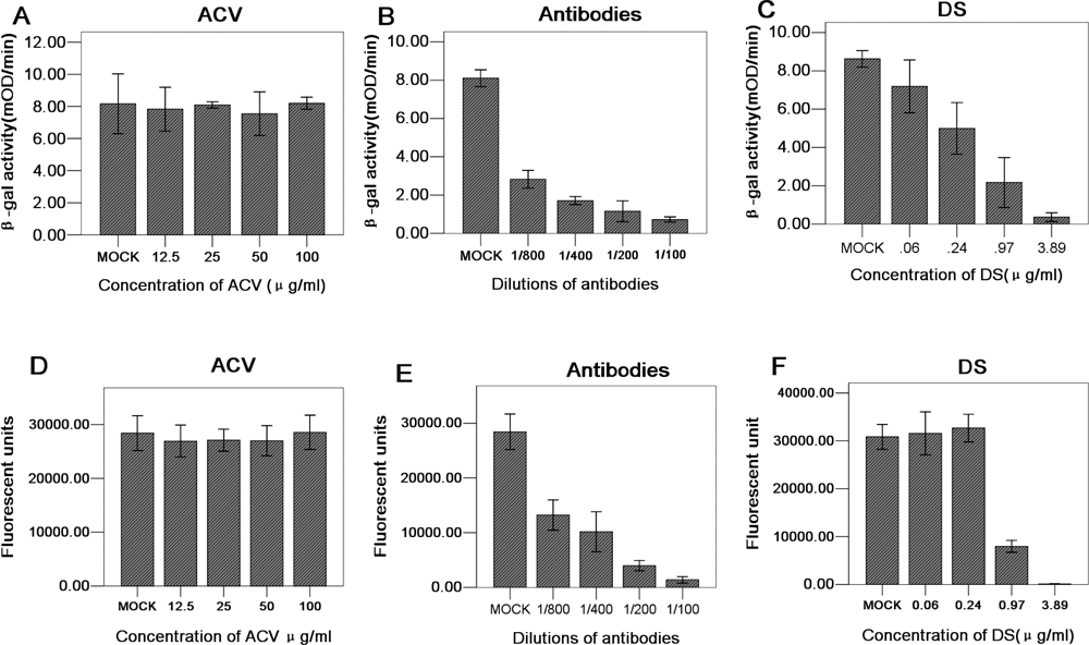
Short assays in HSV-1/Blue Assay (HBA) and Vero-ICP10-Luc assay (VILA) for herpes simplex virus (HSV) entry inhibitors. For
Antiherpetics found by the long assay may target any stage of HSV infection, whereas those found by short assay only target the early stages. When 2 assay versions are performed in parallel, different result combinations suggest the possible targets of the inhibitor. The inhibitory activities displayed by the testing inhibitor in both long and short assays indicate that the inhibition target was at the early stages of infection. However, inhibitory occurrence in the long assay alone suggests that the target was after the early stages of infection. In addition, combining the long and short assays in anti-HSV activity screening presents 2 other major advantages. First, the screening can efficiently select virus entry inhibitors or IE inhibitors without missing the potential inhibitors against other targets. Second, when the long assay is performed, a colorimetric cytotoxicity assay can be conducted in parallel to avoid false positives due to the cytotoxicity of the testing compound.
Aside from being cell-based antiviral screening assays, HBA and VILA are suitable for the rapid analysis of the potential target of an antiherpetic agent. As illustrated in Figure 1 , when the virus is allowed to mix with cells for attachment before a candidate agent is added ( Fig. 1A ), the assay helps show if an attachment is required. An antiviral effect observed only in this case indicates a postattachment target for the candidate agent. Similarly, when the candidate agent is mixed with cells before a testing virus is added ( Fig. 1B ), the assay helps show if the agent functions before virus attachment to cells. If the agent functions, then a cellular preattachment target is observed. On the other hand, when the candidate agent is mixed with a testing virus before adding to the cells for infection ( Fig. 1C ), the assay helps show whether virus exposure to the agent inactivates the virus before infection. If the virus is inactivated, the candidate agent obviously has a viral preattachment target. Similar information could also be obtained from VILA, when the Vero cells in Figure 1 are replaced with Vero-ICP10-Luc. In this case, HSV-1/Blue can be replaced with any laboratory (or primary isolated) strains of HSV, whereas the inhibition efficacy could be quantified with the measurement of luciferase activity.
Two screening assays specialized for antiherpetics targeting the early stages of HSV infection were established based on HSV IE promoter-directed reporter systems. HBA was capable of evaluating HSV entry inhibition activity against different host cell types, whereas VILA was capable of evaluating HSV entry inhibition activity against different HSV isolates. Both assays are quantitative with higher sensitivity and shorter assay time. Furthermore, both are adaptable to robotic high-throughput assay for antiherpetics. These assays also are efficient in determining the antiviral mechanisms of antiherpetic agents.
Footnotes
Acknowledgements
We thank Drs. Gary H. Cohen and Dr. Roselyn J. Eisenberg for providing HSV-1/Blue and the antibodies and Dr. Wang Yifei for HSV-1(F).
This work was supported by National Basic Research Program (973) (grant no. 2009CB522300, 2010CB530100); Chinese Academy of Sciences (grant no. KSCX1-YW-10); the Science and Technology Program of Guangdong Province, China (grant no. 2007B030501012); and the Science and Technology Program of Guangzhou, China (grant no. 2007Z1-E0111).