
Abstract
Obesity occurs with a low-grade inflammation as a result of the imbalance between energy intake and expenditure. This physiopathological status is accompanied by alterations in the gut microbiota. Living microorganisms called “probiotics” can affect the gut microbiota and can heal this imbalance, thus obesity. The aim of this study is to evaluate the changes in the gut microbiota in high-fat-diet-induced rats and to investigate whether any difference may be observed with the supplementation of Bifidobacterium infantis 35624 as a probiotic. The rats received different feeding regimens such as normal fat diet, high-fat diet, and B. infantis 35624 supplemented high-fat diet for 8 weeks. Weights gain was evaluated. Glucose and insulin levels, as well as gut microbiota, were examined at the end of the study. The high-fat diet group exceeded the criterion for obesity, and there was a significant increase in body weight over the study period (p < 0.001). A significant difference was found between the groups in terms of glucose and insulin levels (p < 0.05). Each group showed a different microbiota, and some bacterial species were found to be significantly associated with the weights of the groups (p < 0.001). Both high-fat diet and B. infantis 35624 supplementation caused changes in the gut microbiota. Beneficial strains associated with obesity may be clarified with species-specific studies to help prevent obesity.
Keywords
Introduction
The human gastrointestinal tract is home to over 100 trillion bacteria. Gut has an anaerobic environment; obligate anaerobes, including Fusobacterium, Bifidobacterium, Eubacterium, Bacteroides, and Peptostreptococcus, are at least 1000:1 more than facultative anaerobes including Enterobacteriaceae, Lactobacilli, and Streptococci. This bacterial community is called “microbiota.” 1 The nutrients were broken down by digestive enzymes and through the intestinal fermentation, ∼10% of the daily energy is provided. 2 The energy balance, including fatty acid oxidation in muscle, triglyceride storage in liver, and body weight, was found to be associated with the capacity of the microbiota to absorb nutrients in intestinal lumen. 3 Hyperphagia and high-fat high-calorie diets lead to obesity and insulin resistance (IR) through various pathways. Related diseases, such as diabetes mellitus, fatty liver disease, and cardiovascular disease, make obesity a frightening problem worldwide. 4 Although the rate of obesity changes by region in the world, the World Health Organization reported in 2024 that one in eight people are now living with obesity. 5 It has reported that 67.5% of deaths related to high body mass index are attributable to cardiovascular disease, and obesity has direct adverse effects on cardiac structure and function and leads to the development of both atherosclerotic and non-atherosclerotic cardiac disorders. 6
Obesity is based on energy imbalance, and it is characterized by chronic “low-grade” inflammation. This inflammation is associated with some changes in intestinal epithelial functions and microbiota composition. 7 Living microorganisms that affect beneficially the host by improving the balance of gut microbiota are called “Probiotics.” Recently, studies have pointed to the relationship between the body weight, fatness, IR, and gut microbiota, and many studies have showed the healing effects of the probiotics on obesity.8,9Bifidobacterium infantis 35624 is clinically used on dyspepsia and irritable bowel syndrome as a probiotic formula. 10 No study has yet been observed on its effect in obesity. The aim of this study is to evaluate obesity-related parameters, such as body weight, glucose, insulin levels, and gut microbiota changes in high-fat diet (HFD)-induced rats, as well as to investigate the effect of B. infantis 35624 as a probiotic supplement.
Significance Statement
There are some studies showing that the gut microbiota is associated with obesity-related disorders.
This study showed the change of gut microbiota both with high-fat diet and with the supplementation of Bifidobacterium infantis 35624.
Studies to be conducted with these observed species may provide clearer results regarding their relationships with the probiotic use against different nutritional regimens.
Materials and methods
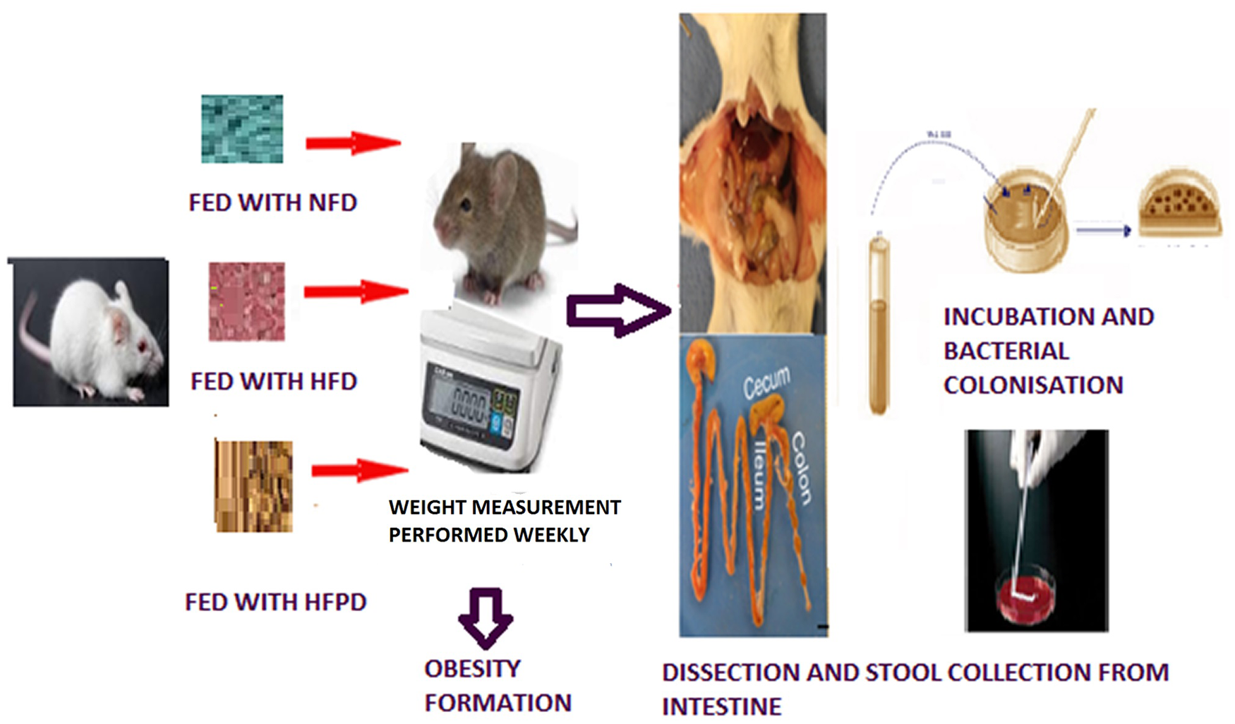
Animals and dietary intervention
This study was performed in Animal Experiments Laboratory of Medicine Faculty of University Ordu, Ordu, Turkey. Twenty-week-old male Sprague–Dawley rats weighing 250–300 g (n = 24) were obtained from the Experimental Animals Application and Research Center of University Ondokuz Mayıs, Samsun, Turkey. All rats were kept in plastic cages as four in one, in an air-conditioned room with 12 h light and dark cycle at constant temperature and humidity (23 ± 1 °C and 55%–65%, respectively) with free access to food and water. After 1 week of adaptation, rats were randomly divided into three groups (n = 8); feeding on normal fat diet (NFD), HFD, and HFD with probiotic supplementation (HFPD). NFD as standard feed diet containing 10% kcal fat and HFD as “cafeteria (CAF) diet” containing 60% kcal fat were obtained from Arden Research and Experiment Company, Ankara, Turkey. The CAF diet consists of high fat and sugar, overly processed, unhealthy, but delicious products such as chocolate, cake, cookies, chips, and biscuits. The diet contains 10% of energy from protein, 30% from carbohydrates, and 60% from fat by dry weight. CAF was used not only to increase the body weight but also to induce hyperphagia and cause metabolic syndrome, severe diabetic symptoms, and other metabolic dysregulations reported in studies. 11
The probiotic containing B. infantis 35624 (Symbiosys Alflorex Probiotic Capsules; Biocodex, Laboratoires Biocodex, Gentilly, France) was obtained from a local pharmacy in Ordu, Turkey. Each capsule contains 109B. infantis 35624 in powder form. For administration, the probiotic powder was suspended in 0.5 mL of sterile water using a syringe. The probiotics were administered daily via oral gavage to rats in the probiotic HFPD group at 0.5 mL/rat (at 10 am) for 8 weeks. 12
The rats were weighed in a tared plastic container with a lid. For weight measurement, an AND FZ 5000İ digital precision scale (AND, A&D COMPANY, LIMITED, Japan) was used. Rat weight was recorded on a weekly basis. The Lee index was used in order to calculate the obesity index. 13 At the end of the 8-week treatment period, after the fasting time of 6 h, all rats were sacrificed by cervical dislocation under deep anesthesia with intraperitoneal of ketamine 90 mg/kg (Ketalar; Eczacıbaşı, Istanbul, Turkey) and xylazine hydrochloride 3 mg/kg (Rompun; Bayer, Leverkusen, Germany) frequently used for anesthesia in rats. 14 The sigmoid column removed and was sent for examination of the intestinal flora bacteria.
Biochemical analysis
Blood samples were collected by entering from the bifurcation aorta with a 10 cc injector, and 4–6 cc of blood was taken from each rat. Blood samples were collected in anticoagulant-free biochemical tubes and transferred to appropriate laboratory parameter tubes. Glucose measurements were made by colorimetric method and insulin measurements were made by ELISA (cat no. 201-11-0708-48t; SUNRED, Shanghai, China). The homeostasis model assessment–IR (HOMA–IR) was calculated for the measurement of IR from fasting glucose and insulin.
Isolation and characterization of bacteria
Rats were fed on their respective diets for 8 weeks. Moreover, on the 0th, 3rd, 6th, and 8th weeks of the feeding period, feces were collected from each rat. Fecal samples taken from rats were brought to the microbiology laboratory under sterile conditions in half an hour. Immediately following collection and weight determination, stool samples were dissolved in filter-sterilized phosphate buffer. (Brine (PBS) solution (per liter dH2O: NaCl, 8.60 g; Na2HPO4, 0.86 g; KH2PO4, 0.40 g, pH 7.2), 0.02% peptone from fortified meat (Roth, cat no. 2366), and 0.05% L-cysteine Tenfold serial dilutions were made and prepared for each sample. Bacterial suspensions (100 µl) were spread on Mueller–Hinton Agar 103872 (Merck Microbiologycal Frankfurt Germany) and CASO (Trypic Soy) agar using sterile glass rods. Counts were determined after 6 days of incubation. Only plates containing 20–200 colonies/plate were obtained. All single colony morphology species observed after 6 days of growth were streaked on to Trypic Soy agar to ensure purity. Culture purity was examined by observing cell morphology after gram staining and colony morphology. Cryostocks (100 µl) bacterial suspensions were stored at −80 °C after mixing equal volumes of Tris-buffered aqueous solution (60 mM) containing 40% glycerol. 15 After gram and spore staining, 130 bacterial colonies were reduced to 50 colonies under the microscope and sent for analysis. Microbial characterization was performed using the automated system VITEK® 2 (BioMérieux, Craponne, France). In order to transfer a sufficient number of bacterial samples, they were previously passaged into blood agar and EMB agar (GBL, Biology Laboratory, Istanbul, Turkey) medium 24–48 h before using the applicator stick. Colonies of the pure culture were suspended and loaded into the device. The test data from the unknown organism were compared with the database of the device, defined according to their proximity to each of the database taxa and Eu cast criteria. 16
Statistical analysis
All statistical analysis was performed using SPSS 26 program (Sosa) IBM. Obtained data were presented as the mean with either standard deviation or standard error of the mean. The comparison between the groups were made using Tukey test, one-way analysis of variance. Differences were considered statistically significant at p < 0.05.
Ethic statement
The animal experiments were conducted with the approval of the Animal Experiments Local Ethics Committee of University Ordu (February 16, 2021, no. 82678388). All methods of this study were performed in accordance with the relevant guidelines and regulations of this committee.
Results
Bacterial flora
It was determined that the feces of rats fed with an HFD contained Leuconostoc mesenteroides spp. cremoris, Staphylococcus pseudintermedius, Kocuria kristinae Gemella sanguinis, Enterococcus faecalis, and Proteus hauseri. The feces of rats fed with HFPD contained E. faecalis, L. mesenteroides spp. cremoris, Escherichia coli, S. pseudintermedius, and Staphylococcus lentus. The feces of rats fed with an NFD contained Staphylococcus spp. urealyticus, Staphylococcus sciuri, L. mesenteroides spp. cremoris, E. coli, S. pseudintermedius, S. lentus, and Enterococcus casseliflavus. Gut microbiota characteristics revealed by different nutritional regimens were shown in Table 1.
Gut microbiota analysis of rats with different feeding regimes.
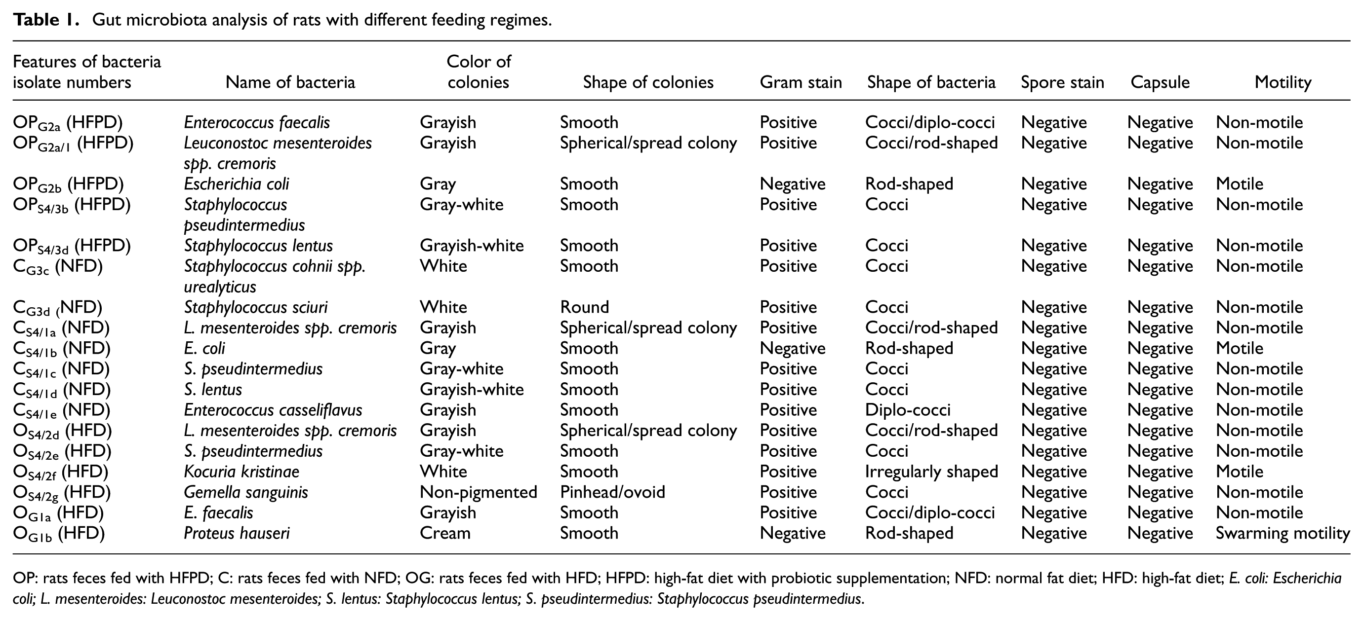
OP: rats feces fed with HFPD; C: rats feces fed with NFD; OG: rats feces fed with HFD; HFPD: high-fat diet with probiotic supplementation; NFD: normal fat diet; HFD: high-fat diet; E. coli: Escherichia coli; L. mesenteroides: Leuconostoc mesenteroides; S. lentus: Staphylococcus lentus; S. pseudintermedius: Staphylococcus pseudintermedius.
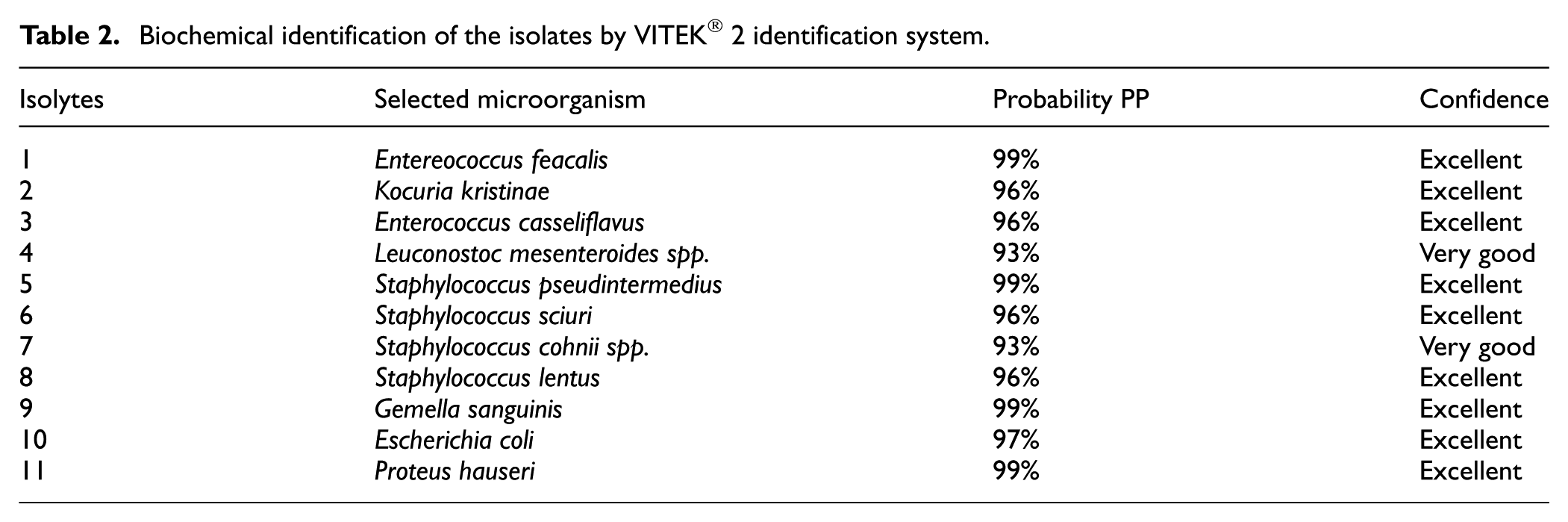
To determine the microbial flora, we analyzed 130 bacterial isolates, and we isolated 50 pure bacterial cultures determined by macroscopic and microscopic observations as gram staining, spore staining, and colony morphology. We tested 50 pure bacterial cultures on the VITEK® 2 (bioMérieux) system for bacterial species identification. Our VITEK® 2 results revealed that 11 of the 50 bacterial isolates were different species. It was observed that E. faecalis 13 times (26%), S. pseudintermedius 12 times (24%), E. coli 17 times (34%), and other species one time each (1%) were detected. The identification rates of these bacterial species were found mostly to be excellent and were shown in Table 2. The effect of dietary bacterial interaction on rat weights was evaluated. S. pseudintermedius, E. faecalis, and E. coli were found to have the effects significantly different between the weights of the groups (p < 0.001). The weight values of the groups according to these bacterial species were shown in Table 3.
Biochemical identification of the isolates by VITEK® 2 identification system.
Results of dietary–bacterial interaction according the groups.
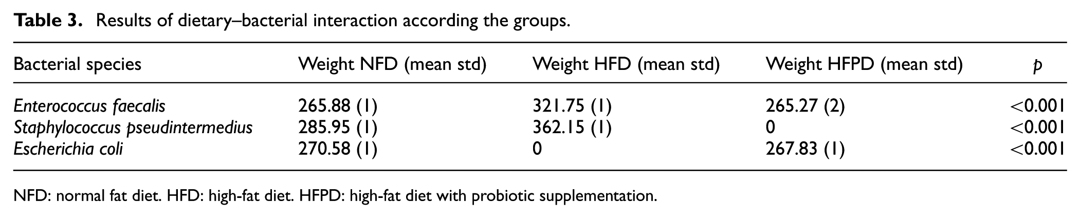
NFD: normal fat diet. HFD: high-fat diet. HFPD: high-fat diet with probiotic supplementation.
Body weight
Weight measurements were taken by animal laboratory technician throughout the study. From the beginning of the study, NFD, HFD, and HFPD groups showed a noticeable body weight gain over the 2.5-week period (time 1). In particular, the HFD group approached the obese criteria with 303.27 g. NFD and HFPD groups were very close to each other, on average 279.5 and 269 g (Figure 1(a)). A significant difference was found between the weight of HFD group compared to those of NFD and HFPD groups (p = 0.002). At the end of the 5th week (time 2) while the average weight in the HFD group was 324.57 g, it was 273.57 and 236.86 g in the NFD and HFPD groups, respectively. It was observed that HFD group weight increased significantly, with a weight gain of 21.50 g. A significant difference was found between the HFD group weight compared to those of NFD and HFPD groups (p < 0.0001; Figure 1(b)). At the end of the 8-week period (time 3), while the average weight in the HFD group was 335.83 g, it was 270.83 and 277.83 g in the NFD and HFPD groups, respectively. A significant difference was found between the weight of the HFD group compared to those of NFD and HFPD groups (p < 0.0001; Figure 1(c)). The rats of the HFD group exceeded the criterion for obesity, and there was a significant increase in body weight over time (all p < 0.0001). NFD and HFPD groups maintained their body weight even at the end of the eighth week. Weight changes of the groups during the study period were shown in Figure 1.
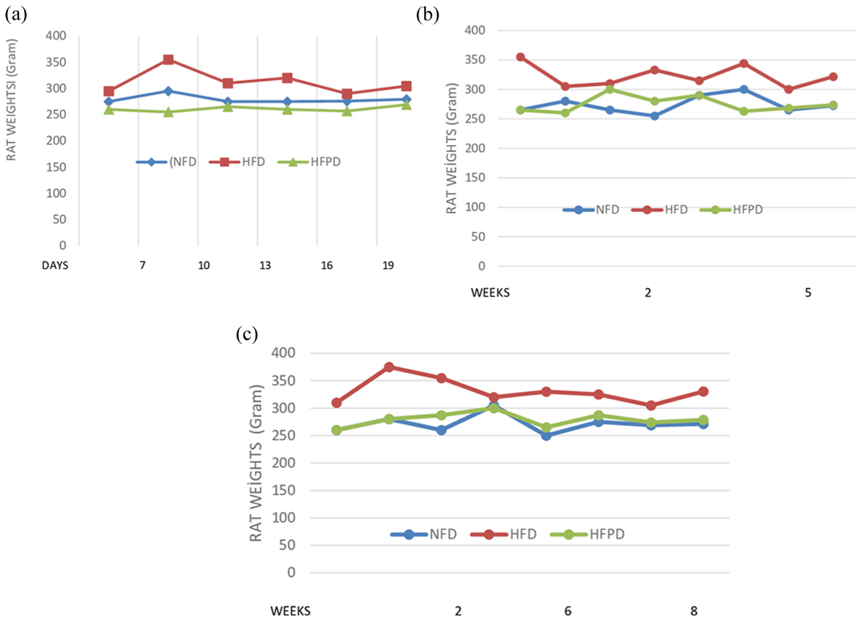
(a) The weight changes of the groups at 2.5 weeks of the study, (b) the weight changes of the groups at 5 weeks of the study, and (c) the weight changes of the groups at 8 weeks of the study.
Insulin and glucose levels
At the end of the study, the mean glucose and insulin levels were found to be 186.4 mg/dL and 4.3 IU, respectively, in the HFD group. However, these values were 129.4 mg/dL and 2.22 IU, respectively, in the NFD group and 139.1 mg/dL and 2.27 IU, respectively, in the HFPD group. HOMA–IR values were found as 0.7, 2, and 0.8 for the NFD, HFD, and HFPD groups, respectively. A significant difference was found between the groups in terms of glucose and insulin values (p < 0.05 and p < 0.05, respectively). Both glucose and insulin levels were found to be very high in the HFD group compared to the NFD and HFPD groups. Changes in glucose and insulin levels of the groups were shown in Figure 2.
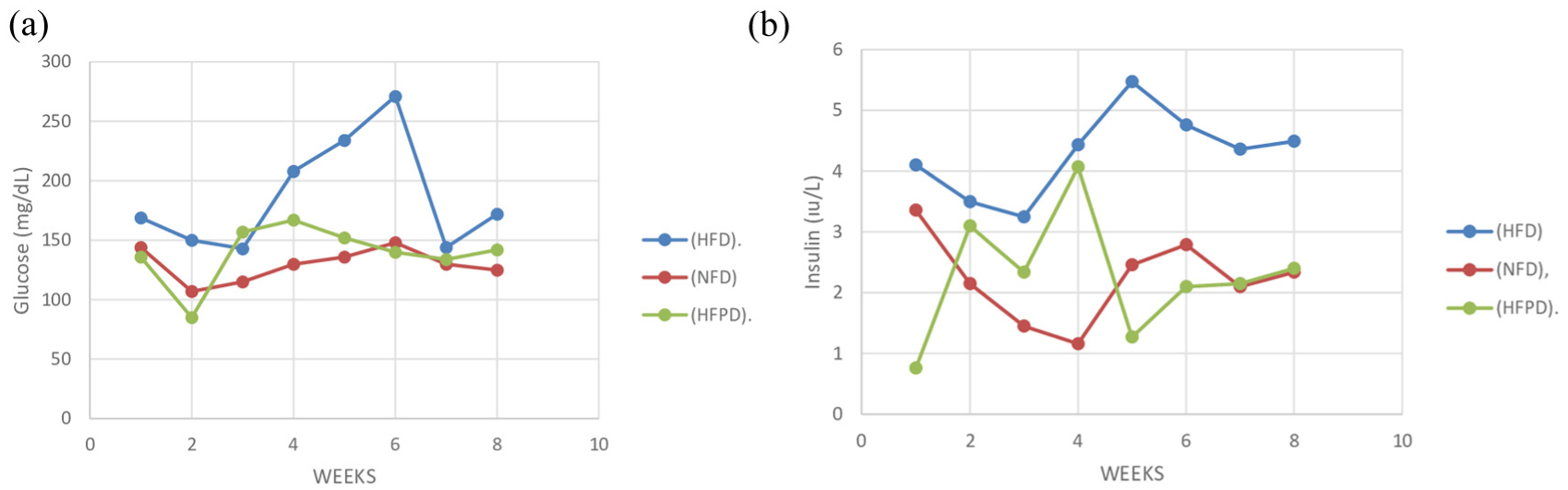
(a) Changes in glucose levels according to the groups and (b) changes in insulin levels according to the groups.
Discussion
The prevalence of obesity is increasing worldwide, primarily due to overeating and high-calorie food consumption. The relationships between the gut microbiota and the obesity are mentioned in many studies. Firmicutes and Bacteroides are two prominent phyla, representing 90% of the species in the gut. The gut microbiota must be balanced in order to keep the host healthy. Imbalanced gut microbiota called “dysbiosis” is associated with pathological disorders such as obesity, metabolic syndrome, and diabetes mellitus. 17 When the intestinal microbiota of obese and thin people is compared, higher Firmicutes and lower Bacteroidetes ratios have been found to be responsible for obesity pathogenesis. 18 The ratio of Firmicutes and Bacteroidetes has been stated as a balance index. If this ratio is above 25, it means sensitivity to obesity. A 20% increase in Firmicutes and a 20% decrease in Bacteroidetes were found to be associated with an additional energy gain of 150 kcal/day. 19 Bifidobacterium and Lactobacillus were considered protective against obesity by increasing the production of lactate and acetate acids, which protect against enteropathogenic agents. 20
In our study, different nutritional regimes caused some changes in gut microbiota. Among these, it was noteworthy that E. faecalis and S. pseudintermedius increased in HFD, while E. coli decreased. Similar to our results, a study found that the genera Staphylococcus was positively associated with obesity. 21 In another study, enterobacteria obtained from obese gut microbiota was given to lean mice, and it was determined that lean mice gained weight. 22 The most frequent sources of dysbiosis are antibiotics, bacteria-destructive medications, infectious diseases, or unhealthy diet. 23 The CAF diet has been linked to gut dysbiosis, local inflammation, and intestinal hyperpermeability. For this reason, this diet formulation is used to demonstrate the harms of an HFD. The relationship between a CAF diet and dysbiosis is not well defined. However, it is argued that this form of diet is composed of ultra-processed acellular nutrients that are rapidly absorbed in the small intestine, thus depriving the colonic microbiota of essential nutrients for growth. 24
HFD significantly increases bile acid production and secretion from the gall bladder. The bile acid emulsifies luminal fat and exposes micelles to lipase-mediated digestion and thus damage the mucosal intestinal barrier and gut microbiota. 25 In addition, luminal dietary metabolites can engage with endogenous ligands, which may differentially regulate intestinal metabolic processes through various nuclear receptor signaling, thus fine-tuning intestinal immune response. 26 Diet-induced gut dysbiosis leads to an increase in mucin-degrading bacteria, weakens the intestinal firewall, allows toxic or undesired substances to penetrate the mucosal and submucosal layers and pass into the bloodstream, resulting in some mucosal immune responses. 27 Through this process, microbiota possibly modulates the gut immune response linked to both local inflammation and some metabolic diseases.
Many studies mention the association of the gut microbiota with some inflammatory diseases. The gut microbiota ferments carbohydrates, proteins, and some fats in the nutrients, thereby providing the host’s energy needs and producing some metabolites. 28 Gut microbiome produces tryptophan metabolites, lipid metabolites, and short-chain fatty acids (SCFAs). Tryptophan metabolites influence immune system cells, SCFAs induce GLP-1, and enteroendocrine cells release peptide tyrosine tyrosine. These mechanisms contribute to regulate the balance between energy intake and expenditure. 29 The intestinal epithelium barrier prevents the passage of toxins and food antigens and protects the host from harmful substances, which activate the immune system. Local and systemic inflammation developed in obesity damage the protective effects of the gut microbiota and intestinal epithelium. 30 Microbial metabolites and components of cell wall, such as lipopolysaccharide (LPS), are the main responsible agents of the host–gut microbiota interaction. The failure of the intestinal epithelial membrane and increased gut permeability lead to bacterial fragments, such as LPS, diffusing through the gut into the bloodstream. This metabolic disorder is called “metabolic endotoxemia.”31,32 LPS levels were found to be higher in people with obesity than in healthy individuals. 33 The Enterobacteriaceae family including Escherichia, Salmonella, Enterobacter, Shigella, Klebsiella, Proteus, and Serratia genera provides LPS production. 34 Palmas et al. found relatively a high amount of bacterial taxa belonging to the Enterobacteriaceae family having endotoxic activity, in the obesity group when compared to the control group. 35 In one study, it was stated that Bacteroides vulgatus and Bacteroides dorei reduced the enteric LPS production in human gut. 36 Many studies stated the association of the gut microbiota and IR. In one study, insulin sensitivity in metabolic syndrome has been amplified by the fecal microbiota transplantation from lean people. 37 In another one, the metabolic endotoxemia was associated with the development of IR. 38 In a recent study, the presence of commensal E. coli with HFD treatment significantly increased body weight, inflammation in the liver, adipose tissue, and intestinal tissue and triggered impaired glucose tolerance. 39
In this study, the probiotic B. infantis 35624 supplementation showed the results with lower body weight and improved glucose and insulin levels. Both the body weight and the gut microbiota changes are affected by many factors. Physical activity, the amount and type of food consumed, lifelong medications, intestinal diseases, and comorbidities affect human health, the gut, and body composition throughout life. Maintaining the native composition as born of the gut microbiota is not possible. While it cannot be claimed that obesity can be completely reversed solely by taking a supplementary food or probiotic intake, a considerable number of studies mention that certain foods and probiotics have the healing effects on gut microbiota balance and obesity. The remedy-oriented studies have been conducted on anti-obesity diets via regulating the composition of the gut microbiota. In one study, when obese individuals reduced their carbohydrate intake, their body weight decreased and the composition of bacteria in their feces also changed. 40 In another study, polyphenol-rich cranberry extract prevented diet-induced obesity in mice, and Akkermansia muciniphila abundance was found to be associated with the reversing IR and hepatic steatosis. 41 In Boudry’s study, the intestinal permeability and inflammation were reduced and microbial dysbiosis was improved by supplement bovine milk oligosaccharides extracted to diet-induced obese mice. 42 These studies indicated the regulatory effects of probiotics in rats with HFD-induced obesity. In this study, it was observed that the gut microbiota changed with different nutrition regimens and the supplementation of B. infantis 35624 as a probiotic may have contributed to these changes. This study had some limitations. The diversity of microorganisms caused a limitation for the interpretation of the results. Again, it cannot predict the longer-term results of different nutrition regimens or probiotic supplementation.
Conclusion
The studies on the identification of beneficial strains associated with obesity will enable the identification of candidate strains. These findings can identify a targetable subset of the microbiota in treating people with metabolic inflammation. To determine both preventive and therapeutic strategies, precise microbiological targets must be identified and their potential clinical effects revealed.
Footnotes
Ethical considerations
The animal experiments were conducted with the approval of the Animal Experiments Local Ethics Committee of University Ordu (February 16, 2021, no. 82678388). All methods of this study were performed in accordance with the relevant guidelines and regulations of this committee.
Author contributions
Conceptualization: Ömer Ertürk. Data curation: Özlem Özdemir. Formal analysis: Selim Görgün. Funding acquisition: Özlem Özdemir and Ömer Ertürk. Investigation: Özlem Özdemir, Ömer Ertürk, and Selim Görgün. Methodology: Ömer Ertürk and Selim Görgün. Software: Özlem Özdemir. Validation: Ömer Ertürk. Visualization: Ömer Ertürk. Writing – original draft: Özlem Özdemir. Writing – review and editing: Özlem Özdemir and Ömer Ertürk.
Funding
The authors disclosed receipt of the following financial support for the research, authorship, and/or publication of this article: This study was supported by Ordu University Scientific Project Coordination Department (ODUBAP, project no. D-2204, April 19, 2022).
Declaration of conflicting interests
The authors declared no potential conflicts of interest with respect to the research, authorship, and/or publication of this article.
Data availability statement
The data generated in the present study are included in this manuscript. The corresponding author can be contacted for further information on the data.