Matthew Piechnik: A 21-year-old male presented with a painful rash on his left foot that he had noticed 2 d previously. He first noticed the rash while showering, but then it became increasingly painful, prompting his emergency department (ED) visit. He had not experienced a fever or taken any medication for his pain. The patient was a trail maintainer and had been hiking along the Appalachian Trail in New Jersey for several days over the previous few weeks. He had an otherwise unremarkable medical history, no known allergies, and took no medications or supplements.
Jeffrey Kalczynski: Considering the patient’s recent activities and the nature of the rash, several potential causes come to mind. Could you provide a more detailed description of the findings from his physical examination?
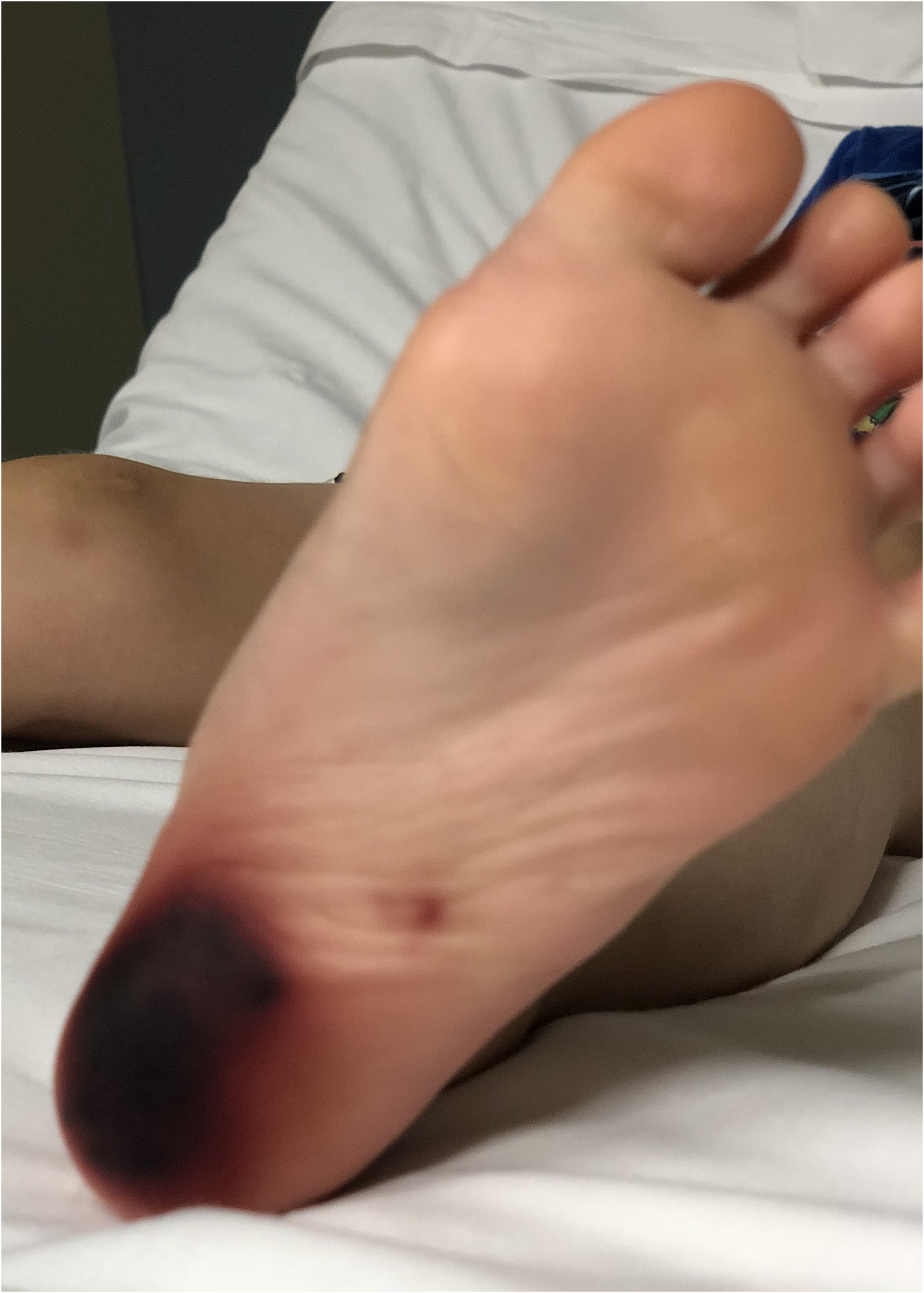
Matthew Piechnik: On examination, there appeared to be an area of brownish-blackish hyperpigmentation on the plantar aspect of the calcaneus region (Figure 1). The patient had full range of motion of his left foot without discomfort, and he had mild pain while bearing weight. There was no visible break in the skin, pain out of proportion, or crepitus noted on exam.
The patient's foot.
Cosimo Laterza: Did the patient suffer trauma or come in contact with poisonous flora, snakes, or insects? Was he wearing proper footwear?
Matthew Piechnik: He reported wearing steel-toed work boots. He denied any trauma or plant exposure. He did remember emptying a millipede out of one of his boots at the end of the day.
Jeffrey Kalczynski: Had there been any spread or change in the qualities of the skin findings after they were first noticed?
Matthew Piechnik: The patient had not observed any spreading, although he did report that the rash had become “darker” since it first appeared.
David Cuthbert: The patient's physical exam makes me believe that it is unlikely to be a bacterial infection given the lesions’ unusual discoloration and hyperpigmented appearance, along with the lack of any swelling, erythema, or warmth on exam. Further, there was no evidence of spreading along a fascial plan or systemic involvement based on his painless range of motion and absence of fever. I would think a localized skin irritant such as an insect bite or sting most likely would have caused this pattern of lesions. Do you think there is a correlation with the millipede?
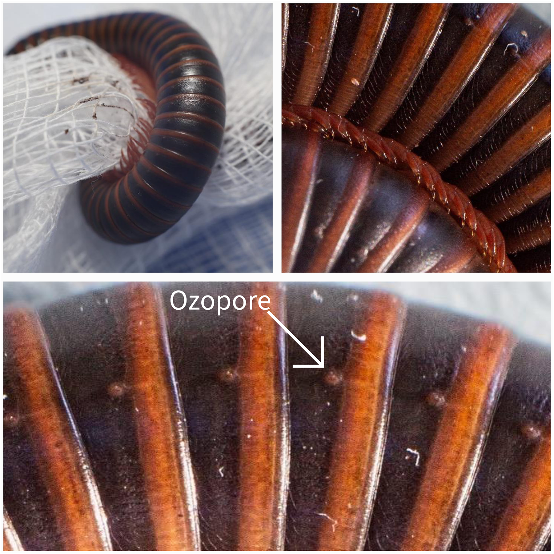
The millipede removed from the patient's boot.
Jeffrey Kalczynski: Yes, while millipedes lack the ability to bite or sting, they secrete skin irritants from their ozopores,1 resulting in a burn-like lesion. The discoloration is often attributed to benzoquinones present within the secretions. These compounds are a defense mechanism in several arthropods, including cockroaches, earwigs, and beetles. They are actually used in the textile and cosmetic industry specifically as tanning agents.2 Typically, millipede exposures result in mild irritation or pigmentation changes that last a few days to weeks.3 Prolonged exposure to a millipede, (Figure 2) as is likely in this case, could result in a more severe reaction.
Cosimo Laterza: Fortunately, the patient noticed the millipede in his boot, which helped narrow the differential. If the history were less clear, what risk factors should we consider that might indicate millipede exposure?
David Cuthbert: Millipedes are most commonly encountered in warm, moist environments such as tropical regions. Children are the most likely demographic to suffer millipede burns.4 Often confused for centipedes, millipedes can be differentiated by their two sets of legs per body segment compared with one set of legs per segment in centipedes.5 In contrast, centipede bites are caused by venom injected by the insects’ forcipules and cause a localized inflammatory reaction similar to other arthropod envenomations such as bee or wasp stings.4
Matthew Piechnik: Yes, and it is noted that millipede burns are primarily a clinical diagnosis. The burns may have a notable odor if significant secretions are still present. The affected area should be cleaned with soap and water for at least 5 min or until symptoms have resolved. Supportive management with topical corticosteroids and/or analgesics may be appropriate.
Jeffrey Kalczynski: There is a low risk for infection secondary to millipede burns. It is important to counsel patients spending time in warm and humid environments to carefully check their shoes and clothing for insects. Avoiding or removing decaying organic material also would further reduce the risk of encountering millipedes. This case involved the feet, but if there is involvement in sensitive areas such as the eyes or face, are there special considerations for managing millipede burns?
Matthew Piechnik: Ocular involvement is the most concerning location for millipede irritant exposure. In some limited case studies, corneal ulcers and blindness have been reported.6,7 Appropriate irrigation and consultation with ophthalmology are important to mitigate injury.
Cosimo Laterza: It is fortunate for the patient that the exposure was confined to an area where complications and treatment are more straightforward. Could you describe the clinical course during the ED visit and the patient's progress during follow-up?
Matthew Piechnik: During the ED visit, the patient was treated with a thorough cleansing of the affected area using soap and water for 20 min. He was prescribed topical corticosteroids and analgesics for pain management. Additionally, he was advised to keep the area clean and dry and to follow up with primary care if there was no improvement.
Jeffrey Kalczynski: How did the patient respond to the treatment?
Matthew Piechnik: The patient followed up via a telemedicine visit 1 wk later and was noted to have complete resolution of his pain. His hyperpigmentation had notably improved following a short course of topical corticosteroids and was completely resolved 2 wk after presentation.
Conclusion
Millipede burns, although uncommon, are a unique form of arthropod envenomation that should be considered in patients with hyperpigmented skin lesions. This case highlights the importance of considering arthropod exposure in patients presenting with unusual skin lesions. Millipede burns are prevalent in warm, humid environments and are often mistaken for other conditions such as chemical exposure or trauma, particularly in the pediatric population. Recognizing the hyperpigmented, discolored, and painful rash is crucial for a clinical diagnosis. Supportive care, including cleansing with soap and water and using topical corticosteroids or analgesics, is essential. The patient's successful treatment and recovery emphasize the importance of proper diagnosis and diligent follow-up care.
Footnotes
Author Contribution(s)
Jeffrey M. Kalczynski: Conceptualization; Formal analysis; Investigation; Writing – original draft; Writing – review & editing.
Matthew Piechnik: Conceptualization; Investigation; Writing – original draft; Writing – review & editing.
The authors declared no potential conflicts of interest with respect to the research, authorship, and/or publication of this article.
Funding
The authors received no financial support for the research, authorship, and/or publication of this article.
ORCID iD
Cosimo Laterza
References
1.
BlumMSWoodringJP. Secretion of benzaldehyde and hydrogen cyanide by the millipede Pachydesmus crassicutis (wood). Science. 1962;138(3539):512–513.
2.
De CapitaniEMVieiraRJBucaretchiFFernandesLCToledoASCamargoAC. Human accidents involving Rhinocricus spp., a common millipede genus observed in urban areas of Brazil. Clin Toxicol (Phila). 2011;49(3):187–190.
3.
HaddadV JrAmorimPC, Haddad WT Jr, CardosoJLC. Venomous and poisonous arthropods: identification, clinical manifestations of envenomation, and treatments used in human injuries. Rev Soc Bras Med Trop. 2015;48(6):650–657.
4.
HaddadV JrCardosoJLCLupiOTyringSK. Tropical dermatology: venomous arthropods and human skin: part II. Diplopoda, Chilopoda, and Arachnida.J Am Acad Dermatol. 2012;67(3):347.e1–9; quiz 355.