
Abstract

To the Editor:
Although typically placid, accidental contact with stingrays has caused numerous injuries to people along the surf zones of the United States, leading to injuries, commonly in the extremities, from barb impalement. 1 Due to the potential risks of a retained barb, imaging and proper removal are vital to patient care. 2 We present a case of a patient suffering from barb injury, highlighting the complicated healing process to help illustrate the importance of foreign body removal and infectious considerations.
A 71-y-old patient with no significant past medical history presented to the emergency department in the summer of 2022 after a stingray barb impalement. The patient was fishing in southern New Jersey, USA, when he accidentally caught a stingray. The barb entered his right wrist and exited his dorsal forearm (see Figure 1). After failing to remove the barb himself, he presented 2 h later at the hospital. The physical exam was notable for prominent edema at the dorsum of the right hand and wrist with notable puncture entry and exit lacerations. He was otherwise neurovascularly intact with stable vital signs. Labs were only significant for a leukocytosis of 12.2 B/L (normal range: 4.0–11.0 B/L). Blood cultures were collected.

Stingray barb impalement of right wrist and forearm.
X-ray films of the right hand and wrist showed a 7.5 cm thin foreign body within the soft tissue, compatible with the known stingray barb (see Figure 2). A local anesthetic was injected, and two small incisions were created at the edge of the exit wound. A hemostat was used to grab the tip of the barb, which was removed without resistance along the direction of entry (see Figure 3). Postprocedure x-ray was negative for retained foreign bodies. Intravenous ciprofloxacin for empiric coverage of Vibrio species and a tetanus, diphtheria, and pertussis booster were administered. The patient was admitted for observation and hand surgery consultation.

Plain radiograph with stingray barb visualized.

Intact barb after removal; comparison image of removed barb and a quarter coin.
On hospital Day 1 (HD1), hand surgery evaluated the patient and recommended conservative medical management. The patient was noted to have worsening weakness, numbness, and decreased range of motion in his fingers, as well as erythema and swelling, which moved proximally up the affected arm; patient was subsequently admitted to hospital medicine. Infectious disease was consulted for antibiotic guidance, and cefazolin was added to empirically cover Staphylococcus and Streptococcus species. An ultrasound of the dorsum of the hand revealed a 3 mm linear echogenic structure within the subcutaneous tissue at the site of injury, which is concerning for a retained fragment of the stingray barb. However, on HD2, the proximal erythema and swelling improved, and the interdisciplinary team deemed the risk of surgical intervention outweighed the benefits at that time.
On HD5, bullae associated with pain and edema erupted from the dorsum of the right hand. Concomitantly, the patient spiked a fever to 38 °C, prompting reevaluation. Cross-sectional computed tomography showed subcutaneous soft tissue swelling and infiltration predominantly along the dorsum of the right hand and wrist but did not reveal an obvious retained fragment. Surgery was reengaged, and after evaluation by a partner hand surgeon, surgical debridement occurred on HD6, given the new clinical findings. The patient was taken to the operating room for excisional debridement and exploration of the dorsal hand wound. Following a curvilinear incision, a small foreign body was visualized and removed from the wound. Nonviable fat and skin edges were sharply debrided, and penetration into the deeper extensor plane was not appreciated.
The patient tolerated the procedure well without complication and was continued on intravenous ciprofloxacin and cefazolin. All blood and wound cultures remained negative. The patient was discharged home on HD9 (POD3) with an oral regimen of cefadroxil and ciprofloxacin, with a plan to follow up outpatient with hand surgery.
On outpatient follow-up, the patient was noted to have improvement in the erythema and swelling but developed central necrosis of the skin flaps. Local wound care included daily cleansing and the use of mupirocin and collagenase ointments. After 2 wk, an additional excisional debridement was performed, resulting in a 2 × 4 cm wound over the dorsal wrist without any exposed structures (see Figure 4). A wound matrix bilayer (Integra) was placed in the operating room and was removed at 2 wk. Local wound care was continued until 5 wk after placement, when the wound was healthy and granulating well. At that time, a full-thickness skin graft from the ipsilateral antebrachium was performed with a standard tie-over bolster. The bolster was removed at POD5. The skin graft went on to heal completely without complications. At the latest follow-up (6 wk after skin grafting), the patient had full active and passive digital range of motion and wrist motion. There was no evidence of infection, and the edema continued to resolve.
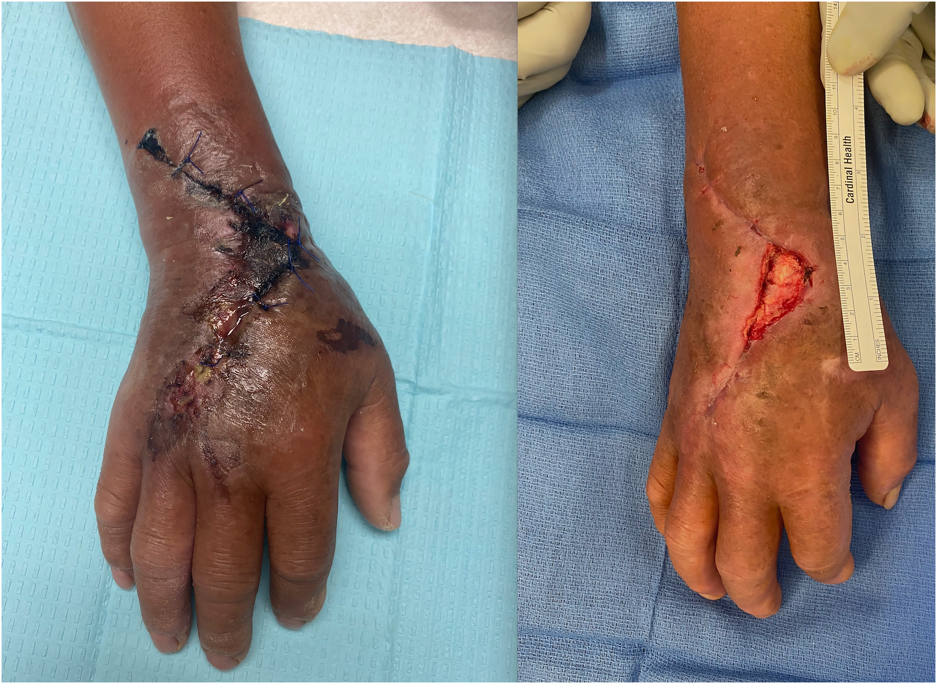
Preoperative image of patient's hand on the left and postoperative image of patient's hand on the right after debridement.
The barb of the stingray acts as a defensive structure via physical trauma and envenomation. 3 Anatomically, the barb is an extension of the stingray's spine, which acts to pierce and attach to subcutaneous tissue. The spine is composed of retroserrated edges, which can cause additional tissue damage when the barb is removed. 4 The venoms of medically important stingray species commonly cause local effects, including erythema and edema; systemic effects from stings delivered to extremities can occur but are rarely documented. 5 Given the possible complications from retained integumentary and glandular tissue, radiographic imaging, MRI, and ultrasonography are often indicated to aid in finding fragments.6,7 Additionally, microbial contamination of the barb can produce infectious complications. 3 Impalement may introduce pathogens from the barb itself and the aquatic environment. Pathogens include Vibrio, Aeromonas, Streptococci, and Staphylococci species. 8
Limitations
Lastly, a limitation of this case includes the lack of identification of the stingray species. Identification can be done via spine morphology and is desirable in understanding its role and treatment.4,9
Conclusion
Stingray envenomation with barb impalement poses several risks to the patient. Prompt and appropriate treatment with barb removal, antibiotic therapy, and high suspicion for retained spines are paramount to ensure recovery.
Footnotes
Acknowledgments
Alaina Todd for her contributions to patient care.
Abstract Presentation
Emergencies in Medicine, December 5, 2022, Maui, Hawaii.
Author Contribution(s)
Study concept and design (AM, TK, AS, CL, PF, LP); data acquisition (TK, AS, CL, PF, LP); data analysis (AM, TK, AS, CL, PF, LP); drafting and critical revision of the manuscript (AM, TK, AS, CL, PF, LP). All authors approved the final manuscript.
Declaration of Conflicting Interests
The authors declared no potential conflicts of interest with respect to the research, authorship, and/or publication of this article.
Funding
The authors received no financial support for the research, authorship, and/or publication of this article.
Statement of Informed Consent
Consent was obtained from the patient for the submission of the case report to the journal.
Statement of Human and Animal Rights
This case study was exempt from IRB review (control # iRISID-2022-1004).