
Abstract
Objectives:
To investigate the expression of β3-adrenergic receptor (β3-AR) in the atrium of rats with chronic heart failure (CHF).
Methods:
The heart failure rat model was established by aortic constriction. Thirty-six male Wistar rats were divided into Sham group (n = 10) and heart failure model group (n = 26), which were further divided into CHF control (CHF group) and BRL group. The rats in the BRL group were treated with a selective β3-AR agonist, BRL-37344 (4.0 nmol/kg, twice weekly) for 4 weeks.
Results:
In the BRL group, the left ventricular end-systolic pressure (83.21 ± 13.0 vs 101.50 ± 12.12 mm Hg) and the absolute values of the maximal rate of rise and fall of left ventricular pressure ([±dP/dtmax] 2.81 ± 0.04 vs 0.35 ± 0.04 and −2.72 ± 0.06 vs −3.33 ± 0.06) were lower than in the CHF group (P < .01). The left atrial mass index (LAMI) in the BRL group (0.4132 ± 0.0306) was higher than that in the CHF (0.3212 ± 0.0136) or Sham group (0.2683 ± 0.0145; P < .01). The levels of the left atrial β3-AR messenger RNA (mRNA) expression in the BRL group (0.932 ± 0.055) was higher than that in the CHF (0.706 ± 0.043) or Sham group (0.310 ± 0.020; P < .01). In all animals, there was a positive correlation between the level of β3-AR mRNA expression and the left or right atrial mass index (correlation coefficient ranged from 0.744 to 0.937).
Conclusion:
There is a significant increase in the β3-AR mRNA expression in the atrium of rats with heart failure. The level of β3-AR mRNA expression was associated with the AMI and was enhanced by a selective β3-AR agonist, BRL-37344.
Introduction
β3-Adrenergic receptor (β3-AR) belongs to the superfamily of G-protein-coupled receptors. 1 Although the intracellular pathway coupling β3-AR stimulation is still incompletely characterized, most studies showed that β3-AR activation decreases cardiac contractility. 1 In the normal heart, β3-AR may exert a negative counterregulation against excessive positive inotropic stimulation, thereby moderating oxygen consumption and preventing calcium overload and ultimately cardiomyocyte toxicity. 1 Recent evidence has shown that β3-AR is involved in the pathogenesis of chronic heart failure (CHF). 2 –5 In the failing heart, β3-AR is upregulated in the ventricular myocytes and selective blocking β3-AR is associated with a reduced ventricular remodeling and dysfunction. 2,4,5 In our previous study, 2 β3-AR activation increased the left and right ventricular mass index in rats with CHF. We also found that there was a significant positive correlation between the level of β3-AR mRNA expression and the ventricular mass index. 2 However, the expression of β3-AR mRNA in the atrium of rats with heart failure is unclear, and the impact of pharmacological β3-AR stimulation on the β3-AR mRNA expression and remodeling in the atrial tissues has not been fully investigated.
In this study, we investigated the levels of atrial β3-AR expression in a rat model of CHF. We also studied the effect of BRL-37344, a β3-AR agonist, on the atrial β3-AR expression and atrial remodeling in the heart failure rat model.
Materials and Methods
Surgical Techniques
This investigation conforms to the Guide for the Care and Use of Laboratory Animals published by the US National Institute of Health (NIH Publication No. 85-23) and was approved by our institutional review board. Adult male Wistar rats weighing 180 to 220 g were provided by the Experimental Animal Center of Guangdong Province. The rats were divided into Sham-operated group (Sham group, n = 10) and model group (n = 26).
The heart failure rat model was established by aortic constriction using a modified method that was previously reported. 2,6 Under general anesthesia (intraperitoneal injection of 20% urethane at 0.3 mL/100 g), the abdominal aorta was isolated and a fine needle (0.8 mm diameter) was inserted into the aorta, which was then tightly ligated at a site about 4 mm above the intersection between the abdominal aorta and the left renal artery. The needle was then withdrawn from the aorta, leaving the ligation in place which leads to a significant aortic constriction and increase in the afterload of the left ventricle. The Sham group received the same operation but without ligating the abdominal aorta.
Rats were then cared for by a curator and the investigators in the animal house facilities for 12 weeks. All rats survived in the Sham group and 24 rats survived in the model group 12 weeks after operation. Twenty-four rats were then divided into CHF control group (CHF group, n = 12) and BRL group (n = 12). The rats in the BRL group received a selective β3 agonist, BRL-37344 via tail vein injection, 4.0 nmol/kg, twice weekly for 4 weeks. 7 The 0.9% isotonic saline, 4.0 nmol/kg twice weekly, was administered in the CHF group and Sham group for 4 weeks. 7 Eleven rats survived in the CHF group and 9 rats survived in the BRL group 4 weeks later.
Hemodynamic Monitoring
The rats were anesthetized with an intraperitoneal injection of 20% urethane (0.3 mL/100 g) 16 weeks after heart failure model was established. A polyethylene catheter was inserted into the left ventricle through the right carotid artery. Another catheter was inserted into the pulmonary artery through the superior vena cava, right atrium, and the right ventricle. The measurements included heart rate, left ventricular end-systolic pressure (LVESP), left ventricular end-diastolic pressure (LVEDP), the maximal rate of rise and fall of left ventricular pressure (±dP/dtmax), and mean pulmonary arterial pressure (MPAP). Every parameter was measured 10 times and the mean values were used.
Pathological Examination
Rats were euthanized with an overdose of urethane and weighed after hemodynamic examinations were completed. The hearts were harvested and the cardiac tissues above the left atrium (including left atrial appendage) and right atrium were excised carefully. Left and right atrial absolute mass were weighed. The index of LAM and RAM (LAMI and RAMI) were obtained by dividing LAM and RAM with body mass, respectively.
The β3-AR mRNA Expression in the Atrium
The β3-AR mRNA expression was detected by reverse transcription–polymerase chain reaction (RT-PCR) which was performed as a 2-step procedure. Total RNA was extracted from frozen atrial specimens with TRIzol Reagent (Gibco BRL, New York, New York) and the concentration was measured in Nucleic Acid and Protein Analyzer (DU 800 Beckman Coulter, Brea, California). Two microliters of total RNA were reverse transcribed with Reverse TranScription System (Promega, Madison, Wisconsin) at 72°C for 60 minutes. β-Actin was used as inner reference. Sense (5′
Statistical Analysis
All data were analyzed and presented as mean ± standard deviation (X ± SD). Statistical significance among groups was performed by 1-way ANOVA test. Bonferroni t test was used for multiple comparisons. Pearson correlation analysis was used to evaluate the correlation between the left and right atrial mass index (RAMI) and the level of β3-AR mRNA expression in the left or right atrium. P < .05 was considered statistically significant. All data were recorded and analyzed using SPSS 16.0 software program.
Results
Changes in Hemodynamics
The heart rate in the BRL (480 ± 31) and CHF groups (456 ± 22) was higher than that in the Sham group (353 ± 25, P < .01), but there was no statistically significant difference between CHF and BRL groups (P > .05). Compared with the Sham group, LVESP and the absolute values of ±dP/dtmax in the BRL and CHF groups were lower (P < .01), whereas LVEDP and MPAP were higher (P < .01). The LVESP and the absolute values of ±dP/dtmax in the BRL group were lower than that in the CHF group (P < .01), whereas the LVEDP and MPAP were higher (P < .01; Table 1).
Changes in the Hemodynamics.
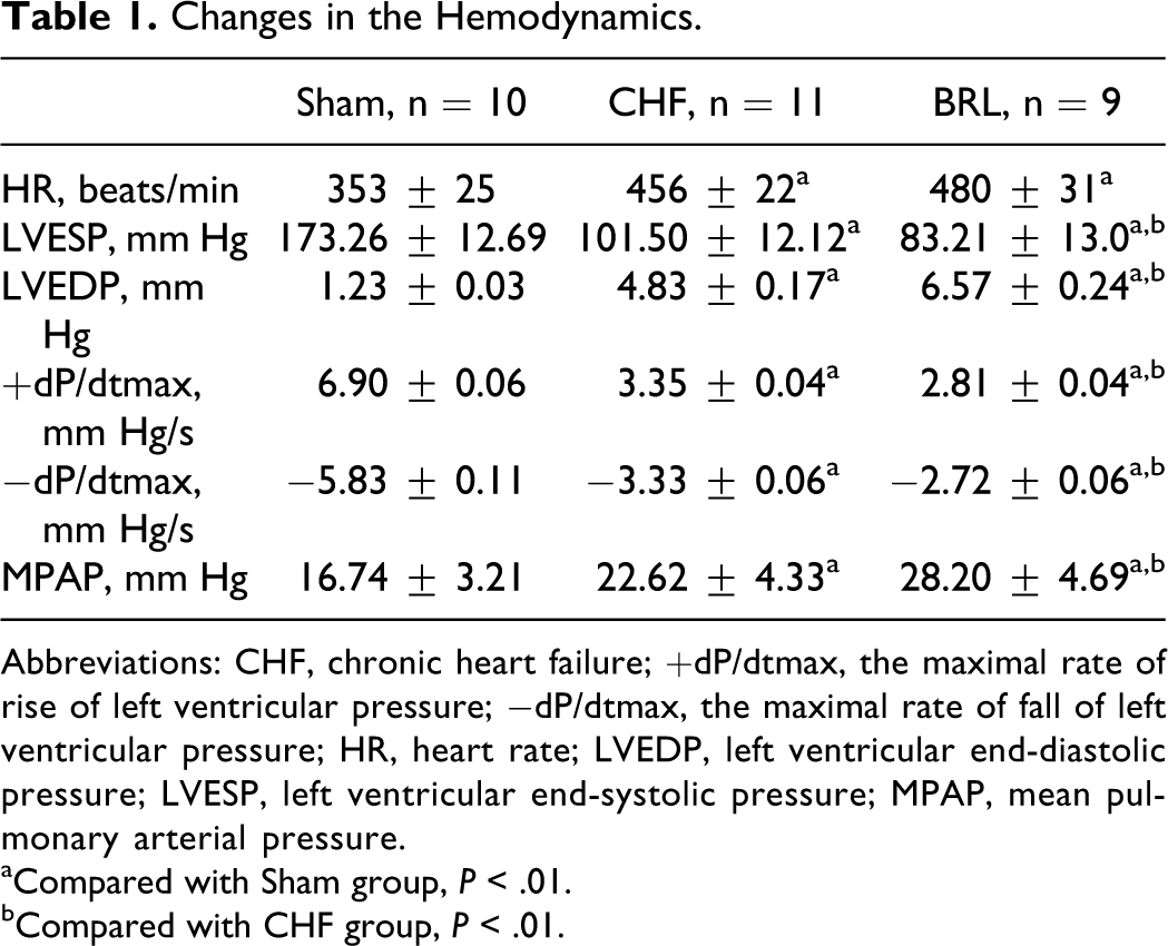
Abbreviations: CHF, chronic heart failure; +dP/dtmax, the maximal rate of rise of left ventricular pressure; −dP/dtmax, the maximal rate of fall of left ventricular pressure; HR, heart rate; LVEDP, left ventricular end-diastolic pressure; LVESP, left ventricular end-systolic pressure; MPAP, mean pulmonary arterial pressure.
aCompared with Sham group, P < .01.
bCompared with CHF group, P < .01.
Pathological Changes
There was no statistically significant difference in the body mass between the 3 groups (P > .05). The LAMI in the BRL group (0.4132 ± 0.0306) was higher than that in the CHF (0.3212 ± 0.0136) or Sham group (0.2683 ± 0.0145, P < .01). The RAMI in the BRL group (0.2806 ± 0.0160) was higher than in the CHF (0.2342 ± 0.0066) or Sham group (0.2046 ± 0.0125, P < .01; Table 2).
Changes in Atrial Mass and Atrial Mass Index.

Abbreviations: CHF, chronic heart failure; LAMI, left atrial mass index; RAMI, right atrial mass index.
aCompared with Sham group, P < .01.
bCompared with CHF group, P < .01.
The β3-AR Expression in Atrium
The expression of β3-AR mRNA in the atrium was demonstrated in Figure 1. As shown in Table 3, the level of left atrial β3-AR mRNA expression in the BRL group was higher than in the CHF and Sham group (0.932 ± 0.055 vs 0.706 ± 0.043 vs 0.310 ± 0.020, P < .01). The level of right atrial β3-AR mRNA expression in the BRL group was also higher than in the CHF and Sham group (0.602 ± 0.025 vs 0.398 ± 0.017 vs 0.172 ± 0.014, P < .01). The level of β3-AR mRNA expression in the left atrium was higher than in the right atrium within each group (P < .01). In each group, there was a positive correlation between the level of β3-AR mRNA expression and the AMI (correlation coefficient ranged from 0. 744 to 0.937, P < .01, Table 4).

β3-Adrenergic receptor (β3-AR) messenger RNA (mRNA) expression in the atrium. M indicates marker. From the left to right, lanes 1-3: right atrium, lanes 4-6: left atrium, lanes 1, 4: Sham group, lanes 2, 5: chronic heart failure (CHF) group, lanes 3, 6: BRL group.
The Level of β3-AR mRNA Expression (β3-AR mRNA/β

Abbreviations: CHF, chronic heart failure; LA, left atrium; mRNA, messenger RNA; RA, right atrium.
aCompared with Sham group, P < .01.
bCompared with CHF group, P < .01.
Correlation Between the Levels of β3-AR mRNA Expression and Atrial Mass Index.

Abbreviations: CHF, chronic heart failure; LA, left atrium; mRNA, messenger RNA; RA, right atrium.
Discussion
The main findings of our study are (1) CHF was associated with an increased β3-AR mRNA expression in the atrial tissues, and the expression of β3-AR mRNA in the left atrium was greater than that in the right atrium; (2) the level of β3-AR mRNA expression was associated with the AMI and was enhanced by a selective β3 agonist, BRL-37344. These results indicate that CHF may enhance β3-AR expression in the atrial tissues, and the enhanced β3-AR expression may lead to atrial remodeling.
Atrial remodeling is one of the pathophysiological consequences of CHF. Atrial remodeling includes atrial electrical remodeling and atrial structural remodeling. 9 Patients with CHF are susceptible to atrial fibrillation, and atrial remodeling is involved in the pathogenesis and maintenance of atrial fibrillation during CHF. The role of β3-AR in CHF-induced atrial remodeling is presently unclear. Previous studies have found that β3-AR may be involved in the ventricular remodeling of failing hearts. 10,11 In rats with cardiac hypertrophy, β3-AR protein density in the hypertrophic left ventricles was higher than that in the nonhypertrophic left ventricles, but no differences were noted between the 2 ventricles in β3-AR mRNA expression. 10 In the isolated hearts, selective β3-AR agonist BRL-37344 decreased LVDP, +dP/dt, and −dP/dt in hypertrophied ventricles, but only reduced +dP/dt in nonhypertrophied ventricles. 10 These results suggest that β3-AR protein density but not mRNA expression was upregulated, and the negative inotropic effects of BRL-37344 were diminished in rats with cardiac hypertrophy.
In dogs with pacing-induced CHF, the β3-AR mRNA and protein levels were increased by 73% and 147%, respectively, in the ventricular myocytes. 11 BRL-37344 produced a dose-dependent depression of myocyte contraction and relaxation which were much greater in heart failure myocytes than in normal myocytes. 11 Selective β3-AR antagonist L-748337, and nonselective β3-AR antagonist bupranolol blocked the negative inotropic effect induced by BRL-37344, while β1-AR antagonist metoprolol and β1-/β2-AR antagonist nadolol had no significant impact on the effect of BRL-37344. 11 These results suggest that the upregulation of β3-AR abundance is parallel with the degree of impaired function of the failing heart.
Studies on the expression and pathophysiological function of β3-AR in the atrium are very limited, and the conclusions from previous studies are inconsistent. In our study, there was an increase in β3-AR mRNA expression in the left and the right atrial tissues in the failing heart. The level of β3-AR mRNA expression was associated with AMI. Selective β3-AR agonist BRL-37344 decreased LVESP and dp/dtmax and increased LVEDP and pulmonary arterial pressure. These results suggest that in rats with CHF, β3-AR is involved in the structural remodeling of the atrium, and β3-AR stimulation aggravates the left atrial or ventricular dysfunction.
The cellular or molecular mechanisms by which β3-AR interferes with the remodeling or function of the atrial tissues are unclear. In human atrial myocytes, at room temperature, selective β3-AR agonist SR58611 and BRL-37344, and CGP12177 produced a dose-dependent increase in L-type Ca++ current (ICa,L). 12 A β1-AR and β2-AR blocker nadolol antagonized some of the stimulatory effects of SR58611 on ICa,L, indicating that the effect of SR58611 was at least partly mediated by activation of β1-AR and/or β2-AR. 12 In the presence of nadolol, CGP12177, SR58611, and BRL-37344 increased the contractility of isolated right atrial muscles. 12 These results suggest that β3-AR may produce a positive inotropic effect through increasing ICa,L in human atrial tissues. 12 However, another study found the positive inotropic and ICa,L responses to CGP12177 were mediated through the low affinity site β1L-AR of the β1-AR. 13,14 In isolated human atrial muscles, BRL-37344 produced dose-dependent increases in contractile force, which was blocked by nadolol or selective β2-AR antagonist ICI118551. 13 Selective β1-AR antagonist CGP20712A did not modify the inotropic effect of BRL-37344. 13
At 37°C, BRL-37344 produced dose-dependent increases in contractile force of isolated human atrial muscles, which was blocked by nadolol or selective β2-AR antagonist ICI118551 but not by selective β3-AR antagonist L-748337. 13 Selective β1-AR antagonist CGP20712A did not modify the inotropic effect of BRL-37344. 13 At 24°C, BRL-37344 and SR58611 did not increase atrial force. 13 BRL-37344 and SR58611 did not increase ICa ,L at 37°C but did at 24°C. CGP12177 increased force and ICa,L at both 24°C and 37°C, which was prevented by bupranolol but not L-748337.
One of the potential clinical significance of the increased β3-AR expression in the atrium is the impact on the electrophysiology of the atrial tissue. In a rabbit atrial fibrillation model, the protein level of β3-AR in the atria was significantly increased. 15 The atrial effective refractory period in atrial myocytes was decreased, and it was further shortened by BRL-37344 treatment. BRL-37344-induced time-dependent shortening of action potential duration, together with a decrease in ICa ,L and increase in IK1 and Ito. All the effects were abolished by selective β3-AR antagonist SR59230A. 15
A limitation of this study is that only mRNA levels of β3-AR were measured in the animal models, and the receptor expression as a protein was not studied. In addition, there were no functional measurements using β3-AR agonists or antagonists at the end of the study, and the acute effect of BRL treatment on hemodynamics was not investigated. These issues may be addressed in future investigations.
In conclusion, there is an increase in the β3-AR mRNA expression in both left and right atrium following heart failure. Pharmacological stimulation of β3-AR further increased the atrial expression of β3-AR in this heart failure rat model. Although the changes in β3-AR expression coincide with atrial remodeling and ventricular dysfunction, the impact of β3-AR expression on the anatomical structure and electrophysiology requires further investigation.
Footnotes
Declaration of Conflicting Interests
The author(s) declared no potential conflicts of interest with respect to the research, authorship, and/or publication of this article.
Funding
The author(s) disclosed receipt of the following financial support for the research, authorship, and/or publication of this article: This study was supported by a Medical, Pharmaceutical and Health Scientific Research Grant from Guangzhou City Council (No. 201102A213198), and a Medical Scientific Research Grant from Guangdong Provincial Government (A2011480).