
Abstract
Introduction
Accumulating evidence suggests that sex differences in ventricular repolarization exist in almost all species, ranging from human to mice, despite marked interspecies differences in the ionic currents underlying ventricular repolarization. Generally speaking, cardiac repolarization is prolonged and repolarization reserve is diminished in female animals and humans, compared to the male. 1,2 The reduced repolarization reserve is demonstrated by a relatively greater increase in ventricular action potential duration (APD) or QT interval and a higher incidence of torsades de pointes (TdP) in the female in response to drugs that block repolarizing K+ currents. 1,2 Many in vitro and in vivo experiments have suggested that the different modulation of cardiac ionic currents by estradiol and dihydrotesterone (DHT) play a major role in the sex differences in repolarization. 1,2
Even though gender-specific differences in ventricular repolarization have gained wide recognition, little information is available concerning the role of the different ovarian hormones in the modulation of repolarization in the female. Several previously published clinical reports suggested a potential impact of estradiol and estradiol plus progesterone replacement therapies on ventricular repolarization in postmenopausal women. In healthy postmenopausal women, hormone replacement therapy (HRT) with estradiol alone usually produces a prolongation of QT interval, while estradiol plus progesterone does not significantly affect the QT interval. 3 –10 Furthermore, it is reported that the degree of QT prolongation in response to ibutilide, a class III antiarrhythmic agent, varies with the menstrual cycle phases: maximum increase in rate-corrected QT interval (QTc) after ibutilide is greater for women during menses and the ovulatory phase, compared with women during the luteal phase. 11 These results suggest that estradiol and progesterone may have different modulating effects on cardiac repolarization which may give rise to a physiological variation in cardiac repolarization and a variable risk of drug-induced TdP in the female during the menstrual cycles. Thus, in the present study, the chronic modulation of female hormones on the repolarization and the susceptibility to class III antiarrhythmic agent was studied in rabbits by ovariectomy and HRT.
Methods
The experimental protocol was approved by the Ethical Committee for Biological and Medical Research in our university (the animal ethical approval document: No. 2009-093) and conforms to International Guiding Principles for Biomedical Research Involving Animals (CIOMS, 1985).
Ovariectomy of Rabbits and HRT
Female New Zealand white rabbits with body weights of 2.0 to 2.5 kg were anesthetized with 3% pentobarbital (30 mg/kg) and underwent ovariectomy under sterile techniques. Two weeks after the surgery, they were randomly divided into 3 groups: the control group (group C), the estradiol supplement group (group E), and the estradiol plus progesterone supplement group (group E + P), in order to mimic the ovarian hormonal states of the menses, follicular and luteal phases in women during the menstrual cycles. 12 These 3 groups of animals were injected intramuscularly each day with vehicle, estradiol benzoate (100 μg/kg per d), or estradiol benzoate (100 μg/kg per d) plus progesterone (5 mg/kg per d), respectively, and the injections were continued for 2 weeks. A time-matched control group was also set up to evaluate the effect of ovariectomy on cardiac repolarization.
Recording and Analyzing of Rabbit Electrocardiogram
Limb-lead electrocardiograms (ECGs) were recorded with rabbits in anesthetized states. Electrocardiogram was recorded on paper at a speed of 50 mm/s using an ECG recorder (ECG-9130P, Nihon Kohden, Japan). The ECGs were coded and randomized to allow blinded measurement of QT interval and other parameters. All ECG recordings were analyzed manually using handheld caliper by a single observer who was unaware of the hormone state of the rabbits. The ECG recordings in which the end of the T wave could not be reliably determined were excluded from the analysis. Each measurement was given as the mean of 3 consecutive beats. The RR interval was measured as the time difference between the 2 consecutive R waves. The intervals from the onset of the QRS complex to the peak and the end of T wave were measured and indicated as QTp and QT, respectively. The Tpeak-end interval (Tpe), usually used as an indicator of the global dispersion of ventricular repolarization, 13 was expressed as the difference between QT and QTp. The ECG parameters (QTp, QT, and Tpe) were corrected for heart rates using the formula described by other investigators 14 (eg, QTc = QT − 0.704 × [RR − 250]) and were indicated as QTpc, QTc, and Tpec, respectively.
Influence of HRT on the Cardiac Sensitivity of Ovariectomized Rabbits to d,l -Sotalol
After 2 weeks of HRT, the rabbits were anesthetized with 3% sodium pentobarbital (30 mg/kg) and then intravenously infused with sotalol hydrochloride (
Drugs
Estradiol benzoate injections, progesterone injections, and vehicle injections were purchased from Shanghai General Pharmaceutical Co., Inc (Shanghai, China). Sotalol hydrochloride injections were obtained from Yangtze River Pharmaceutical Group Co, Ltd (Taizhou, Jiangsu Province, China) and were diluted with normal saline to the desired volumes.
Statistical Analysis
Data were expressed as mean ± standard deviation (SD). Analysis of variance (ANOVA) was used to calculate critical differences among multiple means, and Student t test was used for comparison between 2 means. Fisher exact test was used to analyze the incidence of sotalol-induced arrhythmias. A P value of less than .05 was considered statistically significant.
Results
Effects of Ovariectomy on ECG Parameters in Rabbits
To evaluate the possible influence of the variation of blood female hormones on cardiac repolarization, 45 female rabbits were ovariectomized, and their ECG parameters measured 2 weeks after the ovariectomy were compared with those of the time-matched control rabbits (Table 1). The parameters of ECG such as RR, QTp, QTpc, QT, and QTc intervals in ovariectomized rabbits were not significantly different from those in the control rabbits (n = 45 and 12, respectively). The value of Tpe in the ovariectomized rabbits was large than that of the control rabbits but the significance of difference disappeared after correction with heart rate.
Comparison of ECG Parameters in Rabbits 2 Weeks After Ovariectomy With Those of the Time-Matched Control Female Rabbits a
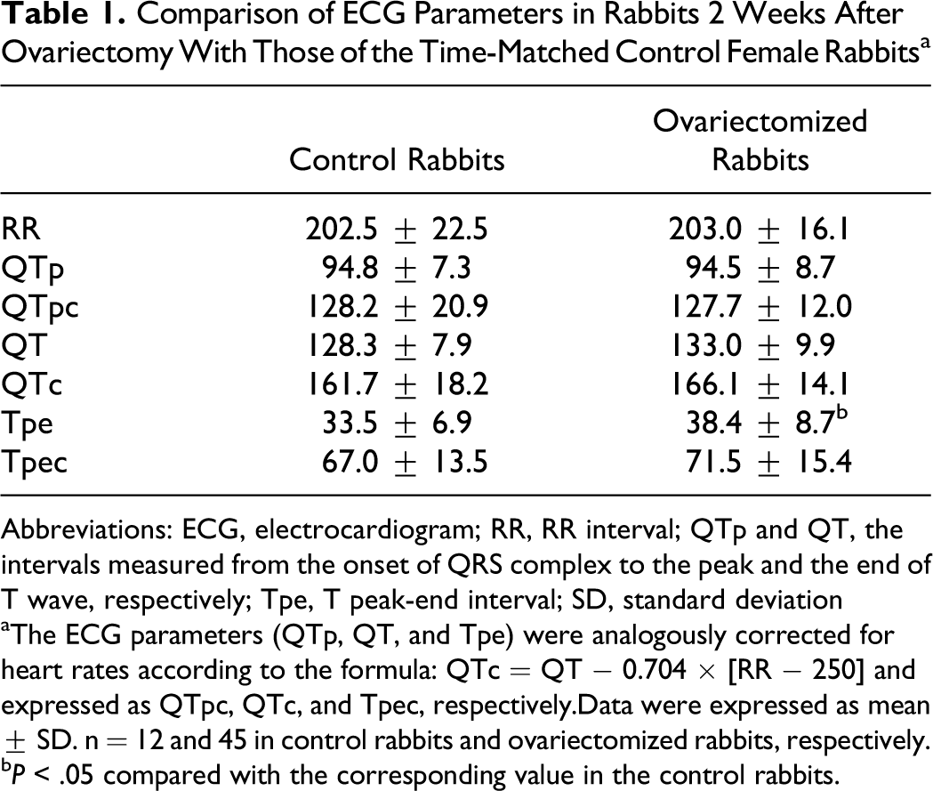
Abbreviations: ECG, electrocardiogram; RR, RR interval; QTp and QT, the intervals measured from the onset of QRS complex to the peak and the end of T wave, respectively; Tpe, T peak-end interval; SD, standard deviation
aThe ECG parameters (QTp, QT, and Tpe) were analogously corrected for heart rates according to the formula: QTc = QT − 0.704 × [RR − 250] and expressed as QTpc, QTc, and Tpec, respectively.Data were expressed as mean ± SD. n = 12 and 45 in control rabbits and ovariectomized rabbits, respectively.
bP < .05 compared with the corresponding value in the control rabbits.
Effects of HRT on ECG Parameters in Ovariectomized Rabbits
Two weeks after ovariectomy, the rabbits were randomly assigned to 3 groups receiving 2 weeks of treatment with vehicle, estradiol, and estradiol plus progesterone, respectively (n = 15 in each group). The comparison of the ECG parameters of the 3 groups was illustrated in Table 2. Most measured and rate-corrected ECG parameters for repolarization (QTp, QT, QTc, Tpe, and Tpec) in group E were not statistically different from those in group C, except that a significantly longer QTpc was observed in group E (P < .05). However, QT, QTc, Tpe, and Tpec in group E + P were significantly shortened when compared with those in group C (P < .05 or P < .01), and QTp, QTpc, and QTc were also significantly decreased in comparison with those of group E (P < .05). The RR intervals were not statistically different among the 3 groups.
Comparison of the ECG Parameters in Ovariectomized Rabbits After 2 Weeks of Hormone Replacement Therapies a

Abbreviations: ECG, electrocardiogram; RR, RR interval; QTp and QT, the intervals measured from the onset of QRS complex to the peak and the end of T wave, respectively; Tpe, T peak-end interval; SD, standard deviation.
aC, E, and E+P indicate the groups treated with vehicle, estradiol, and estradiol plus progesterone, respectively. The ECG parameters (QTp, QT, and Tpe) were analogously corrected for heart rates according to the formula: QTc = QT − 0.704 × [RR − 250] and expressed as QTpc, QTc, and Tpec, respectively. Data were expressed as mean ± SD. n= 15 in each group.
b P < .05 and c P < .01 compared with the corresponding value in group C, respectively.
d P < .05 compared with the corresponding value in group E, respectively.
Modulation of HRT on the Cardiac Susceptibility of Ovariectomized Rabbits to d,l -Sotalol
To investigate whether different HRTs can modulate the cardiac susceptibility of ovariectomized rabbits to
Changes in ECG Parameters Induced by

Abbreviations: ECG, electrocardiogram; RR, RR interval; QTp and QT, the intervals measured from the onset of QRS complex to the peak and the end of T wave, respectively; Tpe, T peak-end interval; SD, standard deviation.
aC, E, and E + P indicate the group of rabbits pretreated with vehicle, estradiol, or estradiol plus progesterone, respectively. The dose of
b
P < .05 and c P < .01 compared with the corresponding value before the application of
d P < .05 compared with the corresponding value in group C, respectively.
e P < .05 compared with the corresponding values in group E, respectively.
The difference in the ECG parameters among the 3 groups was also enlarged by
The

An episode of ventricular tachycardia induced by
Arrhythmias Induced by

Abbreviations: PVC, premature ventricular contraction; NSVT, nonsustained ventricular tachyarrhythmia; SD, standard deviation.
aC, E, and E + P indicate the groups pretreated with vehicle, estradiol, or estradiol plus progesterone, respectively. The dose of
b P < .05 and c P < .01 compared with the corresponding value in group C, respectively.
d P < .05 and e P < .01 compared with the corresponding values in group E, respectively.
Discussion
In the present study, to evaluate the possible influence of ovarian hormones on cardiac repolarization during the different phases of the menstrual cycle, the effect of ovarian hormone deficiency as well as HRT with estradiol alone or with estradiol plus progesterone on ventricular repolarization was investigated in ovariectomized rabbits. It was found that the ECG parameters including RR, QTpc, QTc, and Tpec in ovariectomized rabbits were not significantly different from those in the time-matched control rabbits. In the literature, the influence of ovariectomy on the ECG parameters of rabbits was rarely reported. Since the female rabbit is an induced ovulator, it has low circulating level of estradiol that is little affected by ovariectomy 15,16 ; thus, our observation that ovariectomy did not influence the ECG parameters is reasonable. After 2 weeks of HRT, estradiol alone had no significant influence on ECG parameters such as QTc and Tpec, except that it significantly increased QTpc. However, when progesterone was included in the HRT, QTpc and QTc were shortened, and Tpec, a measurement that reflects the global dispersion of ventricular repolarization, was reduced. Those results suggested that progesterone may accelerate the process of ventricular repolarization and may play an important role in homogenizing the global heterogeneity of ventricular repolarization, whereas estradiol’s modulation of cardiac repolarization is minimal in this model.
Gonadectomy and HRT are the common methods employed in the study of the mechanisms underlying the gender differences in cardiac repolarization. Until now, most of these studies were conducted in ovariectomized or orchiectomized animals supplemented with either estradiol or DHT. It is found that DHT attenuated the QT interval compared with the placebo-treated controls in orchiectomized male rabbits.
17
Whereas in ovariectomized rabbits, the ECG parameters did not differ among the placebo-, estradiol-, or DHT-treated group.
18
Regarding the modulation of estradiol or DHT on the cardiac sensitivity to the repolarization-prolonging agents, it is found that both DHT-treated ovariectomized and orchiectomized rabbits displayed less QT prolongation in response to quinidine challenge compared with placebo controls.
17,19
On the other hand, estradiol enhanced the sensitivity of the cardiac preparations isolated from the ovariectomized rabbits to class III antiarrhythmic drugs. Hara et al
18
reported that in papillary muscles from ovariectomized rabbits, E-4031-induced prolongation of APD90 and the incidence of early afterdepolarization (EAD) were significantly greater in the estradiol-treated than the DHT- and placebo-treated groups. Different from the aforementioned studies, our study compared the HRT with estradiol alone or with estradiol plus progesterone on the modulation of the cardiac sensitivity to
It is important to note that although the chronic treatment of estradiol potentiated the drug-induced QT prolongation associated with proarrhythmic effects, estradiol itself has little influence on baseline QT interval in the present study. This phenomenon was also observed in several previous studies aforementioned.
11,18
Thus, it may be the nature of the process underlying repolarization (ie, repolarization reserve) rather than the baseline duration of ventricular repolarization itself that determines the hormone state-related differences in drug-induced effects. The ionic basis of repolarization reserve is supposed to be IKr and other outward potassium currents (ie, IK1 and IKs) contributing to ventricular repolarization. The chronic treatment with estradiol may mainly reduce the repolarization reserve and thus increase the susceptibility of female rabbits to
Although it is well known that estradiol or the female gender may exacerbate the cardiac sensitivities to K+-channel-blocking agents in animal models, little information is available for progesterone. In the present study it has been revealed that in HRT with both progesterone and estradiol, the resulting shortening of QT interval and the decreased global dispersion of ventricular repolarization by progesterone may account for the lower sensitivity of the female rabbits to drug-induced repolarization prolongation and arrhythmias, thus counteracting the effect of estradiol. Since there is a physiological change in circulating levels of both estradiol and progesterone in women, with a high level of progesterone during the luteal phase, these findings may suggest a protective effect of progesterone against the drug-induced arrhythmias in women. Thus, our observation with progesterone on the risk of
It is worth mentioning that in most in vivo and in vitro studies evaluating the drug sensitivities in rabbits, TdP is the most common form of arrhythmas induced by repolarization-prolonging drugs. However, the typical tachyarrhythmias induced by
In summary, our study provided strong evidence for a different role of estradiol and progesterone in modulating ventricular repolarization and the cardiac susceptibility of the female rabbits to class III antiarrhythmic drugs. Estradiol can increase the susceptibility of female rabbits to
Footnotes
The author(s) declared no potential conflicts of interest with respect to the research, authorship, and/or publication of this article.
The author(s) disclosed receipt of the following financial support for the research, authorship, and/or publication of this article: This research is supported by Program for Changjiang Scholars and Innovative Research Team in University (No. IRT0642) and by Yangfan Project of Tongji University (No. 2010YF01).