
Abstract
Background:
Several radiographic and cadaveric studies have suggested that specific anatomical features of the Lisfranc joint complex, including a shallower medial mortise of the second metatarsal (M2), shorter M2 length, or pes cavus alignment, may be associated with ligamentous Lisfranc injuries. These associations have largely been derived from 2-dimensional imaging or post-injury assessments. Weightbearing computed tomography (WBCT) enables 3-dimensional evaluation of midfoot anatomy under physiologic load and allows a more robust reassessment of these proposed anatomical associations.
Methods:
This retrospective matched case-control study compared WBCT scans of the uninjured contralateral feet of 34 patients with low-energy ligamentous Lisfranc injuries to those of 34 matched controls. Groups were matched 1:1 for sex and side, and within a tight caliper for age and body mass index. Manual WBCT-based measurements included M2 length, M2 base height, medial and lateral mortise depths, foot length, and derived ratios. Overall foot and ankle alignment parameters were assessed using semi-automatic and automated WBCT tools. Paired statistical analyses were performed with correction for multiple comparisons.
Results:
No significant differences were identified between groups for M2 length (74.5 ± 7.1 mm vs 75.2 ± 6.8 mm, P = .42), medial mortise depth (7.1 ± 1.5 mm vs 7.3 ± 1.4 mm, P = .31), lateral mortise depth (6.9 ± 1.6 mm vs 7.0 ± 1.5 mm, P = .48), or any calculated ratios. Of 23 additional alignment parameters, the sagittal first tarsometatarsal angle showed a nominal unadjusted difference with lower values in the Lisfranc group (8.33° vs 9.63°, P = .02); this difference did not persist after FDR correction (P_FDR = .53) and is of uncertain clinical relevance. Intraobserver reliability was good to excellent across all manual measurements, whereas interobserver reliability ranged from moderate to excellent.
Conclusion:
WBCT did not identify anatomical features involving the second metatarsal, Lisfranc mortise morphology, or global foot alignment that were consistently associated with ligamentous Lisfranc injuries. These findings challenge previously proposed anatomical associations and underscore the value of standardized, weightbearing 3D imaging when evaluating midfoot anatomy.
This is a visual representation of the abstract.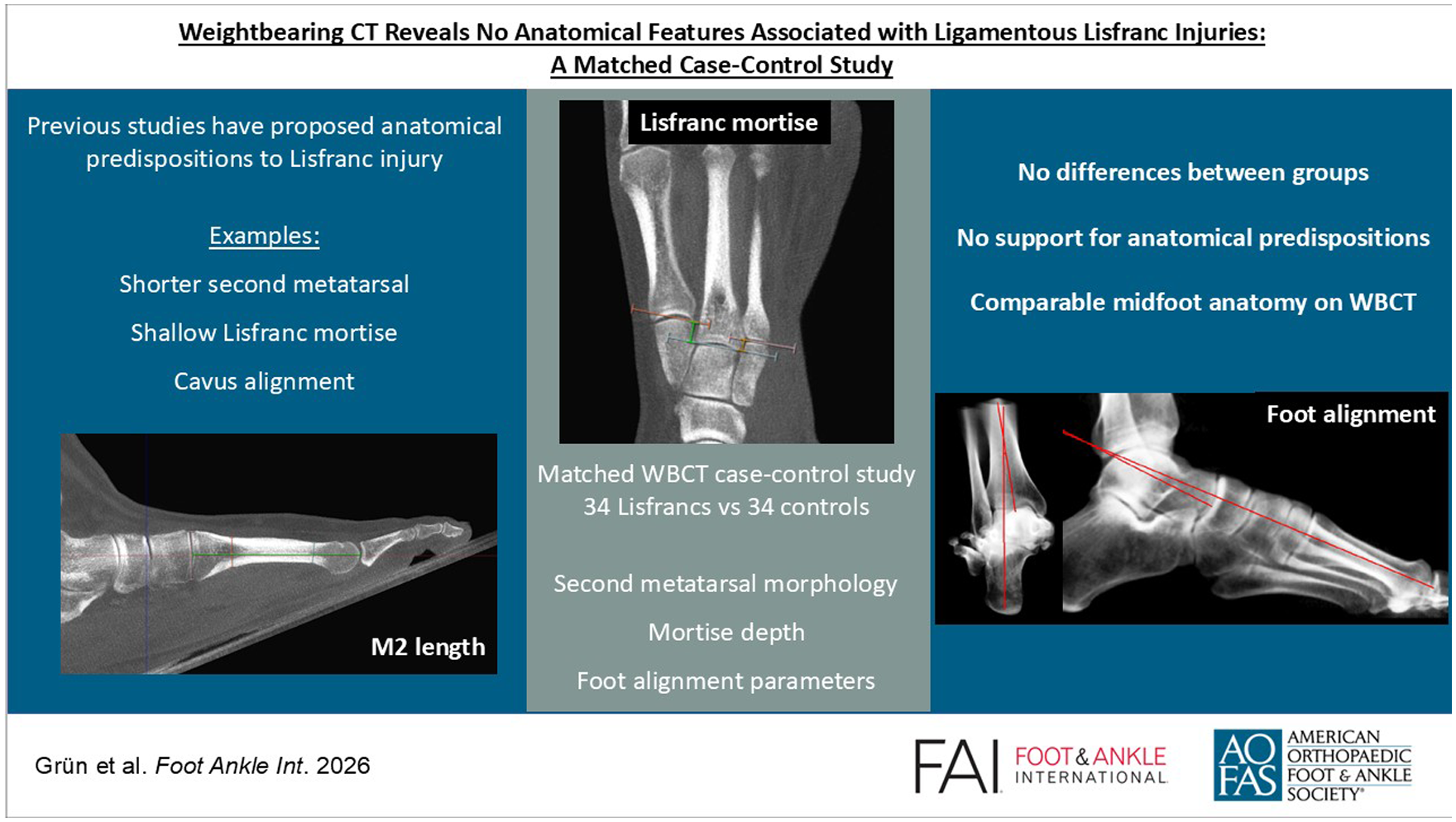
Keywords
Get full access to this article
View all access options for this article.
References
Supplementary Material
Please find the following supplemental material available below.
For Open Access articles published under a Creative Commons License, all supplemental material carries the same license as the article it is associated with.
For non-Open Access articles published, all supplemental material carries a non-exclusive license, and permission requests for re-use of supplemental material or any part of supplemental material shall be sent directly to the copyright owner as specified in the copyright notice associated with the article.