
Abstract
Background:
Patients with progressive collapsing foot deformity (PCFD), hallux valgus (HV), and midfoot arthritis (MA) could benefit from a bone-block tarsometatarsal (TMT) arthrodesis. The allograft wedge, producing distraction and plantarflexion, provides a stable medial column while correcting the deformity, but with increased risk of pseudoarthrosis. The objective of this study was to report the clinical nonunion and results of the bone-block first-TMT arthrodesis in collapsed feet.
Methods:
In this short-term prospective cohort (mean follow-up 17 months) we evaluated patients with PCFD, HV, and MA who underwent allograft bone-block first-TMT fusion between August 2020 and November 2022. Patients were kept nonweightbearing for 6 weeks and followed, having a WBCT at 3 months and subsequent follow-ups. Bone healing was determined by at least 50% of bone trabeculae crossing both graft interfaces. Complications were documented. Forefoot arch angle (FAA), Meary angle, talonavicular coverage angle (TNCA), middle facet subluxation (MFS), and foot and ankle offset (FAO) were obtained, as well as PROs.
Results:
Sixty-four patients (67 feet) were included, mean age 54.0 (18-77), body mass index 31.6 (29.6-33.6), 17.3 months (6-33) follow-up. Median allograft size was 8 mm (5-19 mm). Clinical nonunion rate was 11.9%, minor complications 2.9%, and major complications 5.9%. Tomographic healing at 3 months occurred in 86.4% and 66.7% at the most recent WBCT (mean: 10.1 months; 6-29). Improvement (P < .001) in FAA of 6.9 degrees (5.8-7.9), MFS of 22.6% (19.8-25.4), TNCA of 13.5 (12.1-14.3), Meary angle of 8.1 degrees (6.7-9.5), and FAO of 3.8% (3.1-4.6) were found. PROs improved for VAS, PROMIS-PH, PROMIS-PI, Pain Catastrophic Scale, and EFAS (P < .001).
Conclusion:
Although the first-TMT bone-block arthrodesis restored many markers associated with foot collapse and alignment, the clinical nonunion rate was 11.9%, which is in the top range of the literature for TMT fusions. The use of allograft wedges can explain our findings. Tomographic healing, initially 86.4% at 3 months, decreased to 66.7% at the most recent follow-up.
This is a visual representation of the abstract.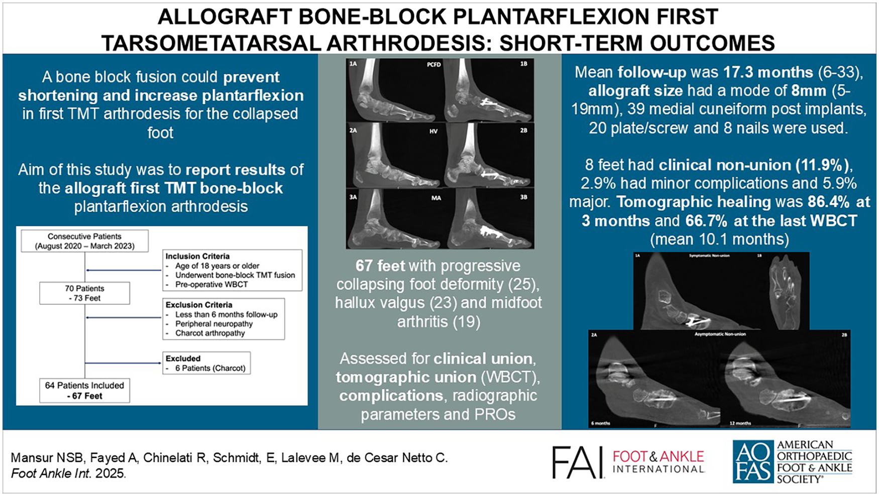
Keywords
Get full access to this article
View all access options for this article.
References
Supplementary Material
Please find the following supplemental material available below.
For Open Access articles published under a Creative Commons License, all supplemental material carries the same license as the article it is associated with.
For non-Open Access articles published, all supplemental material carries a non-exclusive license, and permission requests for re-use of supplemental material or any part of supplemental material shall be sent directly to the copyright owner as specified in the copyright notice associated with the article.