



Abstract

Lymph node (LN) metastasis in canine mast cell tumor (MCT) is a negative prognostic factor that affects post-surgical treatment recommendations.1,2 The use of metachromatic stains, including toluidine blue (TB) and Giemsa, is recommended for proper histologic evaluation of the number and arrangement of mast cells in LNs, which is required for the assessment of the histologic node (HN) status according to recently proposed criteria.4,5
In our experience, the examination of histologic sections of LNs stained with TB for MCT staging frequently leads to the identification of a variable number of cells with metachromatic intracytoplasmic granules in the medullary sinuses, either isolated or arranged in sheets (Fig. 1A). At low magnification, these cells may resemble mast cells, but on closer view they appear larger (20–30 µm), with vesicular, indented nuclei and coarser cytoplasmic granules, uneven in size and color (Fig. 1B).
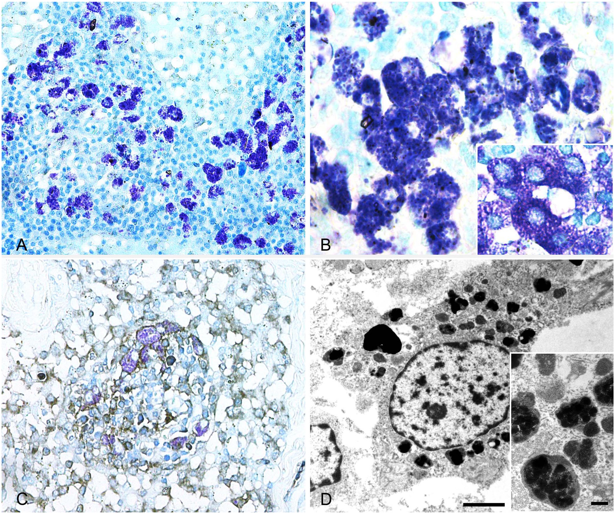
Metachromatic macrophages in lymph nodes of mast cell tumor–bearing dogs.
Immunohistochemical analysis performed with TB counterstain revealed that these cells are KIT-negative and CD18-positive, revealing a histiocytic origin (Fig. 1C). Transmission electron microscopy of selected areas of medullary sinuses identified groups of electrodense, 200-nm granules, admixed with other particles in the cytoplasm of cells that are consistent with macrophages (Fig. 1D).
The presence of histiocytic cells with metachromatic granules in LNs has been reported previously in MCT-bearing dogs undergoing chemotherapy, and it has been suggested that this could be the result of activation of sinus macrophages consequent to treatment. 3 At our institution, the histologic examination of LNs removed surgically from dogs with MCT resulted in the identification of these cells in 27 of 71 (38%) cases, with no significant difference in their prevalence according to the Weishaar status 5 (HN0/HN1: 39%; HN2/HN3: 36%). None of the dogs had received chemotherapy prior to LN extirpation.
When MCTs are manipulated during surgical preparation and removal, physical pressure on the mass can induce mast cell degranulation, and some of these granules may be conveyed to the regional LN, where they may be phagocytized by sinus macrophages. Granules may also be an expression of physiologic mast cell turnover. Alternatively, particles other than mast cell granules contained in macrophage phagosomes may induce a metachromatic reaction if stained with TB. When 10 LNs of dogs without MCT were stained with TB, these cells were detected in a similar proportion (n = 3; 30%) although in lower numbers, thus leaving all hypotheses open.
Speculation aside, the presence of metachromatic macrophages on TB-stained sections of canine LNs must be acknowledged, given the concrete risk of overestimating the presence of nodal metastases in dogs with MCT. As this phenomenon is equally frequent in LNs with a low mast cell infiltrate, it could ultimately lead to the misdiagnosis of non-metastatic (HN0/HN1) LNs as early or overtly metastatic (HN2/HN3), with significant impact on the clinical management of patients.