
Abstract
Johne’s disease (paratuberculosis) is an economically important disease of cattle worldwide. The disease is caused by Mycobacterium avium subsp. paratuberculosis (MAP), a fastidious gram-positive bacterium. PCR is increasingly used in diagnostic laboratories for the detection of MAP in fecal samples given the rapid test turnaround time and sensitivity and specificity comparable to fecal culture. However, efficient extraction of DNA for sensitive detection of MAP by PCR is affected by the complex lipid-rich cell wall of MAP and the presence of PCR inhibitors in feces. We evaluated a high-throughput nucleic acid extraction method (MagMAX core nucleic acid purification kit with mechanical lysis module) in conjunction with an hspX gene PCR for the detection of MAP from bovine fecal samples, which resulted in correct identification of all negative (13 of 13) and positive (35 of 35) proficiency test samples obtained from the National Veterinary Services Laboratories. In addition, all 6 negative and 50 of 51 positive diagnostic specimens tested were categorized correctly.
Johne’s disease (paratuberculosis) remains an economically important disease of cattle worldwide because of production losses, and costs associated with testing, culling, and mortality.6,10 The disease is caused by Mycobacterium avium subsp. paratuberculosis (MAP), an acid-fast, facultative, intracellular bacterium. The disease manifests in most ruminant species as chronic granulomatous enteritis with intermittent or chronic diarrhea, weight loss, edema as a result of hypoproteinemia, and death. 10 National Animal Health Monitoring System studies estimated that MAP-infected animals occur in ~68% of dairy herds 17 and 5–10% of beef herds in the United States. 18
Antemortem laboratory diagnosis of Johne’s disease includes demonstrating pathogen-specific antibodies in serum or milk samples, bacterial culture of fecal samples, and PCR detection of MAP-specific DNA in fecal samples. 3 Detection of antibodies using ELISA has several advantages including low cost, high throughput, and rapid turnaround time. However, the sensitivity of ELISA is reported to be 30% compared to autopsy, given the lack of production of detectable antibodies until late in infection.11,14,15 Isolation and identification of MAP from diagnostic specimens is considered the gold standard for diagnosis of Johne’s disease.9,19 A positive culture confirms the presence of viable bacteria in the sample and allows estimation of bacterial burden in the sample. The sensitivity and specificity of fecal culture are estimated to be ~60% and ~99%, respectively, compared to autopsy. 3 Given the slow growth of MAP, fecal culture is labor intensive and can take 6–8 wk for isolation of MAP in liquid media, and up to 16 wk on solid media. 14 PCR is increasingly used in diagnostic laboratories because of rapid test turnaround time and sensitivity and specificity comparable to fecal culture. 1 Similar to fecal culture, real-time PCR provides an estimate of MAP burden in samples based on the cycle threshold (Ct) value, which is indirectly correlated with the amount of MAP present in the sample. 14
Successful amplification and detection of DNA by PCR requires efficient extraction and purification of DNA from clinical specimens. The complex lipid-rich cell wall of MAP, in addition to the presence of PCR inhibitors in feces, renders isolation of high-quality DNA in sufficient quantities difficult for sensitive detection of MAP by PCR. 9 Previous studies have evaluated several nucleic acid extraction methods that included various combinations of bead beating, heating, or chemical lysis for disruption of MAP cells, followed by nucleic acid purification by the binding of DNA to paramagnetic beads or silica membranes in spin columns or ethanol precipitation of DNA.2,7,9 The efficiency of DNA extraction has been assessed by detecting MAP DNA using different PCR methods. 9 A high-throughput nucleic acid extraction method is desirable to support Johne’s disease control programs and would enable laboratories to test large numbers of samples and thus expedite testing and reduce the cost per sample. Therefore, our objective was to evaluate a high-throughput nucleic acid extraction method for the detection of MAP in bovine fecal samples by PCR.
We used the MagMAX core nucleic acid purification kit with mechanical lysis module (Thermo Fisher) for extraction of DNA from bovine fecal samples, following the manufacturer’s instructions for low-input workflow. The mechanical lysis module involves a bead-beating step for efficient lysis of hard-to-lyse bacteria such as MAP in complex sample matrices such as feces. Briefly, bovine fecal samples, 0.3–0.4 g, were suspended in 1 mL deionized water, vortexed vigorously for 3 min, and centrifuged at 100 × g for 30 s. Then, 175 μL of the fecal supernatant was added to a bead-beating tube containing 400 μL of clarifying solution; the sample was disrupted using a bead beater for 10 min at a speed setting of 30 Hz (TissueLyser II; Qiagen). Subsequently, the sample was centrifuged at 15,000 × g for 3 min to clarify the lysate, and 300 μL of supernatant (clarified lysate) was combined with 10 μL of proteinase K in a 96-well plate, mixed by pipetting, and incubated for 2 min at room temperature. Then, 720 μL of lysis–binding mix was added to each sample. The samples were processed on a magnetic particle processor (KingFisher Flex system; Life Technologies) following the manufacturer’s instructions.
Real-time PCR was performed on all extracted samples using a commercial kit (VetAlert Johne’s disease real-time PCR; Tetracore) for the detection of MAP DNA following the manufacturer’s instructions. The target gene for the PCR is hspX and is specific for MAP. 5 Real-time PCR was performed (CFX96 touch real-time detection system; Bio-Rad). Each PCR run included a positive extraction control, a positive amplification control, and a no-template control. The positive amplification control included in the kit is a synthetic template comprising a portion of the target hspX gene sequence. The kit also includes an inhibition control for monitoring PCR inhibition in each reaction. A sample was considered positive if the Ct value was ≤38.
Experimental data were analyzed by one-way analysis of variance (ANOVA, Prism for Windows; GraphPad Software). Statistical significance was determined at 95% (p ≤ 0.05). Significant differences between means were determined by a Tukey post-test.
Initially, the efficacy of DNA extraction was assessed by the MAP-specific hspX gene PCR using the bovine fecal samples from the 2018 and 2019 Johne’s disease proficiency test (PT) panels obtained from the National Veterinary Services Laboratories (NVSL). The PT samples were blinded for analysis. The samples analyzed included 13 negative, 5 low-shedding, 5 moderate-shedding, 7 moderate-to-high shedding, and 18 high-shedding samples. Four low-shedding samples excluded by NVSL in scoring the PT based on consensus results were not included in the final analysis. The average MAP loads in the PT samples reported by the participating laboratories were 0.5–8.5 colony forming units (CFU) per Herrold egg yolk media tube for low-shedding, 5.1–23.3 CFU per media tube for moderate-shedding, 23.8–36.4 CFU per media tube for moderate-to-high shedding, and as few as 2.5 CFU to too-numerous-to-count per media tube for high-shedding samples.12,13 The MagMAX extraction method in conjunction with the hspX gene PCR correctly categorized all non-shedding samples (13 of 13) as negative, and all low-shedding (5 of 5), moderate-shedding (5 of 5), moderate-to-high shedding (7 of 7), and high-shedding (18 of 18) samples as positive. Overall, there was 100% agreement between the shedding status and PCR categorization of the samples. Cycle threshold (Ct) values were 23.4–30.5 for high-shedding samples (mean Ct = 26.7, SD 2.2; Fig. 1); 30–36.1 for moderate-to-high shedding samples (mean Ct = 32.7, SD 2.6), 31.3–36.1 for moderate-shedding samples (mean Ct = 33.6, SD 1.7), and 33–37 for low-shedding samples (mean Ct = 35, SD 1.6). The mean Ct value for high-shedding samples was significantly (p < 0.0001) different from low-shedding, moderate-shedding, and moderate-to-high shedding samples. There was a considerable overlap of Ct value ranges among low-, moderate-, and moderate-to-high shedding samples, and the mean Ct values did not differ significantly among these samples. These results were similar to Ct values reported for the PT panels by the participating laboratories.12,13 The combined method of MagMAX core nucleic acid extraction and the hspX gene PCR correctly classified all of the PT samples as positive or negative.
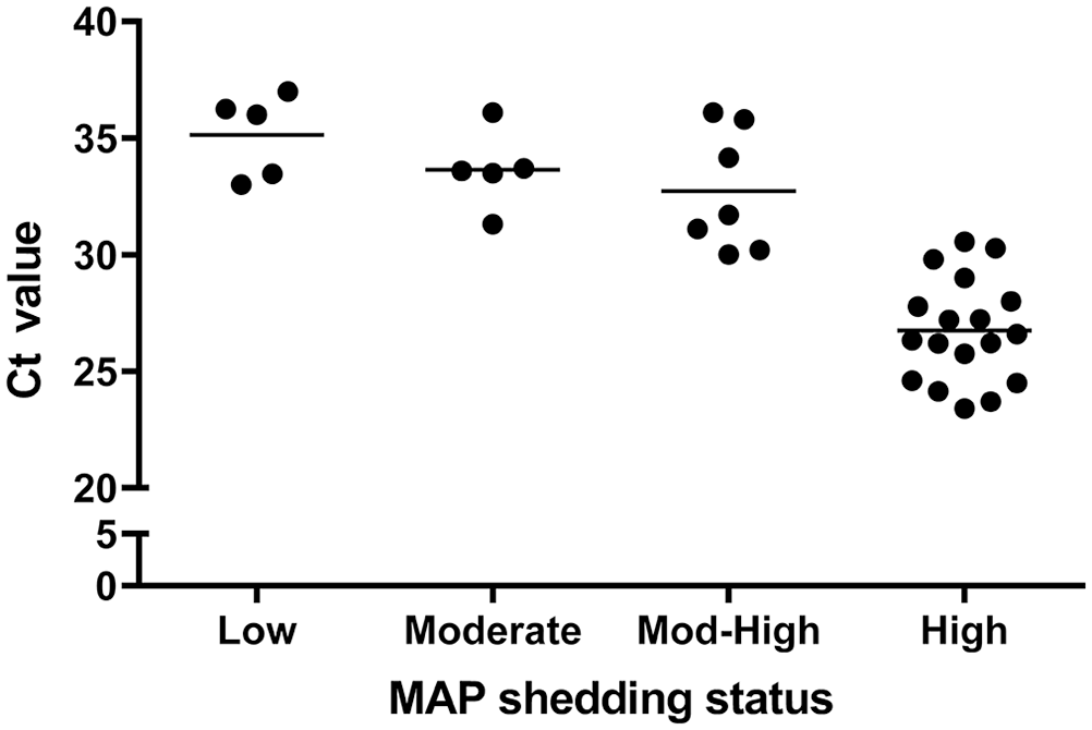
Cycle threshold (Ct) values for Mycobacterium avium subsp. paratuberculosis (MAP) positive proficiency test bovine fecal samples analyzed by the hspX PCR following nucleic acid extraction using the MagMAX core nucleic acid purification kit with mechanical lysis module (Thermo Fisher). The samples were from the 2018 and 2019 Johne’s disease proficiency test panels from the National Veterinary Services Laboratories (NVSL), and shedding status (low, moderate, moderate-to-high, high) of samples was provided by NVSL. The horizontal lines represent the mean. The mean Ct value for high-shedding samples was significantly (p < 0.0001) different from low-, moderate-, and moderate-to-high shedding samples. The mean Ct values did not differ significantly among low-, moderate-, and moderate-to-high shedding samples.
Furthermore, we verified the performance of the MagMAX core kit for the extraction of MAP DNA from fecal samples using diagnostic specimens submitted to the Pennsylvania Veterinary Laboratory (Harrisburg, PA). Most of the diagnostic specimens (~90%) were of bovine origin and included 8 culture-positive, 43 hspX PCR-positive, and 6 PCR-negative samples. The culture-positive samples were tested using para-JEM liquid medium and an automated microbial detection system (VersaTREK; TREK Diagnostic Systems); the presence of MAP in positive cultures was confirmed by acid-fast staining and IS900 PCR as described previously.8,16 PCR testing of the diagnostic specimens had been performed previously by the MAP-specific hspX PCR after extraction of DNA using an in-house method (MagAttract DNA mini M48 kit, BioRobot M48 workstation; Qiagen). Briefly, 0.25 g of fecal sample was suspended in 625 µL of buffer G2 by vortexing for 15 s. The sample was centrifuged at 82 × g for 5 min, and the supernatant was transferred to a tube containing glass beads. The sample was briefly vortexed and disrupted using the TissueLyser II for 10 min at a speed setting of 30 Hz. The sample was centrifuged at 16,000 × g for 10 min, 200 µL of the supernatant was transferred to a tube, and 1 µL of carrier RNA was added. The sample was processed (BioRobot M48 workstation, Genomic Tissue protocol; Qiagen). Initially, we performed direct comparison of the performances of the M48 kit and the MagMAX core kit using the 2018 Johne’s PT samples. The samples were blinded for the analysis. Nucleic acid extracted from the samples using either the M48 kit or the MagMAX core kit was subjected to the hspX PCR. The Ct values did not differ significantly (p > 0.05) among the extraction methods for low-, moderate-, and high-shedding samples, indicating that the methods had comparable performance (Suppl. Fig. 1).
Next, we evaluated the performance of the MagMAX core kit in the identification of known MAP-positive and -negative diagnostic samples (Table 1; Suppl. Table 1). All 6 PCR-negative, 43 PCR-positive, and 7 of 8 culture-positive samples were correctly categorized by the hspX PCR in conjunction with the MagMAX core kit extraction. One culture-positive sample, which was classified as negative by PCR, was previously tested culture positive with a 38 days-to-positive, suggesting the presence of low numbers of MAP in the sample. The average difference in the Ct values for the PCR-positive samples between the M48 kit and the MagMAX core kit was 2.6.
Identification of Mycobacterium avium subsp. paratuberculosis (MAP)-positive and -negative bovine fecal samples by the hspX PCR following nucleic acid extraction using MagMAX core nucleic acid purification kit (Thermo Fisher) with mechanical lysis module.
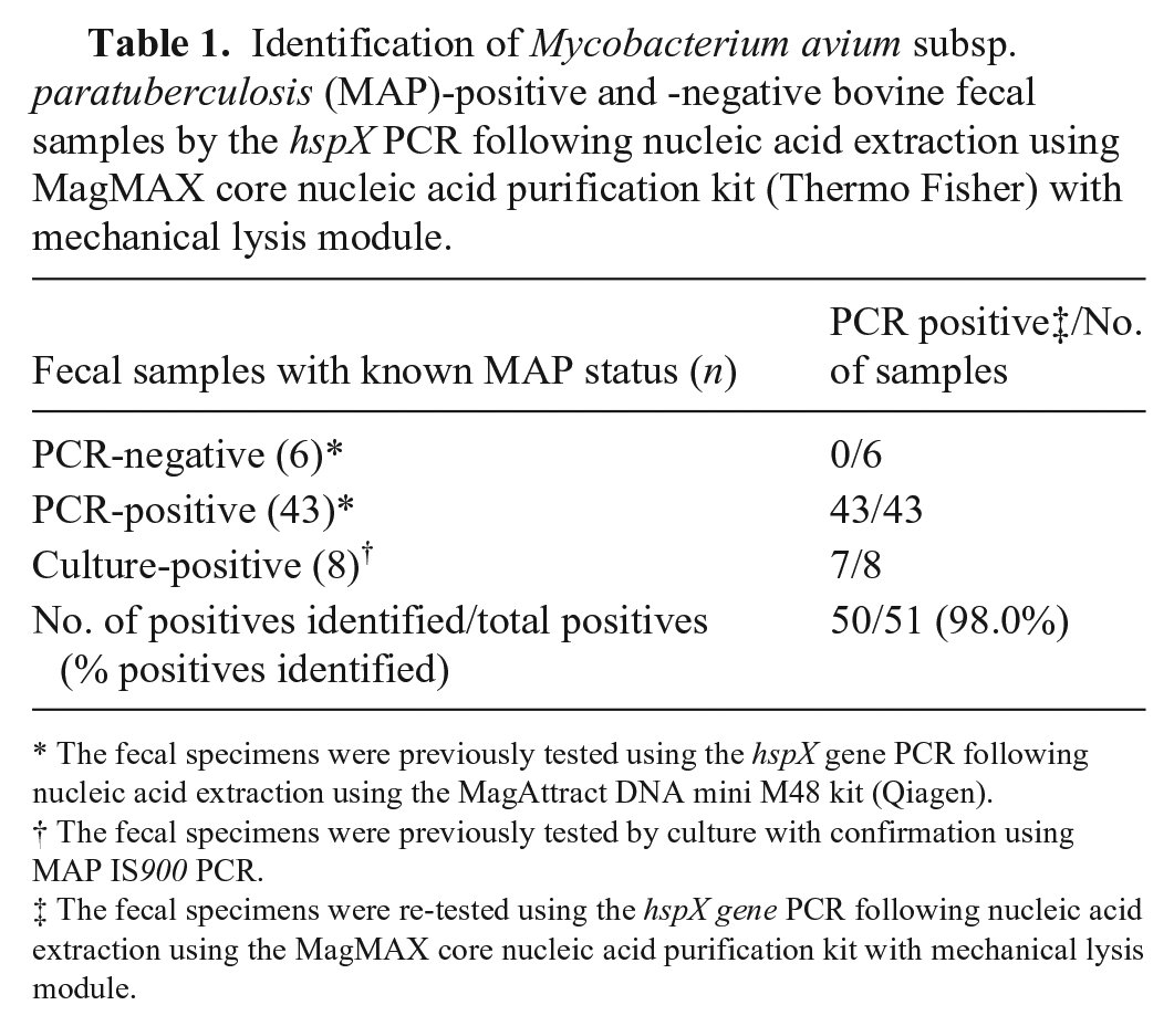
The fecal specimens were previously tested using the hspX gene PCR following nucleic acid extraction using the MagAttract DNA mini M48 kit (Qiagen).
The fecal specimens were previously tested by culture with confirmation using MAP IS900 PCR.
The fecal specimens were re-tested using the hspX gene PCR following nucleic acid extraction using the MagMAX core nucleic acid purification kit with mechanical lysis module.
It has been reported that mechanical cell lysis using bead beating results in efficient extraction of MAP nucleic acid from fecal samples, resulting in greater sensitivity of MAP detection by PCR. 4 A previous study described a modified guanidine lysis DNA extraction method that resulted in accurate categorization of 128 of 130 positive NVSL PT fecal samples (98.5% sensitivity) and 49 of 49 negative fecal samples (100% specificity) by a real-time IS900 PCR. 19 However, the modified guanidine lysis extraction protocol is very laborious, low-throughput, and involved multiple procedures including chemical cell lysis followed by mechanical lysis by bead beating, Chelex matrix absorption, and mini-column purification.
Another study evaluated 6 different commercial DNA extraction kits for MAP DNA extraction from bovine feces using a panel of 25 samples from the NVSL that included 7 negative and 18 positive bovine fecal samples. 9 The study reported significant differences in sensitivities (17.6–94.1%) between the extraction methods when used in conjunction with an IS900 real-time PCR for identification of known positive samples. 9 Notably, use of a different PCR, one that targeted the MAP ISMAP02 gene, resulted in different sensitivities for 5 of 6 extraction kits evaluated. 9 In addition, improved performance was observed for 2 extraction kits when the respective manufacturer-recommended PCR kits were used. Overall, this prior study demonstrated that the sensitivity and specificity of MAP DNA detection in fecal samples are influenced by both the DNA extraction and the PCR methods employed. 9 The availability of rapid, high-throughput, and cost-effective methods for the detection of MAP is expected to increase testing and thereby help control the spread of Johne’s disease in dairy herds and other livestock.
Supplemental Material
sj-pdf-1-vdi-10.1177_1040638721991118 – Supplemental material for Evaluation of a high-throughput nucleic acid extraction method for the detection of Mycobacterium avium subsp. paratuberculosis in bovine fecal samples by PCR
Supplemental material, sj-pdf-1-vdi-10.1177_1040638721991118 for Evaluation of a high-throughput nucleic acid extraction method for the detection of Mycobacterium avium subsp. paratuberculosis in bovine fecal samples by PCR by Nagaraja R. Thirumalapura, Willard Feria, Eric Hue, Corey Zellers and Deepanker Tewari in Journal of Veterinary Diagnostic Investigation
Footnotes
Declaration of conflicting interests
The authors declared no potential conflicts of interest with respect to the research, authorship, and/or publication of this article.
Funding
The authors received no financial support for the research, authorship, and/or publication of this article.
Supplementary material
Supplementary material for this article is available online.
References
Supplementary Material
Please find the following supplemental material available below.
For Open Access articles published under a Creative Commons License, all supplemental material carries the same license as the article it is associated with.
For non-Open Access articles published, all supplemental material carries a non-exclusive license, and permission requests for re-use of supplemental material or any part of supplemental material shall be sent directly to the copyright owner as specified in the copyright notice associated with the article.