
Abstract
We describe and illustrate lesions in an outbreak of lead arsenate poisoning in beef cattle that ingested pesticide residues stored in an abandoned building of a former orange orchard. Of 70 exposed cattle, 14 had diarrhea, paresis, ataxia, recumbency, and/or seizures. Ten of the affected animals died after a clinical course of 12–18 h. Pathologic findings in 3 steers included extensive necrohemorrhagic, ulcerative rumenitis, omasitis, and abomasitis; lymphocytolysis in lymphoid organs; and nephrosis. Hepatic arsenic and lead levels in cases 1–3 were 20, 24, and 31 ppm, and 8.3, 25, and 9.4 ppm, respectively. Lesions in the forestomachs and lymphoid tissues have been rarely reported in cases of lead arsenate poisoning. In southern South America, these lesions are indistinguishable from those produced by Baccharis coridifolia, a toxic plant that contains macrocyclic trichothecenes, thus these conditions should be considered in the differential diagnosis of necrotizing lesions in alimentary and lymphoid organs.
Arsenic is a ubiquitous metal that can be found in soil and water in both organic (aliphatic or phenylarsonic) and inorganic (trivalent or pentavalent) forms.1,4,5,10,15 Arsenic can reach high concentrations in the environment, mainly as a result of industrial activities.5,15 Acute and chronic arsenic poisoning has been reported in many animal species and humans.10,13,16,18 Lead arsenate (LA) is an arsenical insecticide that was used extensively throughout the world in the early 1900s; to date, LA is considered an environmentally hazardous contaminant.1,10 In 1988 in the United States, formulations based on inorganic compounds such as LA were banned from the market given their persistence in the environment and potential risk to public health. 10 Although spontaneous and experimental cases of acute LA poisoning have been reported in ruminants,12,19 description of the pathologic findings is limited in the scientific literature. We report an outbreak of acute LA poisoning in cattle, focusing on the description and illustration of gross and microscopic lesions.
The outbreak occurred in September 2015 at a beef cattle ranch in the department of Canelones, Uruguay. The animals were grazing a pasture of tall fescue (Festuca arundinacea), white clover (Trifolium repens), and bird’s-foot trefoil (Lotus corniculatus), supplemented with red clover (syn. rotklee; Trifolium pratense) hay. In the paddock in which the animals grazed, there was an abandoned building, within which there was a metal bucket containing a finely granular gray-white powder. This powder had remained in the building for more than 20 y, but the animals had not had access to it because the building had been a closed storage site, restricted from animal access. The farm had been recently purchased by a new owner, who partially cleared the building, making it accessible by the cattle grazing nearby. The previous owner had used the farm as an orange orchard, and presumably used this compound as an insecticide.
Of a herd of 70 steers and heifers with an average body weight of 310 kg, 14 sickened and 10 of these died spontaneously within 21 d after a clinical course of 12–18 h. Clinical signs included fetid diarrhea, paresis, ataxia, recumbency, and terminal seizures.
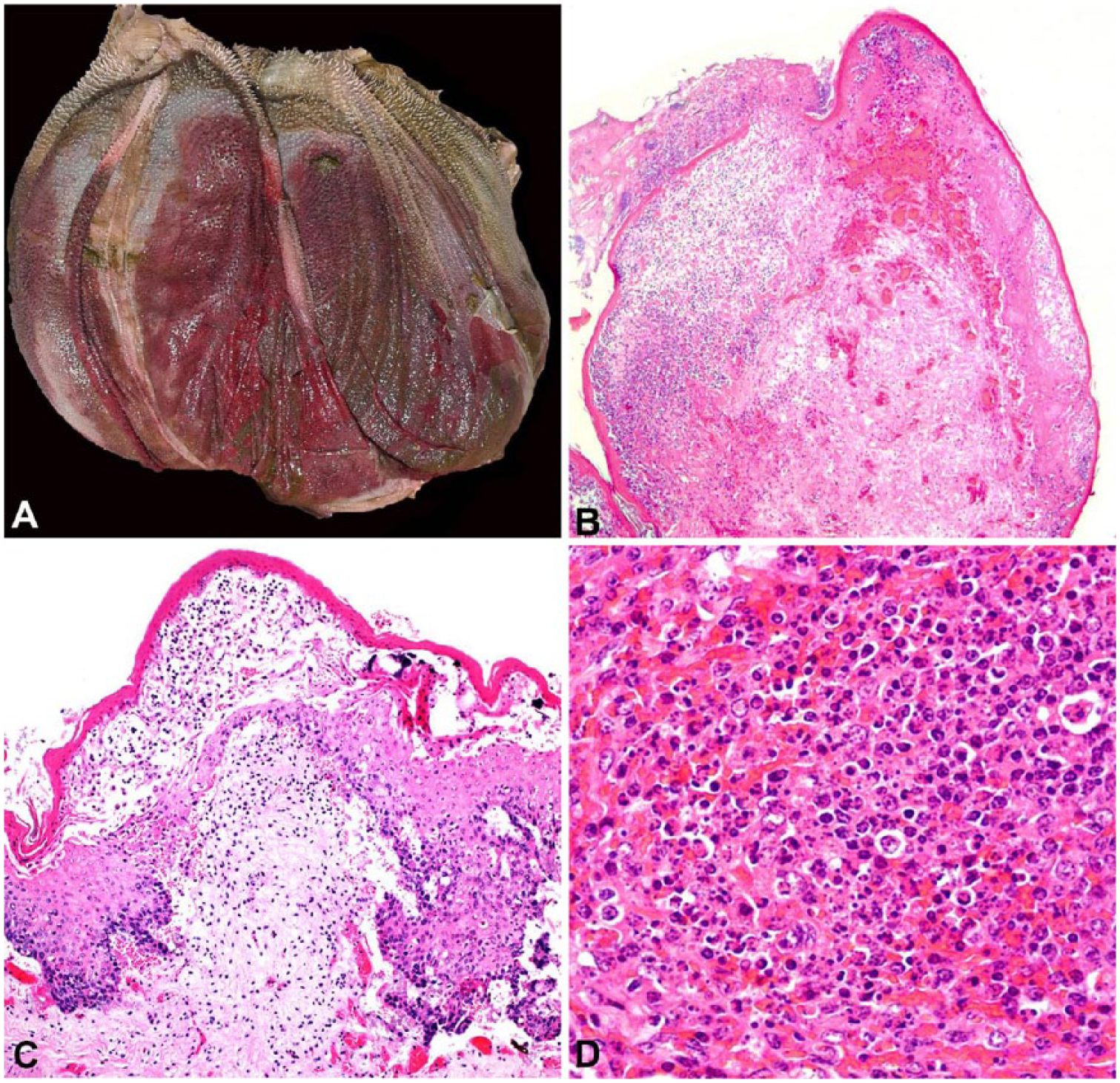
Autopsies were performed on cases 1–3. The carcasses were in good body condition and good state of postmortem preservation. In all cases, the rumen, omasum (Fig. 1A), and abomasum had extensive-to-diffuse reddening of the mucosa, erosions, ulcers, and multifocal epithelial sloughing. Additionally, the abomasum had extensive submucosal edema. In the small intestine and cecum of cases 2 and 3, there was petechiation and diffuse reddening of the mucosa. In case 3, the liver parenchyma had diffuse orange discoloration. In case 1, both kidneys had multifocal-to-coalescing areas of cortical pallor delineated by a red halo. No gross anatomic lesions were observed in any other organs.
Acute lead arsenate poisoning in cattle.
Tissue samples, including tongue, esophagus, rumen, omasum, abomasum, small and large intestine, liver, gallbladder, kidney, urinary bladder, spleen, ruminal and mesenteric lymph nodes, trachea, lung, heart, brain, and skeletal muscle, were fixed in 10% neutral-buffered formalin, processed routinely, and stained with hematoxylin and eosin. Microscopically, the mucosa of the rumen and omasum in all cases was extensively ulcerated and covered by necrotic cellular debris, including necrotic keratinocytes, fibrinous exudate, edema, and hemorrhage (Fig. 1B). Multifocally in the most conserved areas, there was swelling, degeneration, and necrosis of the basal keratinocytes, with separation of the basilar epithelium from the lamina propria, and pustules with abundant intraepithelial neutrophils (Fig. 1C). There was submucosal edema, fibrin exudate, hemorrhage, and infiltration by numerous degenerate neutrophils. Submucosal blood vessels were congested, lined by hypertrophic endothelial cells, and/or occluded by fibrin thrombi. The tunica muscularis and the adventitia were expanded by viable and degenerate neutrophils and edema, and lymphatic vessels were ectatic.
In extensive regions of the abomasum in cases 2 and 3, there were superficial erosions and hemorrhages with fibrin extravasation in the lamina propria, and parietal and chief cells had nuclear pyknosis or karyorrhexis. Gastric glands were occasionally ectatic and filled with necrotic epithelial cell debris. The lamina propria was infiltrated by eosinophils, neutrophils, and lymphocytes. The submucosa was diffusely expanded by edema, and blood vessels in the mucosa and submucosa were congested.
In all cases, the germinal centers of ruminal and mesenteric lymph nodes had multifocal lymphocyte necrosis, parafollicular hemorrhage, and edema. Lymphocytolysis was also seen in the white pulp of the spleen in cases 1 and 2 (Fig. 1D). In the cortical renal tubules of cases 1 and 3, there was scattered epithelial cell necrosis with occasional necrotic cellular debris within the lumen. Intranuclear inclusions were not seen in renal tubular epithelium. In the liver of case 3, there was scattered necrosis of individual hepatocytes and Kupffer cells. No microscopic lesions were observed in other organs, including brain.
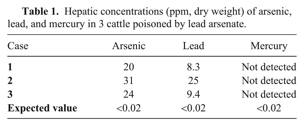
Formalin-fixed liver samples from cases 1–3 were sent to the Toxicology Section of the California Animal Health & Food Safety Laboratory (Davis, CA). Samples were digested with nitric acid and subsequently analyzed to detect and quantify arsenic, lead, and mercury by an inductively coupled argon plasma–optical emission spectrometer (ICAP 6500, Thermo Fisher Scientific, Waltham, MA; Table 1). The detection limit for all analytes was 0.2 ppm (dry weight basis).
Hepatic concentrations (ppm, dry weight) of arsenic, lead, and mercury in 3 cattle poisoned by lead arsenate.
A sample of the powder present in the bucket found in the abandoned building was sent to the Laboratorio Tecnológico del Uruguay for identification and semiquantitative analysis by energy-dispersive X-ray fluorescence. Elemental lead and elemental arsenic were identified in the sample. Based in the presence of these 2 metals, the history of the farm, and the presence of arsenic and lead in livers of affected animals, it was inferred that the toxic substance was LA.
The clinical and pathologic findings in the autopsied cattle, and the determination of arsenic and lead in the livers, allowed confirmation of the diagnosis of LA poisoning. In formalin-fixed tissues, the concentration of minerals may vary. Arsenic concentration may increase with storage time, but changes are minimal in the first 18 mo. 9 In the outbreak reported herein, less than a month elapsed between formalin fixation and analysis, suggesting that the values obtained corresponded to the values of the samples before fixation. On the other hand, lead is less changeable over time than is arsenic in formalin-fixed tissues. 9
Lead exposure was confirmed in all cases; however, only one sample had toxic levels of lead in the liver (>10 ppm), 5 which may have contributed to the mortality. In acute lead poisoning, there is typically no gastroenteritis,4,5 as observed in our cases. In cases of acute LA poisoning, the gastrointestinal lesions are attributed to the effects of arsenic rather than lead. 4
LA is a pentavalent arsenical, which has been used as an agricultural insecticide in the production of fruits and vegetables.1,10 In our outbreak, the cattle had no access to the abandoned building until just before the beginning of the mortality event. After the diagnosis was confirmed, access of the animals to this building was restricted, and no more cases occurred. Although there are no published reports of this poisoning in Uruguay, to our knowledge, there is anecdotal data of its occurrence in the department of Salto in 2001 wherein 15 of 100 Holstein heifers died after having access to metal containers containing LA, presumably used as insecticide in an old vineyard (Carmen Garcia y Santos, pers. comm., 2018). Although acute arsenic poisoning is uncommon, the presence of abandoned structures that are used to store pesticides is an epidemiologic factor to be considered. The fact that the animals have ingested the toxicant despite having good forage availability could be explained by the good palatability of this poison.13,14,18
Most reports of poisoning by different forms of inorganic arsenic describe lesions in the abomasum and intestines without lesions in the forestomachs.3,4,7,8,11,13,15,17,20 In contrast, in our outbreak, severe lesions were also present in the rumen and omasum. These lesions have occasionally been described in ruminants poisoned by LA. Hemorrhages and rumenitis with shedding of the ruminal mucosa have been reported in an outbreak of LA poisoning in cattle, 12 and hemorrhagic erosions of the ruminal wall were reported in sheep poisoned experimentally by LA. 19 However, histologic lesions of the forestomachs were not described in either report.12,19 Mild hemorrhagic and ulcerative lesions in the forestomachs were reported in cattle poisoned by the organic compound monosodium methanearsonic acid and inorganic arsenic trioxide.1,6,17 It has been suggested that the lesions in the digestive mucosa in arsenic poisoning are the result of endothelial damage.1,4 Nevertheless, the extensive necrosis of keratinocytes of the ruminal epithelium and the presence of pustules suggests chemical gastroenteritis caused by direct action of the compound on the mucosa, possibly because the pentavalent inorganic arsenical compound is poorly absorbed. 1
Another lesion found in our cases that has been reported only rarely is necrosis of lymphoid tissue in the lymph nodes and spleen. Marked lymphocytolysis of the lymphoid follicles of the lymph nodes and necrosis of mesenteric lymph nodes and gut-associated lymphoid tissue are reported in poisoning by inorganic trivalent arsenic.8,14 Our results and literature reports suggest that different arsenic compounds may cause different histologic lesions.1,3,5,8,12,14,17,20
In this episode of LA poisoning, the main lesions were in the forestomachs, thus these toxicants should be considered in the differential diagnosis of ulcerative and necrotizing lesions of these organs. In Uruguay, Argentina, and Brazil, poisoning by Baccharis coridifolia, a toxic plant that contains macrocyclic trichothecenes, causes clinical signs and lesions very similar to those described in LA poisoning, including severe necroulcerative lesions in the forestomachs and lymphoid necrosis. 2 Differentiation between poisoning by B. coridifolia and LA should be based on the history of exposure to the plant or pesticides, and arsenic and lead determination in the liver and/or kidney in cattle with compatible lesions. In the outbreak described herein, there was no history of exposure to B. coridifolia. Forestomach ulcers, hemorrhage, and necrosis are typically associated with rumen acidosis with secondary fungal and bacterial infections. We ruled out ruminal acidosis based on feeding exclusively of pasture and hay.
Infectious diseases that can result in necrotizing and ulcerative lesions in the forestomachs include mucosal disease, caused by bovine viral diarrhea virus, and bovine herpesvirus 1 infection. These infectious agents were ruled out in the cases presented herein by specific immunohistochemistry tests performed on rumen and omasum at the University of Minnesota Veterinary Diagnostic Laboratory (Saint Paul, MN; data not shown). Also malignant catarrhal fever may cause ulceration of the digestive tract; however, lymphocytic arteritis and fibrinoid change, typical of this disease, was not observed in our cases.
Footnotes
Acknowledgements
We thank Yisell Perdomo from INIA, Uruguay, for technical assistance with histologic processing of samples, and Jan Shivers from the University of Minnesota Veterinary Diagnostic Laboratory for technical assistance with immunohistochemistry.
Declaration of conflicting interests
The authors declared no potential conflicts of interest with respect to the research, authorship, and/or publication of this article.
Funding
This work was funded by grant PL_015 N-15156 from the Instituto Nacional de Investigación Agropecuaria (INIA), Uruguay.