
Abstract
Ventricular septal defects are one of the most common congenital cardiac malformations in animals, and most often affect the membranous portion of the septum. These defects may rarely close spontaneously. An adult male black-tailed prairie dog (Cynomys ludovicianus) had a smooth shiny botryoid red mass arising from the area of the septal cusp of the right atrioventricular (tricuspid) valve and membranous interventricular septum, and bulging into the right ventricular lumen. Histology and special staining demonstrated a membranous ventricular septal defect closed by the adherence of the septal cusp of the tricuspid valve to the muscular septum (so-called membranous ventricular septal aneurysm or aneurysm of the [peri]membranous ventricular septum). This is a rare finding in animals, and the histologic appearance has not been documented previously, to our knowledge.
Development of the 4-chambered heart from a simple tube is a complex process, and congenital malformations resulting from abnormal development are well described in animals.2,4,10 Ventricular septal defects (VSDs) are among the most commonly reported defects in domestic animals, and usually affect the upper (basilar), membranous portion of the septum near the annulus (“high” VSD). The defect is described as perimembranous if it involves the muscular portion of the septum as well. If left open postnatally, there is left-to-right shunting of blood resulting in increased right ventricular pressures and biventricular hypertrophy. Pulmonary hypertension and right ventricular hypertrophy can lead to right-to-left shunting (Eisenmenger complex). Although membranous VSDs are reported to frequently close spontaneously in people, closure has rarely been reported in animals.1,9 Closure can occur by adherence of the free edge of the septal cusp (leaflet) of the right atrioventricular (tricuspid) valve to the interventricular septum (so-called membranous ventricular septal aneurysm [MVSA] or aneurysm of the [peri]membranous ventricular septum), or less commonly by de novo fibrovascular proliferation.1,3 The higher systolic pressures in the left ventricle generally cause the cusp of the adherent valve to bulge into the right ventricular lumen, creating the appearance of an aneurysm by gross examination or imaging studies, although the term “aneurysm” is indeed a misnomer. 3
Cardiac disease is not well documented in captive or wild black-tailed prairie dogs (Cynomys ludovicianus). One large case series described several cardiac neoplasms in this species, including atrial hemangiosarcoma, multicentric lymphoma, and metastatic adenocarcinoma of undetermined origin, as well as splenic cavernous hemangioma. 8 Additional lesions reported but not described in that survey included myocardial necrosis, myocardial fibrosis, myocarditis, coronary artery thrombosis, and “degenerative cardiac disease.”
An adult male black-tailed prairie dog from a large colony at a zoological park was found dead with no past medical history or premonitory signs. Individual animals are not identified specifically, and therefore the exact age was unknown. The animal was in excellent body condition. Gross autopsy findings included multiple antemortem and postmortem skin lacerations, predominantly affecting the head and genitals (consistent with conspecific aggression). There was significant myocardial pallor and fibrosis affecting ~25% of the left ventricle. A 2-mm diameter dark red botryoid mass with a smooth and shiny surface arose from the septal cusp of the tricuspid valve and membranous interventricular septum, and bulged into the right ventricular lumen (Fig. 1). Tissues were fixed in 10% neutral-buffered formalin, processed routinely, and sections were cut at 6 µm for routine hematoxylin and eosin staining. Additional slides were stained with Masson trichrome and Alcian blue–periodic acid-Schiff.
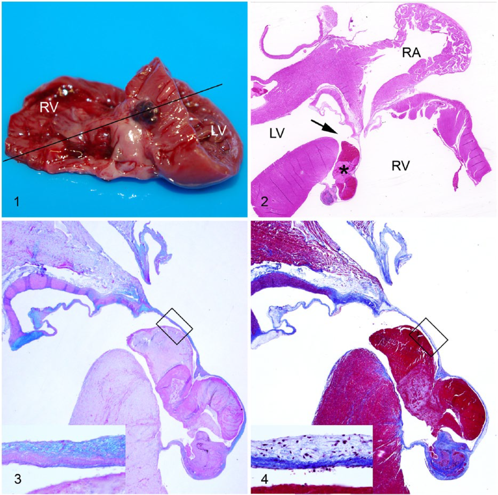
Membranous ventricular septal aneurysm in a black-tailed prairie dog.
Microscopically, the basilar (membranous) portion of the interventricular septum was absent, creating a 0.9-mm long septal defect (Figs. 2–4). In the right ventricle, the defect was covered by a convex thin membrane that bulged into the right ventricular lumen and contained a large recent non-adherent, non-organized thrombus. The majority of the aneurysmal wall was bi-layered and resembled normal tricuspid valve morphology, with a loose myxomatous and alcianophilic matrix on the side facing the right ventricle (atrial surface, lamina spongiosa), and a dense collagenous matrix on the aspect facing the left ventricle (ventricular surface, lamina fibrosa; Figs. 3, 4). Both sides of the resulting aneurysmal space were covered by mature quiescent endothelium. The free edge of the tricuspid septal cusp was adherent to the muscular part of the interventricular septum. The distal portion of the cusp, up to and including the site of adherence, was nodular and densely fibrous with multiple mature small blood vessels. Within the stroma of the nodule, there were moderate numbers of macrophages and lymphocytes with frequent non-degenerate neutrophils. There was extracellular and intrahistiocytic hemosiderin as well as fine mineralization of the stroma. Additional histologic findings included acute pulmonary and coronary artery thromboembolism with gram-negative rods, subacute and chronic renal infarcts affecting ~25% of the parenchyma, and suppurative submandibular lymphadenitis with mixed bacteria.
Based upon the histology and special histochemical staining, the lesion was diagnosed as membranous VSD with aneurysm of the septal cusp of the tricuspid valve (MVSA). MVSA (also called aneurysm of the membranous ventricular septum) has previously been reported as an incidental echocardiographic finding in 17 dogs and 3 cats. 9 The aneurysm was identified as involving the septal cusp of the tricuspid valve in 2 dogs, based on identification of chordae tendineae. The aneurysm was perforated with left-to-right shunting in 8 of those dogs and 1 cat, and 7 dogs had additional congenital cardiac defects. Aneurysm of the ventricular septum has also been reported within the spectrum of hereditary congenital conotruncal defects and tetralogy of Fallot in Keeshond dogs. 5 In humans, MVSA is rare at birth but increases in frequency with age, and is a documented cause of spontaneous closure of membranous VSDs.1,6 Spontaneous closure of VSDs is reported to be far less common in dogs and cats. 9 Although generally considered benign, MVSA in humans has been associated with aortic valve prolapse or regurgitation, tricuspid insufficiency, right ventricular outflow tract obstruction, aneurysmal rupture, and bacterial endocarditis. 11 Left ventricular–to–right atrial shunts are occasionally present. 6 There was no indication of shunting (right atrial or right ventricular dilation or hypertrophy, endocardial jet lesions) in this case. It is unclear whether or to what extent the aneurysm may have contributed to thromboembolic lesions in this animal.
Gross differentials for an intracardiac valvular and/or septal mass lesion would include tricuspid valve hemocyst, valvular telangiectasis, hemangioma, hemangiosarcoma, Purkinjeoma or cardiac glycogenosis, and aortic root and/or coronary sinus aneurysm.4,7,10 Valvular endocarditis or thrombosis generally results in a dull and shaggy, rather than smooth and shiny, surface. Valvular lymphocysts, endocardiosis, and Purkinjeoma are not red. Adhesion of the tricuspid valve cusp to cover a congenital defect of the membranous ventricular septum has been reported as a cause of spontaneous VSD closure. Although uncommon, this lesion should be included in the differential diagnosis for valvular mass lesions, with careful dissection and histologic evaluation of the lesion.
Footnotes
Acknowledgements
Thanks to Gretchen Snavely and Ellen Mullady for histologic specimen preparation.
Declaration of conflicting interests
The author declared no potential conflicts of interest with respect to the research, authorship, and/or publication of this article.
Funding
The author received no financial support for the research, authorship, and/or publication of this article.