
Abstract
In cats, larvae of the dipteran fly, Cuterebra, sometimes cause severe disease by their migration through the tissues of the larynx, pharynx, nasal sinuses, brain, and spinal cord; such infected cats may die without the maggots ever reaching the subcutaneous tissues where they would typically mature. The current study examines the ability of an indirect enzyme-linked immunosorbent assay (ELISA) using crude Cuterebra antigen from maggots to detect parasite-specific immunoglobulin (Ig)G in cats with known (n = 42), suspected (n = 25), or no known exposure to the infection (n = 68). The probability of a given optical density (OD) predicting the infection status of a given animal was determined using logistic regression, and both 1:20 and 1:80 serum dilutions were highly predictive of the potential of a cat being infected with a larval Cuterebra. In 5 cases where 2 samples were collected 1–2 weeks apart, there was a mean OD increase in the second sample for both the 1:20 and 1:80 dilutions, but it was significant (P = 0.044) only at the 1:20 dilution. Sex of the sampled cat was not a significant contributor to the ability of the OD to predict the presence of a larva, but the age of the cat added significantly to the predictive value of the generated curves, with the only exception being with the 1:20 serum dilution with the curve being generated only using the cats known to be positive for larval presence. This ELISA should aid in ruling cuterebriasis in or out in suspect systemic and, specifically, neurologic cases and provide information on kinetics of antibody presence postexposure.
Introduction
Cuterebra, a genus of dipteran obligate parasites of rodents and lagomorphs in North America, has a larval stage (a maggot) that undergoes an obligatory deep migration through the tissues of its host before the third-instar larva appears in a subcutaneous boil (the warble), where it undergoes rapid growth before dropping to the soil to pupate. 7 Surprisingly, in the rodent and lagomorph hosts, there is actually only minimal loss of fitness of the host from infections with these parasites, which are strikingly large relative to the size of their small rodent hosts. 10 There are 34 species of Cuterebra throughout North America, 7 and dogs and cats are known to sometimes have the larvae develop into mature larvae within the lesions in their skin, nasal cavity, or eyelid. 8
In the northeastern United States, there is the seasonal appearance of neurologic disease in cats that has been associated with the migration of Cuterebra larvae through the spinal cord or brain; it is believed that this represents the effect of the maggot entering the incorrect host and undergoing an erratic migration. 3 Cuterebra larvae cannot be identified to species morphologically, and while third-stage larvae can be identified to subgenus, first and second larval instars cannot be identified morphologically beyond the generic level. 7 Thus, it is unknown if the disease in cats seen in the northeastern United States is due to 1 or more of the 6 species of Cuterebra that occur in this area: 3 species in the subgenus Trypoderma that use lagomorphs as their typical hosts, Cuterebra abdominalis, Cuterebra buccata, and Cuterebra cuniculi, and 3 in the subgenus Cuterebra that use rodents as the typical hosts, Cuterebra emasculator, Cuterebra fontinella, and Cuterebra americana. In this region of the United States, the flies all have univoltine life cycles with the females laying eggs on blades of grass in spring or early summer near the entrance to the host’s burrow; the larva then leaves the egg to get onto the host when it is passing by and enters the host through 1 of the body’s orifices. The migratory pattern in mice experimentally infected with C. fontinella revealed that whether the larvae entered the host via the nares or anus, they migrated first to the trachea and thoracic region before migrating through the abdomen to the site of development in the subcutaneous tissues of the posteroventral abdominal region. 2 A mature larva in the skin of a rodent or rabbit requires approximately 3–8 weeks including the migratory phase of the life cycle to reach the stage where it is ready to drop to the ground to pupate. 1
In cats in the northeastern United States, the disease usually presents between late June and the first killing frost in October. In most cats, and in dogs, it seems that the larvae ultimately reach the subcutaneous tissues and mature (although they seem incapable of producing viable adults after pupation), and in these cases the diagnosis of infection is simply the finding of the larva in the subcutaneous boil. Unfortunately, some cats in late summer to early fall develop an acute onset of neurologic disease that may be preceded by upper respiratory signs 1–2 weeks previously. Such cats can present with depression, blindness, and behavioral changes. Lesions may be in the cerebrum or cerebellum in association with feline ischemic encephalopathy, 11 but in some cases the larvae are found within the spinal canal. Diagnosis is typically based on clinical signs, response to treatment with high doses of ivermectin and corticosteroids, imaging with computerized axial tomography or magnetic resonance, or by necropsy. The present report describes the development of an enzyme-linked immunosorbent assay (ELISA) to assist in the diagnosis of systemic cuterebriasis by the detection of parasite specific immunoglobulin (Ig)G within the serum of cats with clinical signs suggestive of this infection to aid clinicians in ruling-in or ruling-out systemic cuterebriasis relative to observed signs in conjunction with other diagnostic tests.
Materials and methods
Antigen
Third-stage larval instars were recovered from naturally infected cats or rabbits via removal from a subcutaneous boil. Instars used for antigen production were stored at −80°C until antigen was harvested. At the time of harvest, instars were removed from the freezer and placed immediately into a mortar that had also been stored at −80°C. Liquid nitrogen sufficient to cover the specimen was added to the mortar, and the specimen was crushed with a prechilled pestle until it reached a powder-like consistency. The material was then transferred to a pestle tissue grinder assembly with a smooth pestle (size A) a and homogenized in a small volume of 0.01 M phosphate buffered saline (PBS). The homogenate was then centrifuged for 10 min at 20,000 × g, and the supernatant filtered through a 0.2-µm filter. The protein concentration of the resulting filtrate was determined using a spectrophotometer b with comparisons being made to a standard curve produced using bovine serum albumin. c Aliquots of the crude antigen extracts were stored at −80°C.
Sera
There were 3 different sources of negative control sera. There was a single sample of pooled serum from 100 normal cats d used as the standard negative reference. Twenty-eight individual serum samples were purchased from a New York supplier of specific pathogen-free cats. e These samples were collected from cats of varying ages: <1 year of age (16), 2 years old (7), 3 years old (2), 4 years old (1), 5 years old (3), and 6 years old (1). Another 39 individual serum samples came from clinically normal cats that passed through the hospital at the Ecole Nationale Vétérinaire de Toulouse (Toulouse, France); this French feline serum was chosen as another negative source because the French cats had been exposed to other pathogens, unlike the specific pathogen-free cats from New York, but should never have had the opportunity to be exposed to Cuterebra larvae, which only occur in the Americas.
The known positive serum samples (N = 42) and samples from suspect cases (N = 25) were submitted by local participating veterinary clinics in Tompkins County, New York, between August 2007 and September 2010. A questionnaire was given to participating animal hospitals to provide a basic clinical history. Included in the history was age, sex, month of presentation, clinical presentation (skin lesion, respiratory or neurologic signs, or other), whether or not a bot larva was recovered (location: skin, nasal cavity, pharynx, or larynx; spinal cord; brain; or other), treatment (larva removed, ivermectin, corticosteroids, other, none), and clinical resolution (yes, no, partial). Known positive serum samples came from 42 cats from which larvae of varying sizes had been recovered (Table 1); any submitted larvae were photographed to record their size. There were also 25 samples submitted by practitioners from feline cases in which cuterebriasis was suspected by the attending veterinarian based on observed clinical signs but from which no larva was recovered (Table 1). An additional serum sample was collected 1–2 weeks after the first sample was submitted in the case of 5 cats from which bots had been removed. The second sample was also analyzed for the presence of Cuterebra-specific IgG, and the results of the 2 tests were compared using a simple paired t-test.
Feline cases of cuterebriasis (with bots) or suspect cases (bot larva not found) from which blood samples were collected. Sizes of bots that were measured from the 42 feline cases from which larval Cuterebra were removed.*
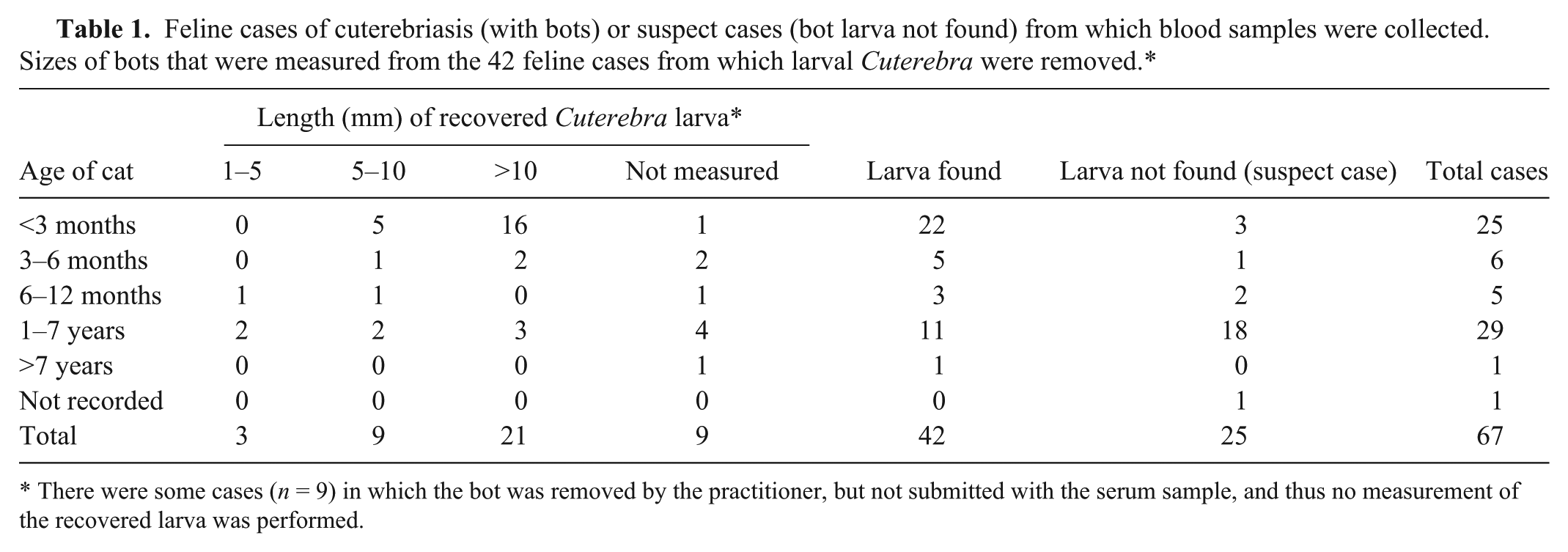
There were some cases (n = 9) in which the bot was removed by the practitioner, but not submitted with the serum sample, and thus no measurement of the recovered larva was performed.
Enzyme-linked immunosorbent assay
The ELISA was run on 96-well plates. f Antigen was coated onto the plates by placing 50 µl of the crude antigen extract diluted to10 µg/ml in 0.01 M carbonate buffer (pH 9.6) into each well. Plates were air dried at 37ºC, and then stored in a desiccator at room temperature until used. Plates were used within 60 days of preparation.
For the assay, each plate was washed 3 times with 0.01 M PBS at pH 7.4 containing 0.05% Tween 20 g and 2% nonfat dry milk powder h (w/v). Each well then received 100 µl of PBS/Tween 20/dried milk as a blocking agent, and the plate was incubated for 30 min at room temperature. After incubation, the plate was again washed 3 times with PBS/Tween 20/dried milk, and 50 µl of diluted test and/or control feline sera was then added to each well; the serum dilutions used for each sample were 1:20 and 1:80 in PBS/dried milk. Similar dilutions of the pooled negative reference serum and a positive reference serum were included on each plate. After all sera were in each well, the plate was incubated for 45 min at room temperature, and then again washed 3 times with PBS/Tween 20/dried milk. Then 50 µl of peroxidase conjugated rabbit anticat IgG i diluted 1:10,000 in PBS/dried milk was added to each well. The dilutions used in the assays were initially determined in a series of titration studies to produce maximum color change with minimal observed background. The plate was then incubated for 45 min at room temperature, and then washed 3 times with PBS/Tween 20/dried milk and once with PBS. To each well was then added 100 µl of a room temperature commercial preparation of tetramethylbenzidine (TMB) substrate. j The plate was gently mixed by tapping and was incubated at room temperature for 10 min. To stop the reaction, 100 µl of 1 N HCL was added to each well, and the plate was read at 450 nm on an ELISA reader. i The optical density (OD) values used in the equations and for comparisons were determined by the subtraction of the average of blank wells that had received only PBS/dried milk rather than negative or positive control sera. The negative and positive controls on each plate were to verify that the results for these 2 numbers were consistent between runs.
Statistical analyses
Due to the small sample size, the populations of cats from which serum samples were collected were only described descriptively without attempts to examine associations between risk factors. In the examination of the ability of the observed OD value to predict an infection within the respective hosts from which samples were collected, the numbers used were the OD values minus the mean of blank wells.
For examination of both the IgG at a dilution of 1:20 and 1:80, all statistics were performed using data generated in the same temporal run using antigen-coated plates that had been prepared at the same time. Chi-square analyses, the paired t-test, and descriptive statistics were calculated using commercial software. l Logistic regression was performed using commercial software. m The significance of association was evaluated between the probability of the presence of the larva in the animal and the serological response in the host using logistic regression analysis. Three responses/dependent variables were analyzed: 1) presence of a larva versus no larva, 2) presence of a larva and suspicion of infection versus no larva, and 3) suspicion of infection (based on clinical evidence) versus no larva. The probability of each of the responses for each dilution was computed as follows,
where α is the constant, β is the change in the probability of a maggot being present due to 1 unit change in the OD (regression coefficient), and γ is the regression coefficient for age. The significance of association between the outcome and the response and either OD or age was evaluated by the significance of the respective regression coefficient. For these comparisons, variables that were also considered were sex (male or female) and the age of the cat in years with midpoint values chosen from the submitted data (0.15, 0.45, 0.75, 3.5, and 10 years). For the graphs where age was a significant contributor to the predictive value of the OD, the overall mean age of the cats (2.52 years) was the value used in the graphical presentation.
Results
Description of cases
Of the 25 suspect cases, 8 (32%) were female and 15 (60%) were male (in 2 cases sex was not recorded on the submission form). Of the 42 cats in which bots had been seen by the veterinarians, 16 (38.1%) of the cats were female, and 24 (57.1%) were male (in 2 cases the sex was not recorded on the submission form). Although a preponderance of both cases with larvae and suspect cases were males (57% and 60%, respectively), sex was not found to be significantly associated with infection. Of the 42 cases with larvae, a significant proportion of the cats (22, 52.4%) were less than 3 months of age (P < 0.000; chi-square goodness-of-fit test). Of the 24 suspect cases with age recorded, only 3 (12%) of the cats were less than 3 months of age, and a significant proportion of the cats in this group (18, 75%) were greater than a year of age (P < 0.000; chi-square goodness-of-fit test).
For 1 of the 42 cats from which a bot was recovered, no presenting signs were submitted with the sample; for 2 of the samples from the 25 cats that were suspect cases, there was no presenting sign recorded. Of the remaining 41 cats with bots identified, 30 (73.2%; 11 females, 17 males, and 2 cats for which sex was not recorded) presented with a skin lesion, 3 (7.3%) with respiratory signs, 2 (4.9%) with a lesion and neurologic signs, 5 (12.2%) with solely respiratory signs, and 1 (2.4%) with a combination of respiratory and neurologic signs. Of the 23 remaining suspect cases, 3 (13%) presented with skin lesions, 6 (26.1%) with respiratory signs, 9 (39.1%) with neurologic signs, 2 (8.7%) with skin and respiratory signs, and 3 (13%) with respiratory and neurologic signs. The months of presentation for the 67 cases with bots and suspect cases, respectively, were July 33.3% and 40%, August 47.6% and 36%, September 16.7% and 16%, October 2.4% and 4%, and November 0% and 4%. Of the 42 cases with bots, of the 33 maggots submitted for measurements, 21 (63.6%) were greater than 10 mm in length.
ELISA results
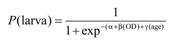
The OD values from dilutions at both 1:20 and 1:80 were elevated in both the cats with bots and in cats that were suspected of having cuterebriasis (Fig. 1). The means and standard deviations for the OD minus the blank for the 1:80 dilution of sera for uninfected, suspect, and infected cats were 0.1013 ± 0.0614, 0.4733 ± 0.4339, and 0.4810 ± 0.4559, respectively. The means and standard deviations for the OD minus the blank for the 1:20 dilution of sera for uninfected, suspect, and infected cats were 0.1599 ± 0.0894, 0.7573 ± 0.4119, and 0.8478 ± 0.3647, respectively.
Box plots (the horizontal line within the box represents the mean optical density [OD] value) and individual data points for OD values obtained using sera from cats with bots, normal cat sera, and sera from cats suspected of having cuterebriasis by submitting practitioners. In the figure are represented enzyme-linked immunosorbent assay results for sera diluted at 1:20 (
The 2 samples collected 1–2 weeks apart from the same cats were compared using a paired t-test. There was a rise in the OD between the means of both the 1:80 and 1:20 serum dilutions. The means and standard deviations for the first and second ODs at 1:80 and 1:20 dilutions were 0.315 ± 0.298 and 0.637 ± 0.228; 0.507 ± 0.416 and 1.029 ± 0.183, respectively. The results of the paired t-test revealed that the difference at the dilution of 1:80 was not significant (P = 0.118); however, at the 1:20 dilution, the mean of the first sample was significantly lower than that made on the second sample (P = 0.044).
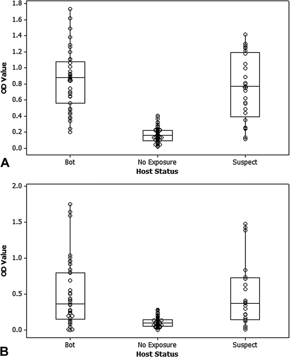
The probability of a given OD predicting the infection status of a given animal was determined using logistic regression. It was found that the OD at both the 1:20 and 1:80 serum dilutions was highly predictive of the potential of a cat to be infected with a larval Cuterebra (data shown only for the 1:20 dilution; Fig. 2). In the generated equations, sex of the sampled cat was not found to be a significant contributor to the ability of the OD to predict the presence of a larva. The age of the cat in years was found to add significantly to the predictive value of the generated curves, with the only exception being the 1:20 serum dilution with the curve being generated only using the cats known to be positive for larval presence.
Graphs generated by logistic regression showing the chance of the cat having a larval Cuterebra based on a given optical density (OD) value at a serum dilution of 1:20. The 3 graphs represent (
For specific clinical cases, the submitting hospital receives a letter describing the testing, results, and interpretation. The contents of the letter include a restatement of the cat’s clinical history as provided on the sample submission form and the raw OD values, the ELISA results for the submitted sample, a known positive sample, and sera from a pool of normal cats at different dilutions. The formula is then applied to the results of the data with the standard curves that are generated with each run, and the clinic is provided with a statement as to the possibility of the cat being infected using the probability function (e.g., “This cat does have minimally elevated levels of anti-Cuterebra IgG present in his blood at the time of sampling, which means he most likely was not infected with a Cuterebra bot. Based on a probability equation generated from serum samples from known positive and negative cats we have examined, the probability of this cat being infected is about 30%.”). Letters may also state that a given cat has no elevation in anti-Cuterebra IgG and has a probability of infection of 0% or a very high level of anti-IgG that indicates “he most likely was/is infected with a Cuterebra bot. Based on a probability equation generated from serum samples from known positive and negative cats we have examined, the probability of this cat being infected is 100%.”
Discussion
The data suggest that this assay can meaningfully differentiate most infected animals from those that are not infected, by performing a standard curve with each assay or set of assays, and using the past results to generate an equation that provides a numerical probability that a given titer indicates an actual infection. The results presented to the clinician includes the generated raw OD standard curves and the curve generated for the specific patient, and the probability that the IgG level obtained supports or does not support an infection with Cuterebra. The test would be more carefully validated and verified if somehow sera could be collected repeatedly from cats at various times postexposure. The development of a species-specific assay is hampered by the fact that it is not known which single species or which of several species from 2 distinct subgenera may be the cause of the majority of neurologic disease seen in cats, and therefore, the use of species-specific antigens from a given rodent or rabbit species may not be an appropriate target for the diagnosis of feline disease unless the antigens are cross-reactive.
Unfortunately, the few animals in the current study that had bots, sometimes fairly large bots, did not yield a positive result in the present assay. There were 16 samples from cats less than 3 months of age that had bots greater than 10 mm in length, and it may be that these young animals do not mount an exceedingly rapid immunologic response to the infection. Perhaps that is the reason why 50% of the cases seen were in animals less than 3 months old. However, it is possible that the crude antigen being used does not have a preponderance of the antigens associated with a given Cuterebra species. Also, it would likely prove useful to have an assay for the detection of Cuterebra-specific IgM with the goal of identifying cases earlier during an infection. This would be especially useful, as cats are likely to be dying in some cases only a few days to a week after infection, and will not have had a chance to mount an IgG response, thus an IgM titer would be valuable.
There are no data on how long a cat will maintain its parasite-specific IgG titer after a Cuterebra infection has matured and the bots have left the body. In the case of infections of calves by the northern cattle grub (Hypoderma bovis), after the larvae leave from their warble in the spring, the IgG levels reach baseline again in approximately 14 weeks. 9 It is hoped that as the present test is used more commonly, it will be possible to generate similar information about antibody kinetics for cuterebriasis in cats. Similarly, work on the common cattle grub (Hypoderma lineatum) has led to the development of an antigen detection ELISA that allows early diagnosis of this infection in cattle. 6 The development of an antigen test for cuterebriasis in cats would possibly be the preferred method for detecting early infections if there is sufficient antigen produced and circulating to allow its detection in blood, serum, or plasma.
It is not possible to determine the species of bot involved based on morphologic criteria, and only a few adult Cuterebra species have been identified and then molecularly characterized. As stated in the introduction, the 6 Cuterebra species occurring in the northeastern United States are C. abdominalis, C. americana, C. buccata, C. cuniculi, C. emasculator, and C. fontinella. Of these 6 species, the sequence of only 1, C. fontinella, appears in GenBank n (along with only 2 other North American Cuterebra species, C. grisea and C. jellisoni).4,5 Ultimately, the goal is to identify the species of individual bots recovered from cats to determine if certain species are the cause of the neurologic disease and to identify the species the bots used for the crude extract preparation. The molecular methods are in place, but properly identified specimens for source DNA have been difficult to attain. The recent loan of leg segments from adult specimens from the Field Museum of Natural History (Chicago, IL), the Department of Biological Sciences of the University of Cincinnati (Cincinnati, OH), and the Department of Entomology of Cornell University (Ithaca, NY) will now allow the further molecular characterization of many species of this genus to occur, particularly C. americana, C. angustifrons, C. apicalis, C. approximate, C. atrox, C. buccata, C. fontinella, C. horripilum, C. princeps, and C. sarcophagoides. However, even after this sequence is completed, there will still be no new information on 2 species, C. cuniculi and C. emasculator, which occur in the northeastern United States where feline neurologic cuterebriasis occurs.
Footnotes
Acknowledgements
The authors would like to thank all the veterinary clinics, veterinarians, and technical and logistic support staff for the effort put into providing the samples and the clinical histories on the cases that make up the bulk of the research presented herein.