
Abstract
During the years 2009–2011, 7 Siberian tigers (Panthera tigris altaica), aged between 2 and 14 years, from the Safaripark of Pombia were referred for necropsy to the Department of Animal Pathology of the University of Turin (Italy). Three tigers, aged 10 (2 animals) and 14 years, had multifocal, irregularly distributed, white, soft, subpleural, 3-mm nodules scattered throughout the lungs. Histologically, there was a marked infiltration of macrophages, with foamy cytoplasm, and multinucleate giant cells interspersed with numerous clusters of cholesterol clefts. A mild lymphocytic infiltration was localized around the lesion. The findings were consistent with endogenous lipid pneumonia, which was considered an incidental finding of no clinical significance.
Lipid pneumonia is a term used to describe the presence of lipid in the lungs. 17 Synonyms for this condition include cholesterol pneumonia, lipoid pneumonia, paraffinoma, 11 and alveolar histiocytosis. 9 Lipid pneumonia can be subdivided depending on the source of the lipids. Exogenous lipid pneumonia occurs following aspiration or inhalation of mineral, vegetable, or animal oils and has been reported in cattle and cats following forced administration of mineral oils. 5
Endogenous lipid pneumonia occurs when pulmonary cell membranes degenerate, causing release of cholesterol and other lipids into the alveolar space. 3 The suspected pathogenesis of endogenous lipid pneumonia is related to pulmonary injury, which causes proliferation of alveolar type II cells, resulting in overproduction of cholesterol-containing surfactant that enters the alveoli and is phagocytosed by macrophages.3,4,13 These macrophages appear as clusters of foamy macrophages associated with cholesterol clefts. The primary cause may be proximal airway obstruction, inhalation of irritating dust particles, pulmonary parasitism, 3 or disturbance of lipid metabolism.3,5
Experimentally, laboratory animals on protein-deficient cirrhogenic and pantothenic acid–deficient diets and those that have had hypophysectomy also have been shown to have an increased prevalence of endogenous lipid pneumonia. 3 The condition is reported frequently in rats 16 and less frequently in other species such as mice, 6 cats,5,10,11 human beings, 1 and dogs.3,15 It has also been described in wild animals such as raccoons (Procyon lotor), 9 Virginia opossums (Didelphis virginiana), 3 a small-spotted genet (Genetta genetta), 13 red foxes (Vulpes vulpes), 7 a llama (Lama glama), 8 and tree shrews (Tupaia belangeri). 2 Clinical signs, if present, are described as nonspecific, such as lethargy, anorexia, and weight loss, or signs of respiratory tract disease.12,14 No report is available in wild felids.
During the years 2009–2011, 7 Siberian tigers (Panthera tigris altaica), aged between 2 and 14 years, were referred for necropsy examination to the Department of Animal Pathology of the University of Turin (Italy) from the Safaripark of Pombia to ascertain the causes of death. Tissue samples for histological examination were fixed in 10% neutral buffered formalin (pH 7), wax embedded, sectioned at 4 µm using a microtome, a and stained with hematoxylin and eosin. Other samples were frozen in Optimal Cutting Temperature embedding matrix, and 4-µm sections were cut using a cryostat b and stained with Sudan III. All tissues were examined by light microscopy. c
At necropsy, 3 of 7 tigers, whose cause of death was renal failure (two 10-year-old animals) and pyloric obstruction (one 14-year-old animal), showed multifocal, irregularly distributed, white, soft, mainly subpleural, 3-mm nodules scattered throughout the lungs (Fig. 1). The nodules were most prominent on the dorsal regions. On cut section, the plaques appeared to be solid and white, and located in the subpleural regions.
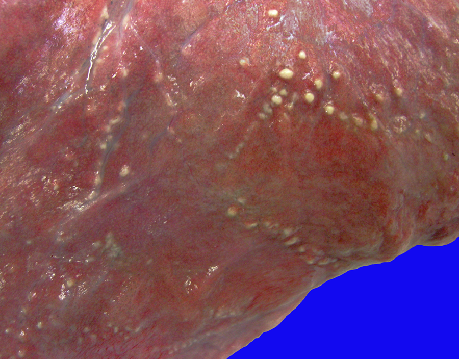
Lung, Siberian tiger (Panthera tigris altaica). Multifocal, irregularly distributed, whitish, soft, subpleural nodules.
Histologically, there was a marked infiltration of macrophages, with foamy cytoplasm, and multinucleate giant cells interspersed with numerous clusters of cytoplasmic clefts that were interpreted as outlines of cholesterol crystals (Fig. 2). Localized around the lesions was a mild lymphocytic infiltration. The majority of the lesions were located under the pleural surface. The same lesions were present to a lesser degree within the parenchyma, usually in the peribronchial or periarteriolar regions. Sudan III staining revealed red-stained, fat-storing cells scattered in the lesions. The outlines and cholesterol crystals remained evident and were not dyed (Fig. 3). These findings were consistent with a diagnosis of endogenous lipid pneumonia, as the necropsy of the animals included in the present study did not reveal any pulmonary obstruction or parasitism. Furthermore, aspiration or inhalation of mineral, vegetable, or animal oils due to oral administration of oil-based drugs or substances was excluded. No pulmonary clinical signs or other macroscopic or histological pulmonary lesions were present. Moreover, there were no cardiac changes compatible with a cor pulmonale. Although the cause of the endogenous lipid pneumonia in the tigers reported herein is not known, it seems reasonable to suggest that it could be associated with the inhalation of irritating dust particles in the animal enclosures open to vehicular traffic of visitors; however, no additional histological findings were detected to support this hypothesis. A genetic predisposition, as described in human beings, cannot be completely ruled out in these tigers.

Lung, Siberian tiger (Panthera tigris altaica). Infiltration of macrophages with foamy cytoplasm, and multinucleate giant cells, interspersed with numerous clusters of cytoplasmic cholesterol clefts. Hematoxylin and eosin. Bar = 50 µm.
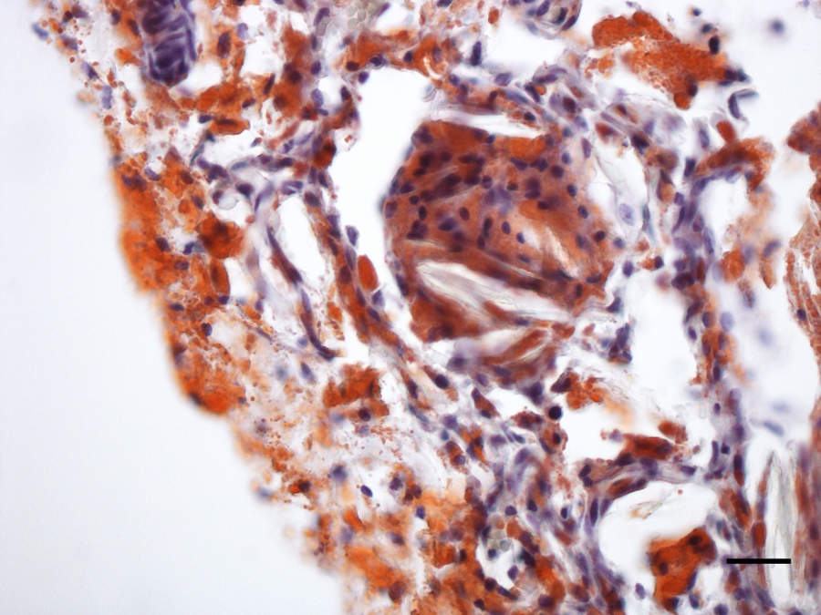
Lung, Siberian tiger (Panthera tigris altaica). Diffusely red-stained, fat-storing cells with outlines of cytoplasmic cholesterol crystals. Sudan III stain. Bar = 25 µm.
Endogenous lipid pneumonia is often incidental and non–life-threatening in the absence of clinical signs or with nonspecific symptoms, radiographic features, and hematological and serum biochemical findings. 10 The endogenous lipid pneumonia in these tigers was considered an incidental postmortem finding of no clinical significance.
Footnotes
Acknowledgements
The authors gratefully acknowledge the Centro di Referenza di Patologia Comparata “Bruno Maria Zaini,” Italy.
a.
Leica Mikrosysteme Vertrieb GmbH, Wetzlar, Germany.
b.
2800 Frigocut N, Reichert Instruments GmbH, Seefeld, Germany.
c.
Leica DM LS2, Leica Mikrosysteme Vertrieb GmbH, Wetzlar, Germany.
The author(s) declared no potential conflicts of interest with respect to the research, authorship, and/or publication of this article.
The author(s) received no financial support for the research, authorship, and/or publication of this article.