
Abstract
Erythema multiforme (EM) was diagnosed in a litter of English Setter puppies. The puppies developed erythematous cutaneous lesions at the age of 2 weeks. Microscopically, there was individual keratinocyte apoptosis associated with lymphocyte exocytosis in all layers of the epidermis. Intranuclear viral inclusions were seen in multiple tissues and organs. Tissues from the tongue, lymph node, spleen, skin, and small intestine were positive for Canine parvovirus-2 (CPV-2) and negative for Canine distemper virus (CDV) and Canid herpesvirus 1 by fluorescent antibody test. Negative-staining electron microscopy detected parvovirus particles in the intestinal contents. The skin and small intestine were positive for CPV-2b and negative for CDV by polymerase chain reaction. The mucocutaneous junctions and small intestines stained positive for CPV by immunohistochemistry. The present report documents CPV-2b–associated EM in a litter of English Setters and substantiates the single previous report associating EM with CPV-2. The finding suggests that CPV should be considered as a possible cause of EM in dogs.
Erythema multiforme (EM) is a cutaneous reaction of multifactorial etiology reported in dogs, cats, horses, and cattle. 6 Erythema multiforme is seen uncommonly in dogs and rarely in cats, accounting for 0.4% and 0.11%, respectively, from cases examined at a university hospital practice. 12 It is believed to be a host-specific T-cell mediated hypersensitivity in which the cellular immune response is directed against various keratinocyte-associated antigens, including those associated with drug administration, infection, neoplasia, and connective tissue disease. 7 In dogs, EM has been reported in association with drugs (antibiotics, anti-inflammatory, antifungal, anthelmintics, and insecticides), staphylococcal folliculitis, feed, a commercial nutraceutical product, and idiopathy. 9,11-13 There is one previous report on Canine parvovirus (CPV)-associated EM in a Great Dane puppy. 4 The present report describes CPV-2b–associated EM in a litter of English Setter dogs.
A 5-year-old female English Setter from a horse farm in south Georgia delivered 12 puppies (11 surviving) through normal delivery (3 puppies) and cesarean section (9 puppies). At 17 days of age, 2 puppies were dead due to unknown cause and 9 puppies survived. Two of the surviving puppies had swollen ear flaps and thickened swollen paws with few pustules. Oral amoxicillin was given to the 2 puppies twice daily. After a week, the 2 puppies developed full body crusts, scabs, intense “itch” response, blistered footpads, and pustules on the lips and the eyelids. The puppies were still in a good body condition and nursing well. Other puppies in the litter were apparently healthy and doing well. Treatment was switched to cefadroxil antibiotic with low-dose prednisone added into the suspension. Heavy growths of Staphylococcus intermedius and beta Streptococcus sp. Group C and moderate Rhizopus sp. were obtained from culture on hair and scab. At 4 weeks of age, a total of 4 puppies were affected and had thickened skin with pustular scabby sores and crusts that were painful to the touch. Cefadroxil and prednisone were given to all 4 puppies with daily antimicrobial shampoos. The lesions worsened irrespective of treatment with oral cephalexin (3 times a day), oral griseofulvin (twice a day), and subcutaneous dexamethasone (twice a day). There was some response to daily dosing of dexamethasone. Skin scrapings were negative for mites. However, ivermectin was given to all puppies and the mother for safety. Two severely affected puppies were hospitalized for more intensive care and diagnostic evaluation. Full thickness skin biopsy was taken from one of the puppies. Given the poor prognosis due to severity of the lesions and deteriorating health status, the 2 puppies were euthanized after 2 weeks of hospitalization (8 weeks of age). The owner reported that more puppies had died at home and that only 3 apparently healthy puppies remained. To the authors’ knowledge, the mother has not been spayed and she has not been rebred. The farm facility had not experienced prior infectious disease problems and another female dog has subsequently raised a litter without complications.
The euthanized puppies were necropsied and examined grossly and microscopically. Microscopic examination was done on the previously received skin biopsy and tissues collected at postmortem in 10% buffered formalin. The tissues were routinely processed, embedded in paraffin wax, sectioned at 5 µm, stained with hematoxylin and eosin, and examined by light microscopy. One set of tissue sections was stained for CPV antigens by immunohistochemistry (IHC) at the Histology Laboratory of the Department of Pathology, College of Veterinary Medicine, University of Georgia (Athens, Georgia). Fresh unfixed tissues from the intestines and skin were tested for CPV-2 DNA by conventional polymerase chain reaction (PCR), as previously described. 3 Positive PCR products, consisting of a 583-bp product, were purified and sequenced in order to determine the subtype of CPV-2 involved as previously described. 8
Grossly, there were extensive skin erythema with alopecia on the face, body, and extremities (Fig. 1a–c). In both puppies, the tongues and oral mucosae were multifocally eroded and ulcerated (Fig. 1d). Microscopically, the epidermis of skin on the lips (mucocutaneous junction), ears, footpads, and body from both puppies was mild to moderately hyperkeratotic (parakeratotic), irregularly acanthotic, multifocally necrotic, and occasionally covered with serocellular crusts. In all layers of the epidermis, there was scattered individual keratinocyte apoptosis with lymphocyte satellitosis (Fig. 2a). In some sections, the lymphocytes and neutrophils formed multifocal intra-epidermal micro-pustules (Fig. 2c). The individual cell apoptosis occasionally extended to the infundibular and upper sections of the hair follicles and associated sebaceous glands. These pathologic findings were consistent with EM.
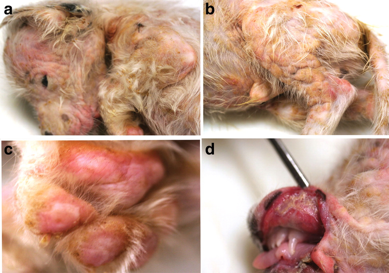
English Setter puppy. Erythematous skin lesions involving the entire body and footpads (
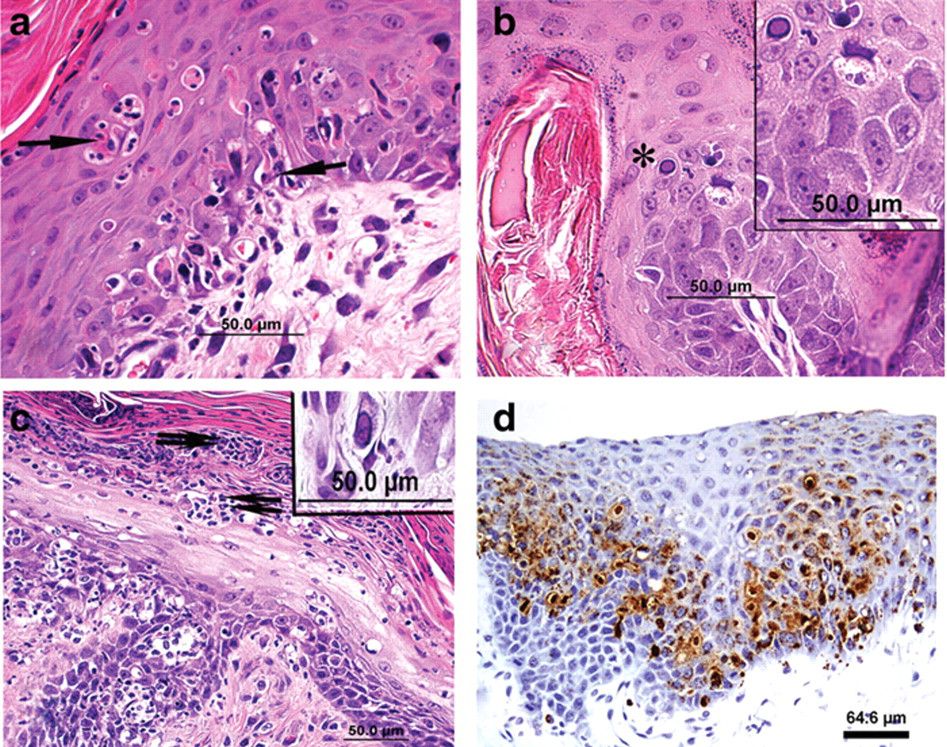
English Setter puppy, skin.
Few basophilic to amphophilic intranuclear inclusions (Fig. 2b) and eosinophilic cytoplasmic inclusions were present in the apoptotic basal cells and the overlying cells of stratum spinosum. Similar intranuclear inclusions were present in a few mast cells in the papillary dermis (Fig. 2c), the mucosal cells of the tongue, the small intestine crypt enterocytes, and the myocardiocytes of the heart in both puppies and in the mucosal cells of the oropharynx overlying the tonsil and in the epithelial cells of the esophageal glands in one of the puppies.
In all sections of the skin, few to several lymphocytes, plasma cells, neutrophils, and mast cells infiltrated the papillary dermis. The microscopic findings in the other tissues besides skin were typical of parvovirus infection. 2,5 The findings in the other examined tissues were unremarkable.
Tissue samples from the tongues, lymph nodes, intestines, and skins from both puppies were positive for CPV-2 and negative for Canine distemper virus (CDV) and Canid herpesvirus 1 by fluorescent antibody test (FAT) staining. Specimens from the skin and intestine from the puppies were also positive for CPV and negative for CDV by PCR. Analysis of the 583-bp PCR product indicated that the virus belonged to the CPV-2b subtype. Negative-staining electron microscopy detected parvovirus particles in the intestinal contents. The mucocutaneous junctions (Fig. 2d) and small intestines stained strongly and mildly positive, respectively, for CPV by IHC.
Erythema multiforme is a cutaneous reaction of multifocal etiology seen uncommonly in dogs and rarely in cats. 7 In dogs, EM is most commonly idiopathic or reported in association with drug administration (antibiotics, anthelmintics, and anti-inflammatory drugs), infections such as staphylococcal dermatitis, and folliculitis, and pseudomonal otitis externa, feed, and a commercial nutraceutical product. 9,11–13 One previous report described CPV-2–associated EM in a dog. 4 A group of chronic and persistent idiopathic EM is seen in older dogs (“old dog EM”) without a history compatible with known triggers. Lesions are more exudative and proliferative, and predominantly involve the face and ears. 7 In human beings, most cases were associated with drug administration and infections such as Herpes simplex virus and Mycoplasma pneumoniae. 10,14
Despite recognition of multiple etiologic and triggering causes, the pathogenesis of EM is not completely understood. 11,12 It is believed to be a host-specific T-cell mediated hypersensitivity reaction in which the immune response is directed against keratinocyte-associated antigens associated with the triggering causes. 7 It is reported that in EM, cluster of differentiation (CD)44 was markedly up-regulated in keratinocytes and infiltrating cells and is involved in T-lymphocyte activation and site-specific extravasation of lymphocytes into tissues. 12 Keratinocyte apoptosis is probably produced by signals from intraepithelial CD8+ T lymphocytes. 13 Lymphocytes bind to the antigenically altered keratinocytes and trigger cell death via apoptosis. 1 Keratinocyte apoptosis is principally seen in the basal cell layer in exfoliative cutaneous lupus erythematous of the German Shorthaired Pointer dog, discoid lupus erythematous, and systemic lupus erythematous. In agreement with the present case, keratinocyte apoptosis in EM is seen at all levels of the epidermis. 7
Erythema multiforme occurs in 2 forms that may overlap to one another and to toxic epidermal necrolysis. Erythema multiforme minor is characterized chiefly by an acute onset of cutaneous erythematous macules and papules. In EM major (Stevens-Johnson syndrome), widespread mucosal lesions, extensive necrotizing and vesiculous skin lesions, and signs of systemic illness such as pain, lethargy, and pyrexia are present. 6 Based on this, the EM seen in the current cases was classified as EM major.
Because of the diversity of triggering factors associated with EM, recognition of the underlying cause for each individual case is important to propose the best treatment regimen. There was some response to daily dosing of dexamethasone in the current cases, which could possibly be due to immune-mediated pathogenesis underlying the lesions of EM as previously suggested. 6 However, the lesions worsened irrespective of treatment. Treatment with immunomodulating drugs would seem to make sense in cases in which elimination of potential trigger factor has been achieved, yet the disease persists. 13 The possibility that anti-inflammatory drugs could increase the severity of the lesions caused by infectious agents should be considered during treatment with immunomodulating drugs.
Age or sex predilection is not documented in dogs and cats. A previous study 12 describing increased frequency of dogs developing EM was recorded in German Shepherd Dogs, Pembroke Welsh Corgis, Old English Sheepdogs, Chow Chows, Cairn Terriers, and Bearded Collies. The report further indicated that increased frequency in the latter 4 breeds should be interpreted cautiously, as only a single dog within each breed was affected. The previous CPV-associated EM was reported in a 2-month-old Great Dane puppy, and the current cases were seen in English Setters. Extensive studies and documentation are needed to determine breed predilection to EM in dogs.
Canine parvovirus infection was confirmed in several tissues and organs from the puppies by FAT, PCR, IHC, and electron microscopy. This indicates that the EM observed in this litter of English Setters was associated with systemic CPV-2b infection involving various tissues and organs with marked cutaneous lesions. It was previously hypothesized that in CPV-associated EM, infection of stem cells and transient amplifying keratinocytes most likely occurred following hematogenous dissemination of the parvovirus. 4 Epitheliotropic viruses are suggested to be common initiators of EM minor in dogs. 7 The present report documents CPV-2b–associated EM in a litter of English Setters, which substantiates the single previous report associating EM with CPV-2 and suggests that CPV should be considered as a possible cause of EM in dogs.
Footnotes
Acknowledgements
The authors would like to thank the staff of the virology and histology sections of the Tifton Veterinary Diagnostic and Investigational Laboratory, College of Veterinary Medicine, University of Georgia, for their technical assistance, and Dr. Uriel Blas-Machado for his help with the IHC.
The authors declared that they had no conflicts of interest with respect to their authorship or the publication of this article.
The authors declared that they received no financial support for their research and/or authorship of this article.