
Abstract
Trichinella spp. can infect various domestic and wild species, including companion animals. Infection occurs because of the ingestion of raw meat (e.g., infected prey). In experimental studies, cats have been found to be a very susceptible host to infection by Trichinella spp.; naturally occurring feline infections have also been reported. However, clinically apparent disease seems to be a rare manifestation of this infection in cats. The skin biopsy of an 8-year-old, neutered, male, domestic cat revealed an inflammatory granulation tissue that surrounded a well-preserved cyst that contained a Trichinella sp. larva. Distinct seropositive reaction against Trichinella spp. antigens was demonstrated by enzyme-linked immunosorbent assay and Western blot. Immunohistochemistry, by using serum from the infected cat as the source of antibody, showed strong immunostaining of Trichinella spp. larvae. During a 1-year follow-up, a postexcisional local tissue reaction was observed. This manifested as a firm, poorly circumscribed subcutaneous mass adjacent to the eye, which demonstrated clinical features and histopathologic findings indicative of chronic inflammation associated with granulation tissue and fibrodysplasia. Digestion of the muscle biopsy revealed one Trichinella sp. larva, which was identified by multiplex polymerase chain reaction as Trichinella nativa. To the authors' knowledge, this is the first documented case of trichinellosis in a cat with a nonhealing ulcerative skin lesion as the main clinical manifestation of the infection.
An 8-year-old, male, neutered Domestic Shorthair cat with an ulcerative skin lesion was presented at a small animal referral practice. At the time of presentation, a 4-mm × 7-mm crusted ulcer was located on the skin below the left lower eyelid. The conjunctiva adjacent to the lesion was hyperemic and slightly swollen. The cat was otherwise healthy. The ulcer had appeared 2 months earlier and had not responded to oral antibiotic therapy with amoxicillin and clavulanic acid or to topical treatment with eye drops that contained fusidic acid. Finally, the skin lesion was excised, with clear surgical margins, with the cat under general anesthesia, and was submitted to a veterinary referral laboratory for histopathology. a Recovery of the cat from surgery was uneventful, and the operative site had healed by the time of suture removal.
The formalin-fixed skin biopsy specimen was routinely processed for histopathology. Sections were cut at 4 μm and stained with hematoxylin and eosin. Histologic examination showed that the biopsy specimen mainly consisted of interlacing bundles of spindle cells (Fig. 1A). The spindle cells were fibroblasts, with an elongated hypochromatic nucleus, dispersed chromatin, small nucleolus, and scant eosinophilic cytoplasm, with indistinct cell boundaries. Mitotic figures were rare. Numerous lymphocytes, histiocytes, plasma cells, and neutrophils were interspersed among the dense spindle-cell population, but none were as abundant as the spindle cells. Moderate perivascular lymphocytic infiltrates with fewer eosinophils were confined to the periphery of the lesion. The previously mentioned tissue reaction surrounded a well-preserved coiled nematode larva within a 400-μm × 250-μm elliptical cyst. The outer layer of the cyst-larval complex consisted of a thick (35 μm) hyaline capsule. The larva was 35-μm wide. The intestine, musculature, stichosome, and hypodermal bacillary bands were identified in transverse tissue sections. A mild lymphoplasmacytic inflammatory reaction was present at the polar ends of the longitudinally sectioned cyst. Because the inflammatory infiltrates and granulation tissue had replaced most structures of the dermis, the exact location of the cyst was not determined. However, at the periocular area, muscle fibers are usually located superficially when compared with other regions of the body. Therefore, the numerous myofibrils observed in the healthy skin adjacent to the inflamed area and located at the same depth as the encapsulated larva, might suggest that the cystic formation had originated within the myofibrils. Furthermore, the epidermis that covered the lesion was ulcerated, and there was moderate serocellular crusting.
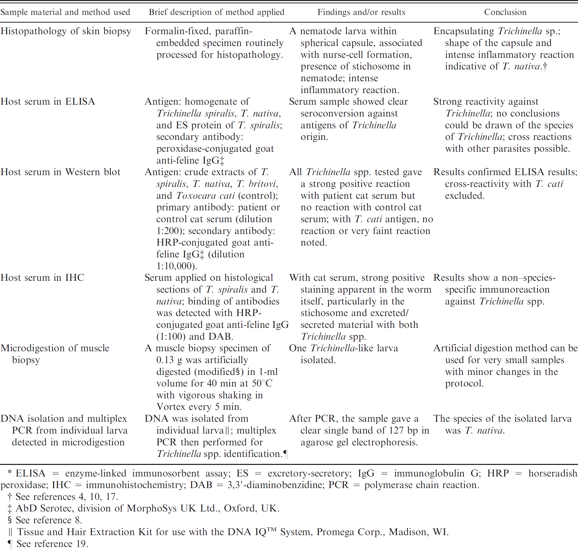
A summary of the diagnostic methods used in the current report is shown in Table 1. Enzyme-linked immunosorbent assay (ELISA) was used to determine host-produced antibodies against Trichinella spp. (Table 1). Serum samples from a young kitten and an uninfected raccoon dog (Nyctereutes procyonoides) served as negative controls. The sera from raccoon dogs infected with Trichinella spiralis or Trichinella nativa were used as positive controls and were derived from a previous experimental infections. 17 The serum sample obtained from the cat showed positive reaction against all antigens of Trichinella spp. origin by ELISA. To confirm the results of serologic diagnosis, a Western blot was performed (Table 1). All the Trichinella spp. tested produced strong positive reactions with the patient cat serum (data not shown), but no reaction with control cat serum. With Toxocara cati extract, no reaction or a very faint reaction was noted with patient serum. There was no reaction with control cat serum.
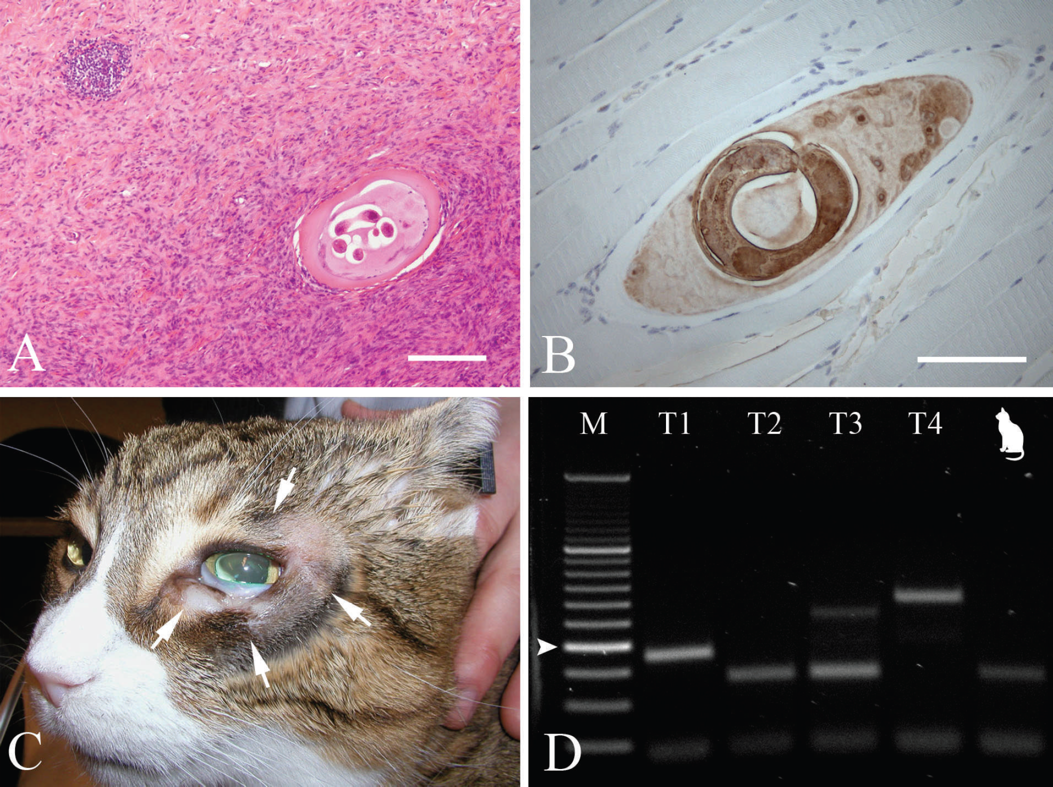
A, biopsy specimen of the original ulcer revealed a well-preserved coiled nematode larva within an elliptical cyst in the center of the lesion. The dermis was severely effaced by interlacing bundles of fibroblasts. Numerous mononuclear inflammatory cells were interspersed among the dense spindle-cell population. Bar = 200 μm.
In immunohistochemical staining (IHC; Table 1), patient cat and control cat serum were applied onto histologic sections of T. spiralis and T. nativa in raccoon dog muscles from a previous study. 17 In the sections treated with patient cat serum, strong positive staining was apparent in the worm itself, especially in the stichosome, as well as in the interior of the capsule (the excreted and/or secreted material) and the nuclei of the host cell (Fig. 1B). Although Trichinella seropositivity was demonstrated by ELISA, Western blot, and IHC, serologic cross reactions with other parasites are a potential confounder in the interpretation of results. This phenomenon is frequently observed in diagnostic serology of Trichinella spp. 1–3,18 Because the cat of the current study was allowed to roam freely, it is likely that it had been concomitantly exposed to nematode parasites, including Trichinella. The most prevalent feline helminth endoparasites in Finland are Toxocara cati and Taenia taeniaeformis. Thus, antibodies against crude antigen of T. cati origin were studied by Western blot; these were negative, and the most likely cause of possible cross reaction was then excluded. However, definite conclusions could not be obtained relative to the specific species of Trichinella based on ELISA, Western blot, and IHC, because current serologic methods cannot provide species differentiation. 9
During the 1-year follow-up, the symptoms recurred; the cat scratched at the eye and squinted. Because of the reappearance and worsening of the clinical signs, the cat was referred to the Small Animal Hospital at the University of Helsinki (Helsinki, Finland). At the time of clinical examination, a V-shaped, alopecic area was observed at the lower eyelid at the site of the previous operation. Subjacent to the affected skin, a firm, subcutaneous mass was present in the periocular area (Fig. 1C). The mass was 1-cm wide and covered the entire lower eyelid and part of the upper eyelid. Ophthalmoscopic and fundic examinations were normal. Clinical laboratory results revealed an increased serum creatine kinase activity (1,689 U/l; reference interval: 60–350 U/l), which could be related to the clinical condition of the cat. Other hematologic parameters were within reference intervals. The cat was also tested for Feline leukemia virus and Feline immunodeficiency virus, with negative results. Contrast computed tomography showed a muscular mass that did not infiltrate the bone. The cat was anesthetized, and several biopsy specimens were taken from the mass and the biceps femoris muscle.
Summary of the diagnostic methods used in the current report.*
ELISA = enzyme-linked immunosorbent assay; ES = excretory-secretory; IgG = immunoglobulin G; HRP = horseradish peroxidase; IHC = immunohistochemistry; DAB = 3,3′-diaminobenzidine; PCR = polymerase chain reaction.
See references 4, 10, 17.
AbD Serotec, division of MorphoSys UK Ltd., Oxford, UK.
See reference 8.
Tissue and Hair Extraction Kit for use with the DNA IQ™ System, Promega Corp., Madison, WI.
See reference 19.
Histopathologic examination of the specimen revealed that the lesion was highly cellular, composed mainly of haphazardly arranged fibroblasts with moderate anisocytosis, anisokaryosis, and rare mitotic figures. Scattered leukocytes were present throughout the lesion. The histopathologic findings were indicative of granulation tissue with inflammation and fibrodysplasia, but the marked proliferation of spindle cells and infiltrative growth warranted the consideration of low-grade fibrosarcoma as a differential diagnosis. Cats are prone to develop sarcomas secondary to chronic inflammation, with feline vaccine-induced sarcoma and feline ocular posttraumatic sarcoma being the classical examples of this kind of carcinogenesis. 14 Interestingly, the role of chronic Trichinella sp. infection as an initiator and promoter of malignant transformation of host cells was discussed in a previous study 15 that described a case of trichinellosis in a cat in association with oral squamous-cell carcinoma.
From the artificially digested muscle biopsy sample derived from the biceps femoris muscle (Table 1), one Trichinella-like larva was isolated and preserved in absolute ethanol at −20°C until analyzed. DNA was isolated from the individual larva, and a multiplex polymerase chain reaction (PCR) was performed according to procedures in a previous study 19 (Table 1). The agarose gel electrophoresis of the PCR products demonstrated a distinct single band of 127 bp, which indicated T. nativa infection (Fig. 1D).
During the present study, the initial diagnosis was based on the finding of a nematode larva identifiable as Trichinella sp. within a skin biopsy specimen and then confirmed by the detection of Trichinella spp. antibodies in the serum of the cat. Later, PCR analysis revealed T. nativa as the causative species. Trichinella spp. have a wide range of host species, and cats have been found to be among the most permissive and susceptible hosts of this parasite. In fact, muscle larvae of Trichinella spp. were detected as early as 1845 in the cat. 11 Cats are effective hunters and become infected upon the ingestion of meat that contains muscle-stage larvae. The cat in the present case had been observed preying on small mustelids. Trichinella nativa is the most common Trichinella sp. in Finnish wildlife, although Trichinella spiralis, Trichinella britovi, and Trichinella pseudospiralis are also present. 16 Furthermore, Trichinella infections that occur in cats have been reported in several countries, including Finland. 12,16 However, Trichinella infection rarely causes clinical signs in their natural hosts, unless they are infected with a very large number of larvae. 7
The clinical effects of feline trichinellosis are primarily based on information obtained from experimental infections. Clinical signs, when present, include mild gastrointestinal disturbances, such as loose stools and vomiting, during the intestinal phase of the parasite's lifecycle. 5,6 Clinical signs related to generalized disease (e.g., dull coat, weakness, muscle stiffness, discomfort, and death) have been reported in severe infections. 5,6 However, clinically apparent natural infections seem to be extremely rare in cats. Before the present case, only 4 cases were described in the veterinary literature. The earliest cases, from Germany (1952), India (1954), and the United States, were summarized in detail. 13 Briefly, the German case was of a heavy infection, characterized by gastrointestinal signs, wasting, weakness, and stiffness of the limbs, which resulted in death. The cat from India suffered from a lack of appetite, vomiting, diarrhea, and lockjaw 2 days before death. The postmortem examination revealed an intestinal phase of Trichinella sp. infection. The first case reported from the United States occurred in Massachusetts and was characterized by transient hemorrhagic enteritis with identifiable adult nematode worms within diarrheic stools. Migrating Trichinella larvae were found in the peripheral blood of the cat with a modified Knott's test. 13 The muscle phase of the infection seemed to be clinically unapparent, although a moderate eosinophilia was present for 3 months. Since then, an additional case of trichinellosis was reported in association with oral squamous-cell carcinoma in a cat from New York. 15
To date, published cases of clinical trichinellosis in cats demonstrated that, although the disease is rare, it can have several manifestations. The present case describes a nonhealing ulcerative skin lesion associated with T. nativa infection. There are several methods that can be used to confirm the clinical diagnosis of a suspected case of trichinellosis. Although, some assumptions concerning the Trichinella sp. involved can be made based on histopathology, techniques that use molecular biology are needed for reliable identification to the species level.
Acknowledgements. The authors thank the cat's owner for cooperation and Kati Holmsten for laboratory assistance.
Footnotes
a.
Patovet Ay, Helsinki, Finland.
b.
AbD Serotec, division of MorphoSys UK Ltd., Oxford, UK.
c.
Tissue and Hair Extraction Kit for use with the DNA IQ™ System, Promega Corp., Madison, WI.