
Abstract
An 8-month-old, intact male Golden Retriever with a history of left forelimb lameness for 2 months was presented to the Veterinary Medical Teaching Hospital of Konkuk University (Seoul, Korea). Results of a physical examination revealed a mass in the left axillary region. A thoracic radiography showed an osteolytic lesion in the scapula and the presence of a soft tissue density from the thoracic wall to the scapula. A computerized tomography revealed a mass invading into the scapula, and small nodules in the lung that suggested metastasis. At necropsy, a pale-yellow, irregular, firm, 8 × 10 × 5 cm mass extended from axillary region and destroyed the scapular. In addition, small nodules were noted in the lung. On microscopic examination, the mass consisted of round-to-oval cells, with eccentrically located hyperchromatic nuclei and eosinophilic cytoplasm in fibromyxoid stroma. Tumor cells were observed in blood vessels in the primary mass. Tumor cells strongly expressed vimentin, desmin, and myoglobin. In phosphotungstic acid-hematoxylin staining, cross-striations were detected in rhabdomyoblasts. In periodic acid-Schiff reaction, only a few cells were detected. The diagnosis was primary rhabdomyosarcoma of the appendicular muscle of a young dog. The tumor presumably originated in the skeletal muscle of the limb, invaded into the adjacent scapular bone, and metastasized to the lung.
Rhabdomyosarcoma is a malignant neoplasm that arises from striated skeletal muscles or muscle progenitor cells. 1,5,9 It is typically aggressive, locally invasive, and can metastasize. 1,2,5,8 There are relatively few reports of this tumor in the veterinary literature. 1,3,9,11,12,14 In humans, rhabdomyosarcoma is a common soft-tissue tumor in children. A predilection for the head and neck, genitourinary areas, retroperitoneum, and extremities is noted. 4,10 In contrast, its reported incidence in domestic animals is less than 1% of all neoplasms. 6 It is most commonly observed in dogs. 5 Rhabdomyosarcoma has no apparent sex, breed, or regional predisposition. It has been reported in the pharynx, gingiva, urethra, urinary bladder, cardiac muscle, greater omentum, larynx, trachea, and tongue. 2,5,6,12 As in humans, in domestic animals, rhabdomyosarcomas are subclassified as embryonal, botryoid, alveolar, and pleomorphic according to histopathologic morphology. 1,2,5,7,14 This report describes the clinical, histopathological, and immunohistochemical data of a dog diagnosed with embryonal rhabdomyosarcoma. Rhabdomyosarcoma occurs in animals with an average age of 6.3 years (range. 6 months to 14 years). 5 The average age of embryonal rhabdomyosarcoma is 8.2 years old, and the youngest animal is 1.5 years old. 5 This case has some distinctive characteristics in that this is a very young age of embryonal rhabdomyosarcoma in a dog, the rhabdomyosarcoma arose at a rare site, the appendicular muscle. The rhabdomyosarcoma involved in scapular bone tissue, and there was the solid evidence of hematogenous metastasis into the lung of the embryonal rhabdomyosarcoma.
An 8-month-old, intact male Golden Retriever with a history of left forelimb lameness of 2 months' duration was referred to the Veterinary Medical Teaching Hospital of Konkuk University (Seoul, Korea). The dog had a protruding mass (8 × 5 × 10 cm) in the left axilla, which enlarged rapidly during the previous month. Results of a physical examination revealed another mass, which was approximately 4 cm in diameter in the scapular region. A complete blood cell count and serum biochemistry analysis showed no abnormalities. Thoracic radiography and computerized tomography (CT) demonstrated a distinct osteolytic lesion, with a geographic pattern in the scapula and extension of a soft-tissue density from thoracic wall to the scapula (Fig. 1). Geographic pattern regard to focal geographic areas of lysis tend to have well-defined margins and are the least aggressive form of lysis. 15 A CT revealed a 0.6-cm in diameter nodule in the lung, in addition to the masses that invaded into the scapula and the thoracic wall (Fig. 2). Also, the masses that invaded into the scapula and extended to thoracic wall were evidently observed. Because the tumor was rapidly progressing and already had metastasized into the lung, a poor prognosis was rendered. The owner requested euthanasia, and the dog was necropsied. At necropsy, a pale-yellow, irregular, and moderately firm mass was found in the left axillary region. Another extensive mass was found in the site from the thoracic wall to the scapula. The mass extensively infiltrated adjacent tissues, including the scapula. The mass in the middle portion of the scapula showed an intramedullary invasion with subscapular fossa and spine destruction. The neoplastic tissues invaded the bone. In addition, small, 0.4–0.6-cm-diameter nodules were found in the lung.
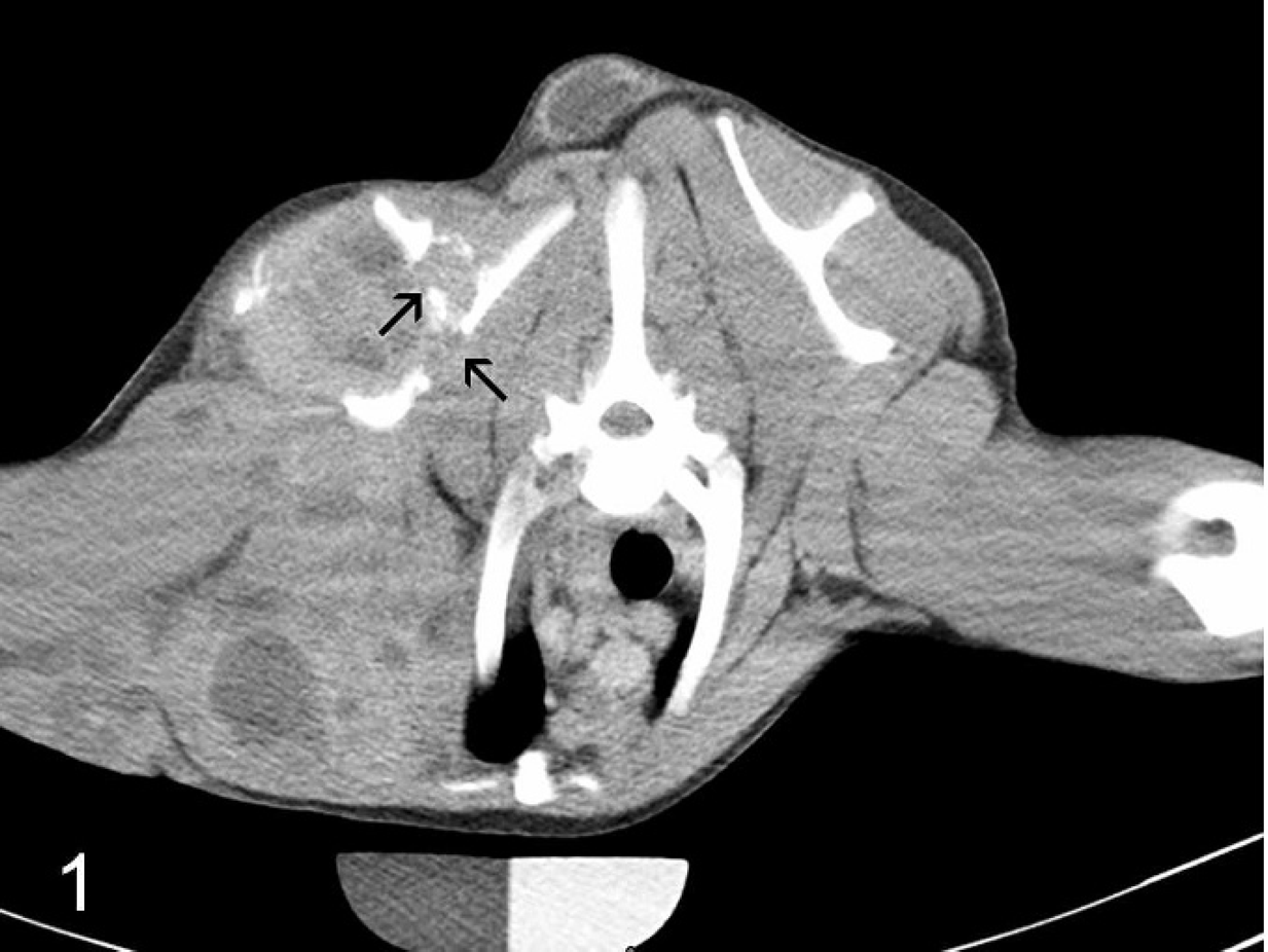
Computerized tomography; dog. The tumor invaded the scapular bone and thoracic wall. The large mass extending from axillary region to thoracic wall invaded the scapula (arrows).

Computerized tomography; dog; lung. A nodular structure (arrow) was found in the left lung. The small nodule suggested lung metastasis of the tumor.
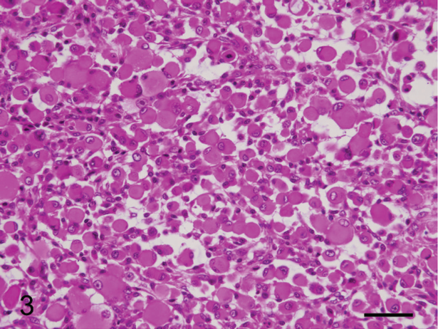
Tissues collected were fixed in 10% neutral buffered formalin, embedded in paraffin, and sectioned at 4 μm. Sections were stained with hematoxylin and eosin (HE). On microscopic examination, the mass consisted of round or oval cells, with eccentrically located hyperchromatic nuclei and deeply eosinophilic cytoplasm in fibromyxoid stroma (Fig. 3). Some cells had scant cytoplasm, without evidence of differentiation. More differentiated cells were identified as rhabdomyoblasts and had abundant eosinophilic cytoplasm with rounded nuclei. Many tumor cells were markedly anaplastic, and moderate mitotic figures suggested that the tumor was malignant. 5 Numerous bizarre cells with multiple nuclei were often present. Based on the histopathologic morphology of tumor cells, it was tentatively diagnosed as an embryonal rhabdomyosarcoma. The tumor cells were observed in the lung (Fig. 4). The scapula-bone tissues were entrapped in the tumor cells and served as a scaffold of anaplastic tumor cell deposition (Fig. 5). Significantly, tumor cells were remarkably observed in blood vessels in the primary mass, providing the evidence for hematogenous metastasis of rhabdomyosarcoma.
To confirm the diagnosis, sections were stained with phosphotungstic acid-hematoxylin (PTAH) and periodic acid-Schiff (PAS). For immunohistochemistry (IHC). slides were incubated with antibodies to vimentin, a muscle specific actin, a desmin, a myoglobin, a cytokeratin, b and S-100. a The antibodies were diluted 1:100 (muscle-specific actin, cytokeratin, desmin, and myoglobin) or 1:400 (S-100 and vimentin). S-100, desmin, and myoglobin were assayed by using the 2-step Envision System-HRP b (horseradish peroxidase), and the others were assayed by using Envision System-AP b (alkaline phosphatase), according to the manufacturer's instructions.
In PTAH-stained sections, cross-striations were detected in the cytoplasm of tumor cells (data not shown). However, in PAS sections, only a few cells showed a weak positive reaction. Tumor cells were negative for cytokeratin and strongly positive for vimentin (data not shown), S-100. desmin, muscle-specific actin, and myoglobin, which is seen exclusively in striated muscle. The neoplastic striated muscles are strongly positive when reacted with an antibody to desmin, as is muscle-specific actin. Myoglobin is expressed later in differentiation. The expressions of desmin and myoglobin were more intense in more differentiated neoplastic cells by showing a close relation to the degree of differentiation (Figs. 6, 7). Cells positive for desmin were found in blood vessels of the axillary mass (Fig. 6). S-100 protein is found in normal skeletal and cardiac muscle cells and varies in degrees of expression. Expression of S-100 was moderately high (data not shown). Based on the clinical, histopathological, and immunohistochemical data, the mass was diagnosed as an embryonal rhabdomyosarcoma.
Rhabdomyosarcoma can occur within striated muscle, as in this case, or in visceral organs, such as the prostate, urinary bladder, and gallbladder, which lack striated muscle fibers. 13 A clear pattern of site prevalence has not been frequently documented in domestic animals, but a few botryoid rhabdomyosarcomas that originated in urinary bladder of young dogs were reported. 2,5 In people, rhabdomyosarcoma arising in large skeletal muscles is most common in adults, and these tumors are typically less aggressive than those arising at other sites in children. Similar to humans, the majority of rhabdomyosarcomas reported in dogs have occurred in tissues that normally do not contain striated-muscle cells, such as the pharynx, gingiva, urethra, trachea, larynx, and the jaw bone. 6,12,14 The reason that rhabdomyosarcomas arise in these sites is not clearly understood, but it is thought that myogenic transformation may be induced in non-muscle cells by a genetic problem, such as genetic lesions, related loss of heterozygosity, genetic fusions, or tumorigenic influences. 13 In the present case, the tumor occurred in the axillary region and involved striated muscles of the shoulder and thoracic limb. This is the second report of such a case that occurred in the axilla, including surrounding muscles. 5 More noticeably, the present case reports a rare bone involvement of rhabdomyosarcoma. A bone lesion of rhabdomyosarcoma was presented as an osteolytic lesion with geographic pattern in the scapula in a radiographic view, and, presumably, tumor cells were seen in the scapular with HE stain as well as by IHC.
Embryonal rhabdomyosarcoma; dog; mass. Tumor cells are variably sized and exhibit marked anisocytosis. Cells are primarily round to oval, with eosinophilic cytoplasm and eccentrically located nucleus. Hematoxylin and eosin. Bar = 30 μm.

Embryonal rhabdomyosarcoma; dog; lung. Neoplastic cells, which are the same in morphology in the mass, are observed in the lung. Hematoxylin and eosin. Bar = 85 μm.

Embryonal rhabdomyosarcoma; dog; scapula. Abundant tumor cells around the scapula bone and several osteoclastic-like, multinucleated giant cells (arrowheads) are observed. Hematoxylin and eosin. Bar = 30 μm.

Immunohistochemistry; dog; mass. Tumor cells positive for desmin (arrow) are present in blood vessel (arrowheads). Envision system-HRP (horseradish peroxidase). Counterstain with hematoxylin. Bar = 30 μm.

Immunohistochemistry; dog; mass. Tumor cells stain positive for myoglobin. Envision system-HRP (horseradish peroxidase). Counterstain with hematoxylin. Bar = 30 μm.
Malignant rhabdomyosarcoma is regarded as one of the most aggressive mesenchymal tumors that often metastasize by lymphatic or venous routes. 1,8 Metastases of rhabdomyosarcoma in the domestic animal were reported in regional lymph nodes, lung, heart, spleen, adrenal glands, kidneys, and other skeletal muscles. 1,5,6 Although the metastasized sites are well known, good evidence describing the route of metastasis does not exist. Only a few previous reports describe the presence of malignant rhabdomyoblasts in pulmonary blood vessels. 1,8 In the present case, tumor cells were present in blood vessels of the axillary mass, which suggests that the primary tumor originated in skeletal muscles in the axillary area and metastasized into the lung by the hematogenous system (Fig. 6).
In summary, an 8-month-old dog was presented with a history of left forelimb lameness and a protruding mass in the left axillary space. Based on the clinical, histopathological, and immunohistochemical results, the mass was diagnosed as an embryonal rhabdomyosarcoma. This embryonal rhabdomyosarcoma is a rare case, occurring at 8 months of early age. Moreover, clinical signs of lameness and protrusion of the mass started 2 months earlier. This case of rhabdomyosarcoma occurred in an axillary region that was rare and showed scapular invasion. Dissemination occurred to the lung via the bloodstream, confirming rhabdomyosarcoma metastasized by hematogenous routes. This report provides good evidence of hematogenous metastases of embryonal rhabdomyosarcoma in a young dog.
Acknowledgements. Ji-Young Yhee and Dae-Hyun Kim contributed equally to this paper and should be regarded as co-first authors. The authors thank Ms. R. H. Jang for excellent technical assistance.
Footnotes
a.
BioGenex, Kormed Corp., Seoul, Korea.
b.
Dako, Fine Life Science Co., Seoul, Korea.