



Abstract
Canine rotavirus was isolated from feces of a Korean Jindo dog with mild diarrhea, and the isolate was genetically characterized. Rotaviral antigen was detected in the feces using a commercial rotavirus antigen detection kit and cytopathic effects were observed in a cell line inoculated with the feces. The virus isolate (GC/KS05) was identified as subtype G3P[3] using reverse transcription polymerase chain reaction (RT-PCR). The strain displayed 98% and 90% identity with the VP7 genes of a canine rotavirus isolate (RV52/96) from Italy and the simian rotavirus strain (RRV) respectively. However, the GC/KS05 isolate exhibited only 83% and 82% identity, respectively, with the G3 serotype canine strains, RV198/95 and K9. Phylogenetic analysis of the VP7 and VP4 genes of GC/KS05 strain led to the classification of VP7 in a different cluster than other canine rotavirus VP7 genes, and VP4 within the cluster of canine rotavirus VP4 genes. The Korean isolate was thus more closely related to the RV52/96 isolate than the other isolates for which sequence data is available. Detailed analysis of the VP7 region revealed 6 amino acid variations between the new isolate and RV52/96. After 5 passages in cell culture, the GC/KS05 strain remained pathogenic for young pups, in which inoculation resulted in diarrhea and virus shedding in the feces.
Keywords
Group A rotaviruses cause neonatal diarrhea in human and many animal species. Rotavirus is a nonenveloped and double-stranded RNA virus with 2 outer capsid-independent neutralizing antibody-inducing proteins, VP7 and VP4, which are used for classification into G and P serotypes, respectively. Both proteins are involved in protective immunity. 8 Canine rotavirus most often causes mild enteritis, especially in pups younger than 2 weeks. To date, only 6 isolates of rotavirus have been reported from dogs, including 3 isolates from the US, 1 from Japan, and 2 from Italy. 5,6,10,12 All 6 isolates have been classified as serotypes G3 and P5A[3]. However, one isolate from Italy exhibited a distinct sequence in the VP7 gene. 2,3,4,11,13 Restriction enzyme analyses, DNA and amino acid sequencing, and phylogenetic studies collectively demonstrate that the Italian isolate (RV52/96) is a G3 subtype canine rotavirus. 11 This study reports the isolation and characterization (using genotyping and sequencing of VP7 and VP4 genes) of a rotavirus in cell culture from the stool of a 3-week-old puppy with mild diarrhea.
A stool sample from a 3-week-old female Jindo dog was submitted to the Green Cross Veterinary Institute in Yong-in, Korea. The pup had exhibited mild diarrhea and anorexia. The dog recovered 5 days after the onset of clinical signs. The animal was diagnosed with rotaviral infection using RT-PCR and a commercial rapid rotavirus antigen detection kit that can detect all group A rotaviruses, and was not tested by bacteriological methods. An attempt was made to isolate the rotavirus using the monkey kidney cell line MA104 in the presence of trypsin, as previously described. 10 Viral growth in cell culture was assessed by examining inoculated cells for cytopathic effects (CPE), detecting rotaviral antigen using a commercial rapid rotavirus antigen detection kit, a and staining the cells for rotaviral antigen by indirect fluorescent antibody (IFA) test, using rabbit antirotavirus polyclonal antibody (data not shown). The virus isolate in this study was passaged 5 times in MA-104 cells. Genomic dsRNA was extracted from infected MA-104 cells and from fecal samples using TRIzol reagent, b according to the manufacturer's instructions. Extracted RNA was resuspended in 30 μl of diethyl-pyrocarbonate c (DEPC)-treated deionized water, and stored at −70°C. G-typing and P-typing of the isolate were carried out following the RT-PCR and sequencing methods previously described for the VP7 and VP4 genes (Table 1). 10 Sequencing of RT-PCR products was performed using an ABI373 automated sequencer through a commercial service. d The sequence of each product was determined twice, and the identity established using the National Center for Biotechnology Information (NCBI) database. The nucleotide and deduced amino acid sequences of the VP7 and partial VP4 genes of GC/KS05 strain were compared with previously published group A rotavirus gene sequences in PubMed. Phylogenetic trees were constructed following the neighbor-joining method 14 using Clustal W, 16 and the bootstrap probability was calculated using 1,000 replications.
Primers used for genotyping and sequencing.

Comparison of nucleotide and deduced amino acid sequences among the VP7 genes of the GC/KS05 isolate and reference rotavirus strains representing the G3 serotype (genotype) in the NCBI database.

For experimental infection with the isolate, 4 six-day-old conventional beagle puppies were divided into 2 groups (I and NI) containing 2 puppies each. Group I puppies were orally inoculated with 1 ml of the isolate, which had a titer of 10 6 tissue culture infective dose 50 (TCID50) per milliliter. The group NI puppies were administered 1 ml of sterile phosphate buffered saline (PBS) orally. Clinical signs of disease were observed for 4 days and viral shedding in feces was detected with RT-PCR during the same period. Four days after challenge, all puppies were euthanized with 0.1 ml of xylazine (Rompun e ). Three segments of small intestine, and 1 section of colon were collected as previously described: 7 a segment of the duodenum was removed at the right lobe or head of the pancreas, and a section of small intestine was obtained approximately 60 cm caudal to the head of the pancreas to represent the jejunum, a colon segment was removed 5 cm caudal to the ileocecal valve.
Comparison of nucleotide and deduced amino acid sequences among the VP4 genes of the GC/KS05 isolate and reference rotavirus strains in the NCBI database.

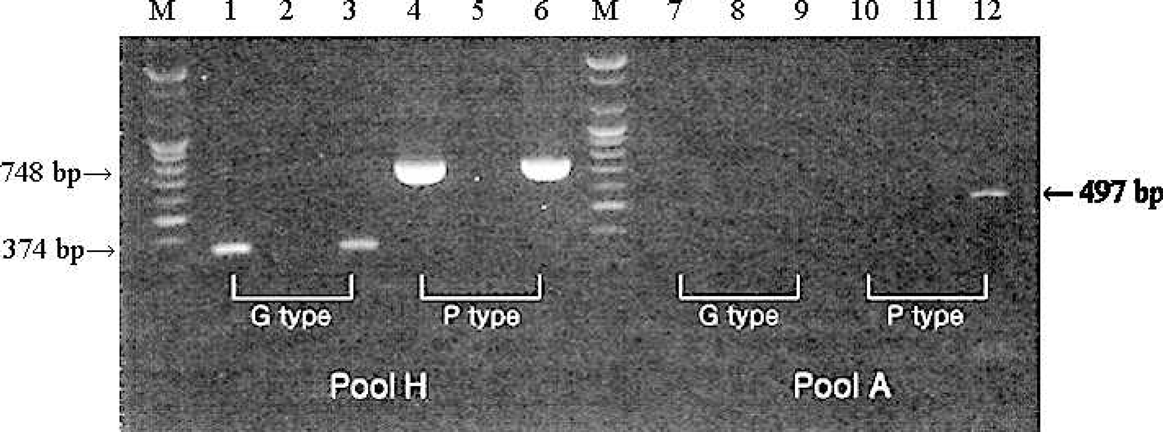
Genotyping of the G and P types by RT-PCR. Lanes 1, 4, 7, and 10: CU-1 strain; Lanes 2, 5, 8, and 11: negative; Lanes 3, 6, 9, and 12: GC/KS05 isolate.

Amino acid sequence of the VP7 protein of the GC/KS05 isolate aligned with that of the RV52/96 strain. The variable regions, A (aa 87–101), B (aa 141–150), C (aa 208–224) and F (aa 235–242) are shown.
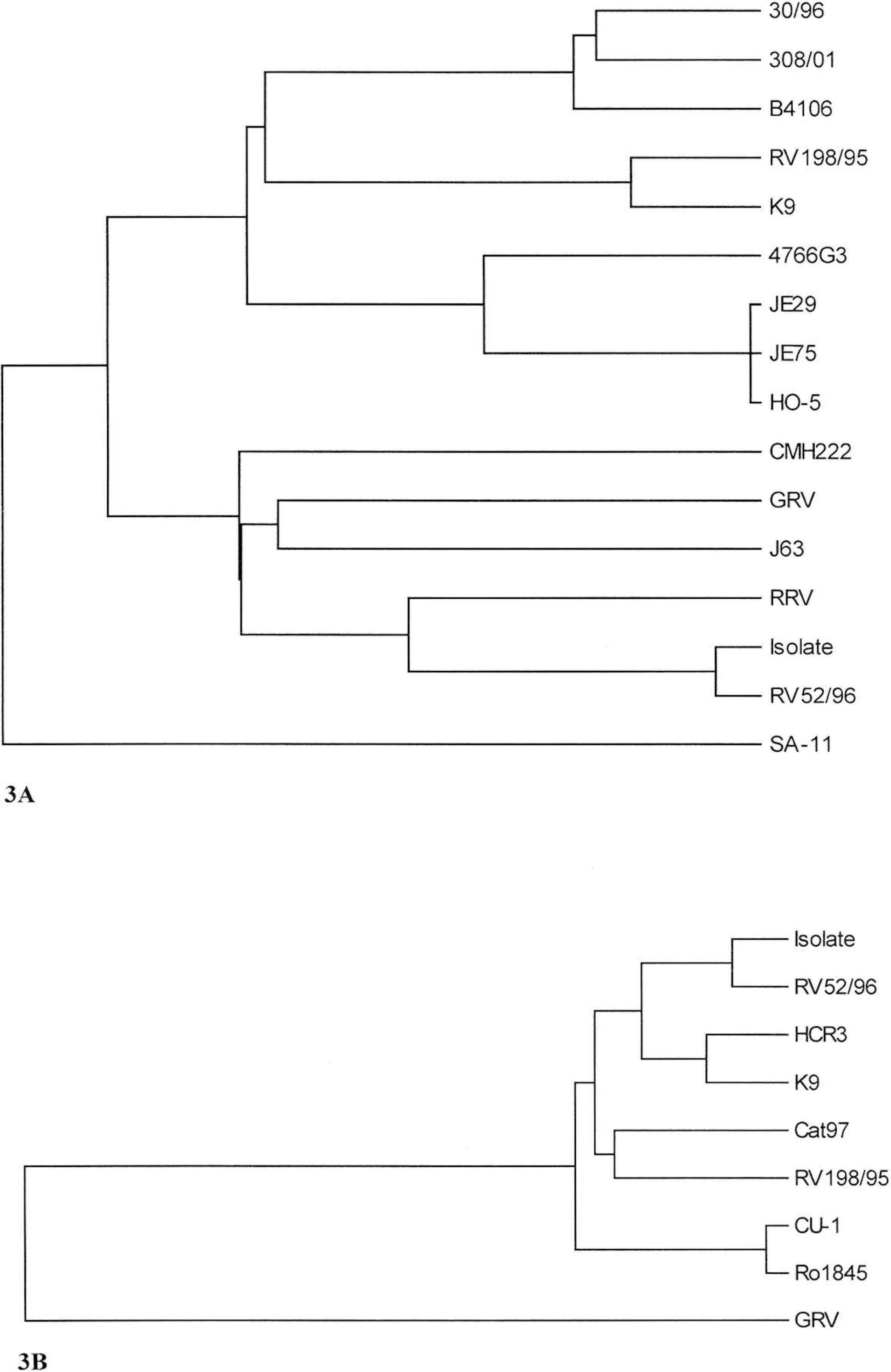
Phylogenetic trees for the VP7
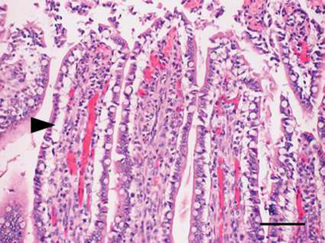
Jejunum of a puppy experimentally infected with GC/KS05 strain at 96 post-inoculation hours. Note that there were mild exfoliation and necrosis of enterocytes on the tip of intestinal villi and also marked epithelial and subepithelial vaculation (arrow head) in the jejunal intestine. Hematoxylin and eosin stain. Bar = 50 μm.
Each of the 3 segments was immersed in 10% buffered formalin and histologically examined. All animal experiments complied with the current laws of Korea. Animal care and treatment were conducted in accordance with the guidelines established by the Seoul National University Institutional Animal Care and Use Committee.
When the fecal sample was inoculated onto MA104 cells, CPE was observed after the third passage and the IFA test was positive for rotaviral antigen (data not shown). The isolate, designated GC/KS05, was characterized as G3 and P[3] (HCR3-like) on the basis of its amplification by the G3/Send9 (producing a 374 bp product) and Con 3/P3 (producing a 748 bp product) primer sets, respectively (Fig. 1). The VP4 gene of this isolate was also recognized by the P[7] (OSU-like) primer (producing a 497 bp product). Sequence analysis of the VP7 and partial VP4 regions also indicated that the GC/KS05 isolate belonged to the G3P[3] genotype. The VP7 and VP4 sequences of GC/KS05 were closely related to those of the canine rotavirus RV52/96 strain (98% nucleotide and amino acid identities) previously identified as G3P5A[3] (Table 2A, 2B). When compared to RV52/96, GC/KS05 had 6 and 5 amino acid differences in the VP7 and partial VP4 regions, respectively. The sequences of antigenic regions A (residues 87–101), B (residues 142–152), and F (residues 235–242) of the isolate were identical to those of the RV52/96 strain, whereas a single amino acid difference (Thr replaced with Ala in the isolate) was observed in region C (residues 208–221) (Fig. 2). Using a neighbor-joining tree (Fig. 3a) based on the nucleic acid sequences of human and animal G3 rotaviruses, 2 groups were formed, and the Korean isolate was clustered together with the canine strain (RV52/96) and Rhesus monkey strain. However, the VP4 nucleic acid sequence of GC/KS05 showed close relatedness to the VP4 nucleic acid of other canine rotavirus strains (Fig. 3b).
Following inoculation of puppies with GC/KS05 rotavirus, diarrhea was observed at postinoculation hours (PIH) 48, and viral RNA was detected from PIH 24 through 96 in the feces of the inoculated pups. One of the inoculated pups sacrificed at PIH 96 contained mild exfoliation and necrosis of enterocytes on the tip of the intestinal villi, and also marked epithelial and subepithelial vacuolation in the jejunum (Fig. 4); however, no villus atrophy was observed. Based on the detection of nucleic acid and antigen in the feces of infected puppies as well the observation of epithelial and subepithelial vacuolation in the villi, it was concluded that the GC/KS05 isolate was pathogenic as in infected villus cells.
The isolated canine rotavirus in Korea was characterized as G3P[3] by RT-PCR and sequencing. VP7 expresses the major neutralizing epitope, and is distinguished by means of both serological and genomic analyses into G type, with good correlation between both analytical methods. Four variable regions, A (residues 87–101), B (residues 141–150), C (residues 208–224) and F (residues 235–242), in the VP7 protein play a major role in neutralization. 1,9 The canine strain, RV52/96, is believed to be the rotavirus G3 subtype, which differs from other canine strains by 1 amino acid alteration in each of regions A and B, and 3 changes in region C. Specifically, in region C, an immunodominant antigenic site, substitutions at residues 212, 213, and 221 had been observed. 11 Because the amino acid sequence of the GC/KS05 isolate differ from that of RV52/96, by only 1 residue in the variable region C (residue 213; Ala for Thr), the Korean isolate might be a regional G3 subtype strain. Sequence analysis of the VP7 sequence of a large number of canine rotavirus isolates is required for identifying the prevalent types. When the partial VP4 nucleic acid sequence of GC/KS05 was analyzed using the RT-PCR product of the P[7] (OSU-like)-specific primer, the isolate was similar to other canine rotavirus strains (except CU-1). Overall, GC/KS05 and RV52/96 (canine) were clustered together with high bootstrap probabilities, whereas K9 (canine), HCR3 (human), RV198/95 (canine) and Cat97 (feline), and CU-1 (canine) and Ro1845 (human) were clustered together. Further genetic and serological studies are necessary to identify genotypic and antigenic relationships among isolates from various species with respect to the VP4 region. The present findings indicate that VP4 is less variable than VP7 for rotavirus isolates from the same species.
In conclusion, this report describes the isolation, identification, and genetic characterization of canine rotavirus in Korea and its use to experimentally infect puppies. The genotype of the isolate was G3 P[3], consistent with previous results. 15 However, this isolate exhibited some nucleotide and amino acid variations in VP7 and VP4 genes, which appear to distinguish it from RV52/96, the previously characterized G3 P[3] genotype. Thus, there appear to be regional variants of the G3P[3] genotype. Further characterization of the isolate in comparison with other canine rotavirus strains, including RV52/96, is warranted to advance knowledge of canine rotaviruses.
Footnotes
a.
Animal Genetics, Inc., Suwon, Korea.
b.
Gibco BRL, Grand Island, NY.
c.
Sigma Chemical Co., St. Louis, MO.
d.
Genotech, Co. Ltd., Daejeon, Korea.
e.
Bayer Korea, Ltd. Seoul, Korea.