
Abstract
The interrogation of materials with X-rays or neutrons to determine structure, energetics, and dynamics is fundamental to advancing physical and chemical materials science and enabling innovative material technologies. A persistent challenge in materials development is that progress depends on understanding structure and dynamics across multiple length and time scales in increasingly complex, multicomponent systems featuring interfaces, heterogeneity, and hierarchical organization. Despite rapidly growing demands on materials characterization, current experimental approaches are almost exclusively based on isolated X-ray or neutron scattering and spectroscopy, reflecting a paradigm largely unchanged for decades. To assess the scientific need for a new experimental paradigm, a 3-day workshop sponsored by the U.S. National Science Foundation (NSF) was held at the SpringHill Suites, San Jose, California, from June 2 to 4, 2022. The workshop brought together 70 national and international experts who critically evaluated opportunities enabled by concurrent neutron and X-ray (NeX) scattering, spectroscopy, and imaging experiments. The participants reached a clear consensus that establishing NeX capabilities is crucial for advancing the science of complex materials in the United States. This report illustrates the scientific drivers for NeX experiments through representative examples spanning biomaterials, energy materials, soft matter, nanomaterials, quantum materials, geoscience, and applied materials research. The complementarity of neutrons and X-rays is essential for robust model development and refinement, particularly in multiphase and multicomponent systems. While joint refinement of data from separate experiments is valuable, concurrent measurements uniquely eliminate uncertainties arising from sample evolution, environmental drift, and irreproducibility associated with experiments performed at different locations and times. Realizing NeX capabilities will require the development of new instrumentation, data analysis frameworks, and robust sample environments compatible with both neutron and X-ray probes. Addressing these challenges will enable unambiguous interpretation of complex materials behavior and open new frontiers in materials research.
Introduction
Addressing significant societal challenges in different areas, including energy, healthcare, sustainability, or security, will require developing novel multifunctional material systems with unprecedented property combinations. These materials will integrate light and heavy elements, feature multiple inorganic, organic, or biological components, and display responsive behavior dependent on transitions on hierarchical time and length scales. A workshop, June 2 to 4, 2022, at Spring Hill Suites, San Jose, sponsored by the U.S. National Science Foundation, NSF, was held to determine the demand for concurrent neutron and X-ray (NeX) experiments. For 3 days, 70 experts discussed the anticipated demand for NeX based on existing scientific cases. Hereafter, the summary of the scientific need for NeX scattering and spectroscopy illustrates that the number of sciences benefiting from concurrent measurements is extensive, especially chemistry, and includes, but is not limited to, soft matter, biology, pure and applied chemistry, electrochemistry, quantum materials, bio and geosciences, life sciences, applied and engineered materials, cultural heritage, counterfeit and contraband detection, nuclear forensics, homeland security, technical and industrial applications. This surprisingly large number of areas affected points to a substantial bottleneck almost exclusively caused by oversubscribed neutron facilities like the NIST Center for Neutron Research (NCNR), which has increased due to the shutdown of many neutron sources in the past 20 years.
The workshop discussions identified a typical pattern in support of concurrent measurements in all the reviewed subjects. First, combining experimental neutron and X-ray data refinement is essential to take full advantage of the differences in interactions with matter. While structural analyses benefit from measurements with X-rays and neutrons in every case, these are typically done at different locations and probing different sample volume elements. Some time may pass between the measurements, leading to different sample histories. Consequently, uncertainties in the data often result, as neither the sample nor the sample environment conditions are necessarily the same in two separate measurements, complicating data analysis and interpretation. Samples can show time-dependent changes. The results may depend on the beam position on the sample and beam size due to heterogeneity in the sample. The differences in sample environment, including temperature, humidity, and pressure, may influence the measurement. Despite the advanced technology available, it is technically impractical, or even impossible, to repeat a single experiment under precisely the same conditions, even with the sample stored in the same sample environment. A surprising number of examples across disciplines show that significant changes result from a slight shift in any of these factors. These considerations underscore the importance of concurrent experiments.
Typical examples include phase transitions, which need concurrent experiments to take advantage of the specific advantages of neutrons and X-rays. A typical scenario is a structural change with a change in the magnetic field. Neutrons are highly sensitive to the magnetic moment, which enables a measurement of the magnetic structure. Both neutrons and X-rays are sensitive to the crystal structure, but with fundamentally different contributions to the diffraction signals from different elements. Due to the complexity of materials, contrast enhancement techniques using resonance X-ray experiments and neutron contrast variation and polarization are essential. Currently, the number of existing opportunities is minimal, including only one combined SANS/SAXS (small-angle neutron scattering/small-angle X-ray scattering) instrument at Instut Laue Langevin (ILL)1,2 and the China Neutron Spallation Source (CNSS). 3 Both the ILL and CNSS concurrent NeX facilities feature perpendicular neutron and X-ray beams. The ILL facility features horizontal SAXS and SANS beams on D-22. This facility was used to characterize the in situ growth of gold nanorods. 4 The CNSS facility features a vertical SAXS/WAXS instrument installed on the very small-angle neutron scattering (VSANS) instrument. There are a few combined neutron and X-ray imaging options at NCNR, ILL, and Paul Scherrer Institut (PSI) (https://www.psi.ch/en).5,7
Importance of neutrons and X-rays for science and society
Hereafter, we use SANS and SAXS to illustrate the importance of science and society based on neutron and X-ray experiments. We outline that these essential tools become increasingly important with the increasing availability of instruments.
2.1 Scientific importance
SAXS has been widely used mainly for structural characterization. Inspecting the literature thoroughly, most studies work with two-phase or pseudo-two-phase systems. These samples are characterized by one phase with an electron density that dominates. In such a case, a two-phase approximation is possible because the contrast, i.e., the squared difference of the average electron densities, is essentially determined by one phase. There are plenty of experiments that take advantage of this approach, including precipitation in metal alloys, structural defects, e.g., in diamonds, pore structures in rocks or fibers, particle size and growth in solutions, catalysts, carbon black/soot, void structures in glasses and ceramics, structural correlations in liquids, polymers, and biological macromolecules. 8 However, samples generally are multiphase systems or contain multiple constituents. This is evident for composite materials, which often include various constituents or polymers, nanoparticles, and surface layers with different properties. 9 The following survey shows the need for the joint analysis of X-ray and neutron scattering. More importantly, a closer inspection reveals the need for isotopic labeling and polarization for neutron experiments and X-ray energy variation for X-ray experiments, which confirms the unique role of research reactors, spallation sources, and synchrotrons for science and society.
Surprisingly, the literature scan shows that SAXS experiments are mainly applied to model materials, but natural multiphase systems are rarely attempted. For such more realistic systems, resonant X-ray scattering techniques introduce elemental sensitivity by changing the energy close to the chemical absorption edge specific to individual elements. The low X-ray energy range is significant for soft materials. In this range, a variety of techniques have been applied: resonant soft X-ray scattering (RSoXS) and reflectivity,10,11 single wavelength anomalous diffraction (SAD), multi-wavelength anomalous diffraction (MAD), X-ray diffraction (XRD), and anomalous small-angle X-ray scattering (ASAXS). Variation of the scattering contrast is crucial to extracting the relevant information, like the size of agglomerates of a specific constituent. 8 Two technologies enable approach (1) RSoXS and (2) a combination of neutron and X-ray scattering. Only a few RSoXS instruments are available because these techniques require variable energy and a high-intensity beam because of the sharp absorption edge. Today, many more SAXS and SANS instruments are available than RSoXS beamlines. Hence, the following statistical analysis of publications and impact concentrates on SAXS/SANS, but with good evidence that results are generically applicable to other synchrotron and neutron instruments.
2.2 Publication count
The first experiments on nanostructures—carbon black and similar materials—by Warren and Krishnamurti, beginning in the 1920s, and the substantial contributions by André Guinier in the 1930s, demonstrated the importance of SAXS.12–17
In the 1930s, Chadwick discovered the neutron, 18 and around 30 years later, the isotopic sensitivity of SANS was exploited to experimentally verify the random coil conformation of a polymer chain in the melt. As hypothesized by Paul Flory, the experiment created knowledge that is standard in polymer textbooks today.19,20 Since these early experiments, neutrons and X-rays have repeatedly validated their importance for structural characterization. A Google Scholar search indicates that over 42,000 articles have been published in the last three decades.
Figure 1 shows that the number of SAXS publications has continuously increased over the past 30 years, while the number of SANS papers appears almost steady. This surprising fact about SANS has been supported by an independent study, including SANS and other neutron techniques from 2005 to 2015. 21
Fischer and Craievich correlate the fast growth of SAXS articles with the increasing number and quality of the new synchrotron sources. 22 This explanation is supported by the fast regional increase of SAXS publications in Argentina and Brazil, which is related to the availability of local synchrotron resources. 22 As Figure 2 illustrates, this trend is not unique to SAXS. It includes all instruments available in the regional X-ray sources. 22
Fischer and Craievich explain the steady numbers of neutrons with a nearly constant number of instruments available because new neutron sources compensate for the shutdown of several neutron sources. 22 The regional increase in neutron users in Asia and Oceania correlates with new sources’ availability. 22 Hence, as illustrated in Figure 3, it is plausible to assume that the downtrend of publications in France relates to the loss of neutron facilities. 22

Number of published articles related to SAXS or SANS vs. year. Reprinted from Fischer and Craievich. Copyright (2019), under the terms of the Creative Commons Attribution License (CC BY). 22
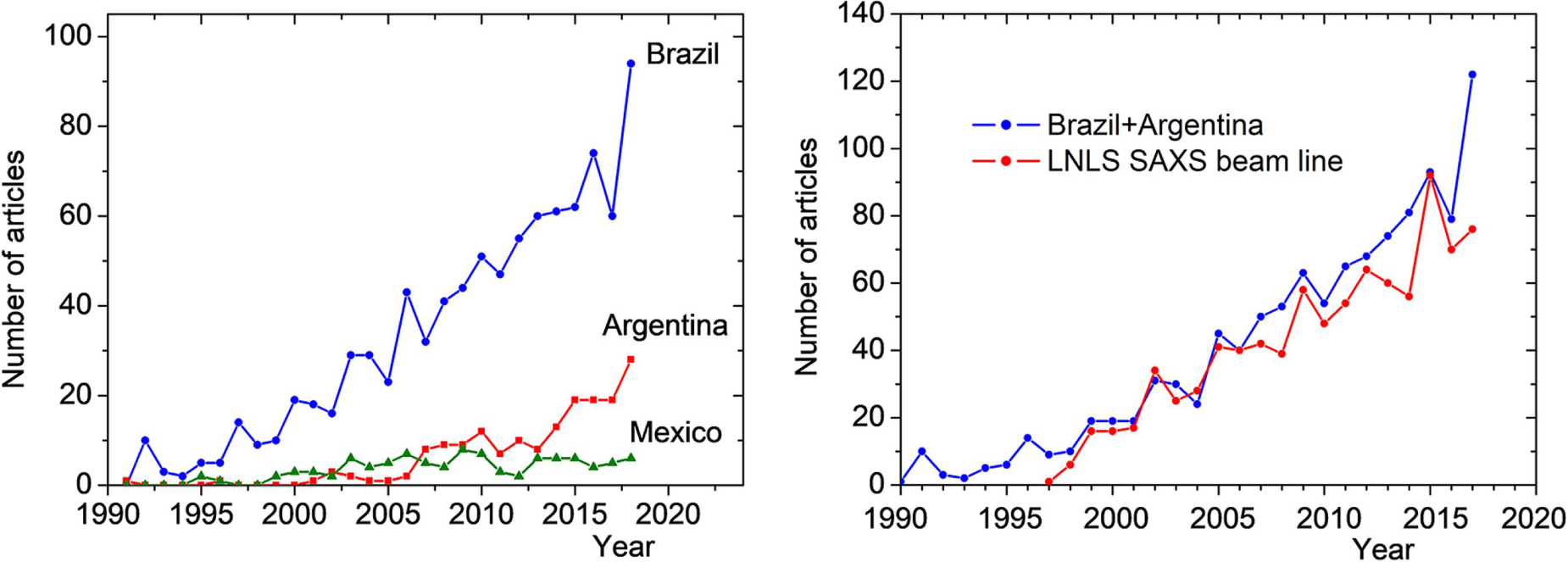
The number of publications from Argentina and Brazil is increasing because of the availability of a synchrotron SAXS beamline. Reprinted from Fischer and Craievich. Copyright (2019), under the terms of the Creative Commons Attribution License (CC BY). 22

Geographical distribution of SANS users. Note: The downward curve around 2010 represents France, while the steep upward curve around 2012 corresponds to China. Reprinted from Fischer and Craievich. Copyright (2019), under the terms of the Creative Commons Attribution License (CC BY). 22
In conclusion, the availability of X-ray or neutron sources creates opportunities that produce a significant number of publications, with the number directly connected with availability. To identify the most urgent needs of the scattering community, it is imperative to identify the interests of the different scientific communities, as mirrored by the number of scientific publications. Figure 4 shows that among 10 popular journals, seven publish only in soft matter, and the other three (Journal of Applied Crystallography, Journal of Physical Chemistry, and Journal of Colloids and Interfaces) publish both soft and hard matter. The authors’ conclusion is plausible that small-angle scattering (SAS) is predominantly used to study soft materials but much less for structural characterizations of hard or inorganic matter. Within the soft matter field, most of the articles, roughly 90%, deal with polymers, which may include polymers in the melt solution but also composites with nanoparticles.
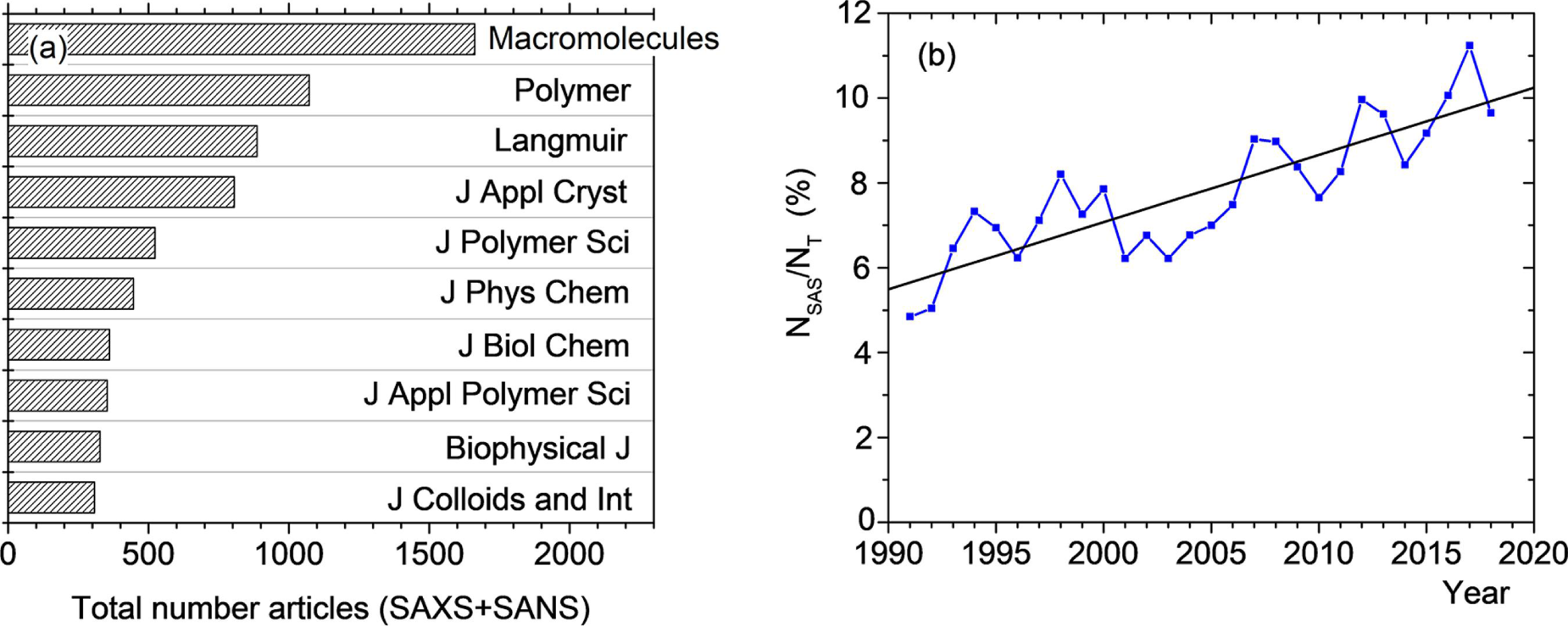
Distribution of publications, including SAXS and SANS, over different journals. Reprinted from Fischer and Craievich. Copyright (2019), under the terms of the Creative Commons Attribution License (CC BY). 22
2.3 Compact sources offer new opportunities
Neutron sources suitable for material characterization beyond radiography are traditionally limited to large-scale user facilities such as reactor or spallation sources. In the past decades, however, compact accelerator-driven neutron sources were developed. These developments led to the formation of the Union for Compact Accelerator-driven Neutron Sources (https://www.ucans.org/) in 2010, and the UCANS proceedings, as well as presentations, are an excellent source for information about such neutron sources. An example of such a neutron source is Japan's RIKEN accelerator-driven compact neutron source (RANS). 23 Since these sources are accelerator-driven, constant wave and pulsed operations are possible. Xu et al. 24 demonstrated time-of-flight texture analysis with the RANS source, requiring ∼6 hours of beam time for a bulk texture measurement of a steel sample. Ikeda et al. discuss the applicability of engineering diffraction (microstructure, residual stress, etc.) of the RANS. 25 In combination with cost-efficient and detection-efficient novel detector technologies such as event-mode neutron cameras, 26 close to 4π steradian coverage with close to 100% efficient detectors for thermal and cold neutrons could be accomplished at a low cost compared to conventional neutron detector systems and without the need to develop and maintain custom-made detector electronics. A new accelerator-driven compact neutron source prototype, RANS-II, was completed in 2021. 27
Laser-driven particle acceleration has also been developed over the past two decades. It utilizes the ability of an intense photon beam to co-emit ions with relativistic electrons from a target, such as deuterated polymer. Strong, annular magnetic fields produced by this process result in the ions and electrons forming highly directed jets along the laser channel, with the electron jet being impeded by the magnetic field, offsetting the two jets. This results in a high, teravolt electric field, resulting in further ion acceleration, particularly of the slower ones in the jet. 28 In the case of a deuterium jet, the ions produce neutrons through deuterium breakup and nuclear stripping interactions in a converter target consisting of, for example, Be or Li. This technique is less than two decades old, and the first commercial units became available, e.g., from Tau Systems (https://www.tausystems.com/). Since the laser pulses are ∼ 100 fs, the resulting neutron pulse durations can be short depending on the neutron energy. This enables time-of-flight methods, including epithermal techniques like neutron absorption resonance-based methods. 29 Shielding requirements for laser-driven and small accelerator sources may allow sample locations only a few meters away, providing acceptable flux on the sample compared to spallation or reactor sources, where the closest sample position from the source is set at 9 m in at least one instance, but is more often considerably longer.
Similarly, compact synchrotron light sources became available over the past decade, including compact X-ray free-electron lasers.30,31 For such sources, a so-called table-top particle accelerator produces electron beams with energies up to 30 MeV that interact with a high-powered infrared laser to produce ultrashort X-ray pulses up to 20 keV photon energy that can be used for material characterization. These developments include commercially available compact synchrotron light sources (however, Lyncean is now out of business). 32
These exciting developments warrant discussion of scientific applications of such sources, including the combination of neutrons and synchrotrons. The workshop summarized in the present article strived to identify such applications.
3.1 Broader scientific impact
The success of X-ray and neutron scattering techniques in soft matter, bioscience, condensed matter materials sciences, and other areas of research, development, and technology has led to the development of yet more powerful sources and advanced scattering and imaging measurement technologies. Examples include SAXS, SANS, ultrasmall-angle X-ray scattering (USAXS), ultrasmall-angle neutron scattering (USANS), inelastic neutron spectroscopy (INS), quasielastic neutron spectroscopy (QENS), neutron spin echo (NSE) spectroscopy, X-ray photon correlation spectroscopy (X-PCS), neutron reflectometry (NR), X-ray reflectometry (XR), grazing incidence, neutron spectroscopy (NS), diffraction, stroboscopic and pulse probe techniques, as well as microscopy and, more recently, tomography. The methods facilitate structural measurements from atomic through millimeter length scales and dynamics from the sub-pico-second through nano-second and even into the minute and beyond time scales.
Over the years, gradual improvement has enabled breakthrough experiments in bio and soft matter and in materials and condensed matter physics. Novel instrumentation has emerged, especially that planned for the future Second Target Station neutron source of the Oak Ridge National Laboratory, which will substantially improve the user experience and accuracy by enabling measurements of an unprecedented range of time and length scales with one instrument.
Here, we concentrate on structural characterization and slow structural changes. These are all currently part of the soft matter and biosciences portfolios. The techniques addressing these issues currently available to researchers through infrastructure provided by national laboratories, synchrotrons, reactors, and spallation neutron sources. SANS represents the most prominent contributions using neutrons at high-flux reactors and spallation sources. Synchrotron, SAXS, likewise, is the most prevalent for contributions using X-rays. The importance of the SAS techniques has led to intense competition among many meritorious proposals for limited resources, which impacts scientific productivity in two ways. First, the acceptance rate of beamtime proposals is roughly one-in-two based, for example, on statistics from NCNR (Figure 5) and more so at HFIR (Figure 6) and SNS (Figure 7). Second, the time between the development of a research project and its execution at the beamline is at least 6–9 months. These factors leave meritorious proposals without or with considerably delayed access. The system would better serve the community if alternative sources were available to supplement the current, large infrastructure by providing fast, flexible access. Smaller X-ray facilities, either commercial lab-based sources or small-scale local university X-ray synchrotron infrastructure, also provide crucial, but more limited, XRD, SAXS, and XR capabilities. Likewise, midscale neutron facilities based on emerging compact accelerator and laser technology will provide additional resources for SANS, NR, and imaging.

Proposal oversubscription statistics for the NIST center for neutron research. The data lists av-erage values over the cycles from 2015 to 2020. The plot was constructed by the authors using data from the National Institute of Standards and Technology. The data was obtained from the NIST proposal statistics webpage, which is regularly updated with new versions. https://www.nist.gov/ncnr/proposal-statistics (accessed 2 February 2026).

Proposal statistics for the High-Flux Isotope Reactor (HFIR) at Oak Ridge National Laboratory (ORNL), Oak Ridge, TN, USA. Data are provided by the User Office of the Neutron Sciences Directorate and are updated regularly. The most recent version is available at https://neutrons.ornl.gov/users/proposal-statistics.

Proposal statistics for the Spallation Neutron Source (SNS) at Oak Ridge National Laboratory (ORNL) in Oak Ridge, Tennessee, USA. Data were provided by the User Office of the Neutron Sciences Directorate and are updated regularly. The most recent version is available at https://neutrons.ornl.gov/users/proposal-statistics.
While we emphasize the importance of SAS in soft matter and biosciences, even the earliest emergence of high-resolution NS resulted in paradigm shifts in soft matter and biology in using neutron energy transfer to measure time- and length scale-dependent dynamic structure factors. This was particularly true with the advent of momentum transfer with high length scale resolution. The success of these techniques will further drive demand, likely saturating planned facilities. While SAS, and as we will see below, imaging, are likely provide the first realization of instruments at compact, small, and midscale sources, the importance of spectroscopy techniques will drive future instrument development in this area.
X-rays have been particularly successful in characterizing materials with high atomic number elements. In contrast, neutron-based characterization is ideally suited for materials comprised of light elements present in bio and soft matter. The emerging trend toward material systems with multiple components and phases, often comprised of organic, biological, and inorganic constituents, present challenges for material characterization methods. While individual constituents can be characterized through sequential X-ray and neutron experiments, challenges arise if the coupling between transitions within constituents is to be determined, for example, to understand dynamical changes within material systems. These considerations argue for simultaneous neutron and X-ray measurements. While the large-scale neutron and synchrotrons are, in some cases, in neighboring facilities, they are not collocated so that the beam from one can be transported to the other.
3.2 Why X-rays and neutrons
Soft matter and bioscience research have shared emphasis on the structure and dynamics of matter containing mostly light elements. Both areas rely extensively on X-ray and neutron measurements. The two fields inform each other using these measurements, commonly used in interpretations that rely on similar concepts. The sensitivity of X-rays and neutrons to distinctly different attributes of materials makes measurements with the two scattering methods highly complementary. Recent advances in X-ray and neutron sources and instrumentation are enabling exciting new capabilities in soft and hard matter sciences, particularly when the two methods are combined. 33
The strength of X-ray techniques relies on their sensitivity to electron density, making them more sensitive to heavier elements. Even so, as discussed above, X-ray methods have made essential contributions to soft matter and biosciences and will show increased importance in materials systems research. Regardless of this success, a crucial limitation is the insensitivity of X-rays to hydrogen and lithium. A second fundamental limitation is the scattering phase problem, the answer to which is essential to solving structure. While the phase problem is addressed with X-rays using the incorporation of metals (isomorphous replacement), its use is primarily confined to crystallography. Instead, the contrast variation of samples in solutions enhances and negates parts of multicomponent systems typical of soft materials and biosciences. This is possible through a solution of electron density variation, such as by adding sucrose or heavy metal salts.34,35 More recently, anomalous 36 and soft X-ray resonant contrast variation X-ray scattering and reflectometry have been introduced.10,11 However, these highly specialized methods only provide viable alternatives for some systems. Thus, these X-ray techniques are neither easily applied nor generally applicable to soft matter.
This is where neutron scattering becomes essential. The nuclear interactions in neutron scattering make the technique more sensitive to light elements, particularly hydrogen. Furthermore, the nuclear scattering of neutrons leads to sensitive discrimination of isotopes that gives an extra dimension to the neutron scattering measurement. The most used one in soft matter and biosciences is the significant difference in scattering amplitude (the scattering length) between deuterium and hydrogen. This difference is used extensively for contrast enhancement, labeling, solvent contrast variation, or contrast matching. The structure of multicomponent systems represents a typical class of problems in soft matter and biosciences. These techniques either enhance or mask the scattering contributions of one or more components to address the scattering phase problem. The chemistry of deuteration is not always straightforward, in which case a national laboratory facility can provide more advanced technology. When applicable, solution contrast enhancement and matching through mixtures of deuterated and protonated fluids is trivial for neutron scattering.
The choice between X-rays and neutrons often results from the measurement requirements. The large flux of X-ray sources makes them the method of choice for short-time-dependent processes. The small beam size of X-ray sources gives better point-to-point resolution than with neutron instruments and is helpful for small sample sizes and for probing sample heterogeneity, e.g., scanning over samples. X-rays are not subject to the large incoherent cross section of hydrogen that limits structural information from neutrons over higher Q-values. Neutron penetrability enables measurements in robust sample environments and bulk samples. There is a good match of thermal and cold neutron energies and wavelengths to atomic and molecular dynamic time and structural length scales. Neutrons do not impart damage to the specimens, enabling long measurement times. The development of neutron-focusing optics37,38 and improvements in neutron guides39,40 have increased available flux on samples and improved measurement intensities. Current developments in neutron spin polarization techniques, such as SESAME (spin echo scattering angle measurement) or SESANS (spin echo small-angle neutron scattering), promise measurements over significantly larger length and time scales, 41 with implementations at TU-Delft,42–44 ISIS, 45 China Mianyang research reactor (CMRR).46,47
3.3 Soft matter
Soft matter and biosciences experienced dramatic advances with the earliest introduction of scattering techniques. Characterization using single-crystal and fiber XRD measurements enabled seminal advances, such as the discovery of the DNA double helix, the molecular structure of many proteins, and the structure of polymeric materials. The development of neutron sources created vast new opportunities, as their introduction provided information that had not been attainable before then. Thus, the impact of the combined use of X-rays and neutrons is substantial, making essential and unique contributions toward developing critical technical advances supporting today's new economies.
There are extensive and far-reaching scientific impacts of X-ray and neutron scattering and imaging methods in soft materials. A nowhere-near-exhaustive list includes: Semi- and fully synthetic polymer materials and self-assembly (Figure 8). Colloidal and nanomaterials, liquid-crystalline materials, and mixtures (Figure 9). Energy materials include polymer photovoltaics, electronics, nanoparticle-enabled solid-state lighting, electrode materials, and polymer electrolyte fuel cell membranes.
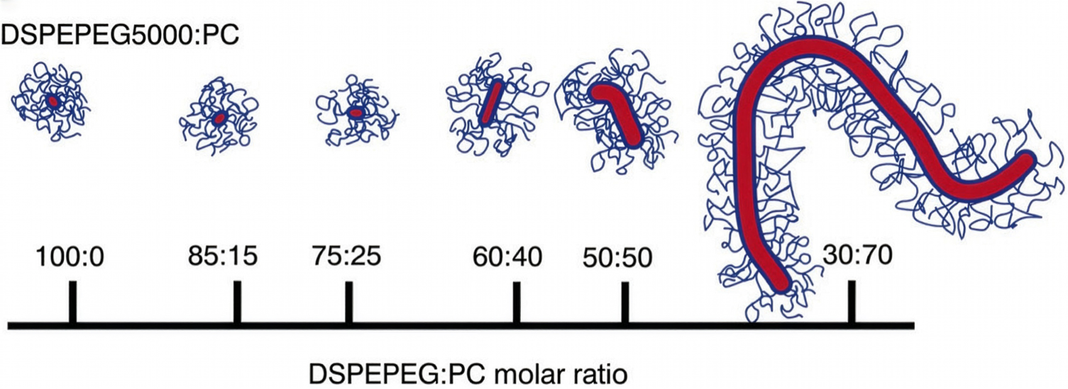
Result from combined SANS/SAXS studies. The hydrophobic core, here acyl chains. The hydrophilic shell is the polar head group and a small amount of water. The outer part of the hydrophilic PEG chains can be described by a Gaussian random walk conformation, with a highly hydrated, loose-structured shell surrounding the core. Structure from co-fits of SANS and SAXS Arleth. Adapted with permission from Arleth et al. (2005), Copyright 2005 American Chemical Society. 48
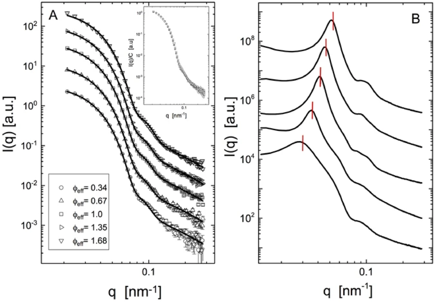
SANS and SAXS data to study the interpenetration of polymeric microgels. Reprinted from Mohanty et al., Copyright (2017), under the terms of the Creative Commons Attribution License (CC BY). 49
Likewise, such work in the biosciences encompasses information that was not attainable by other means: Structural biology includes protein assemblies, nucleic acid conformations, lipid membranes, functional interactions, and viruses. Biofuel production, including algae, grasses, and other plant-based materials.
There are vital materials science and bioscience crossover areas involving highly productive research and development: Medical devices, pharmaceutics, ophthalmic materials, Bio-inspired and bio-derived materials, Biofuel processing, Sustainable materials (e.g., nanocellulose-/lignin-based polymer hybrids), Agricultural materials (e.g., soils) and bio-based environmental remediation.
3.3.1 Impact on emerging research
Further advances and emerging research in soft matter will depend on the ability to characterize the structure and dynamics of multiphase materials and the kinetics of their changes across a range of time and length scales spanning orders of magnitude. Emergent phenomena from the behavior of complex material systems derive from transitions of the coupled transitions in structure and dynamics across a range of constituents and length scales. 50
To meet the challenges implicit in this transformative research, a new generation of infrastructure is needed to advance the understanding of soft material systems and biosciences, allowing for the concurrent characterization of organic and inorganic constituents and their respective interfaces. This would be enabled by the design of experimental infrastructure capable of concurrent X-ray and neutron analysis, NeX (both scattering and imaging modes would be desirable).
Examples of how such infrastructure would enable transformative progress across various systems include: Clarifying how microstructure features (such as grain boundaries) contribute to dendrite formation in solid Li-metal batteries, enabling novel fabrication strategies for next-generation high-performance Li-ion batteries with enhanced cyclability. The combination of X-ray and neutron contrast characteristics in imaging systems would enable an understanding of the evolution of embedded cell clusters, which is a bottleneck for advancing tissue engineering methods. Understanding how transitions in surfactant systems can drive the organization of colloidal building blocks would enable the fabrication of mutable hybrid materials that adapt to changing environments. Identifying the role of structural changes of soil organic matter on the soil's absorption and transport characteristics would promote the development of technologies toward more sustainable agricultural processes. Similarly, understanding the coupling between microstructure changes in rock formation and carbon release during extraction could encourage the development of more sustainable extraction processes.
The combination of X-ray and neutron scattering can also be critical for studies of polymers with dynamic bonds (e.g., vitrimers) that can be easily recyclable, supporting the circular economy, and demonstrate unique self-healing properties, which combined with their extreme toughness and strong adhesion properties, underpin the development of advanced, tailored, durable polymer materials. Stimuli-responsive soft materials are another fascinating class of examples of emergent properties, where systems change, react, and adapt to changing environments without human interference. Simultaneous NeX analysis of the structure and its variations with dynamic studies will enable a much deeper understanding of these materials’ underlying bond and structure rearrangement phenomena.51–61
Materials composed of multiple phases and constituents with properties that are sensitive to the composition, proportion, and distribution of constituent phases, as well as the structure and dynamics of the interfaces, often show properties that emerge by coupling of constituent transitions. These materials’ physical properties, which occur over small time and length scales, drive the macroscopic evolution of the structure and properties of the material. One example is colloidal crystal alloys, whose properties derive from the packed organization of nanoparticles and polymer or surfactant constituents into superlattice structures. Phase transitions within the confined polymer or surfactant layer surrounding each nanoparticle alter the interaction between nanoparticle building blocks. The result of the combined changes in particle interactions and confinement triggers polymorphic transitions and changes in the material's physical properties.62–83
The development/design of bio-inspired materials would benefit from concurrent NeX methods. For example, self-assembling lipid materials in unilamellar (e.g., liposome) or multilamellar structures have rich phase behavior as a function of composition, temperature, and pH. These materials, as lipid nanoparticles, have been employed in lipid-assisted drug delivery. Key recent examples are the delivery of mRNA and siRNA vaccines.84,85 Liquid-crystalline materials present over higher concentrations exhibit tunable mechanical and electro-optical properties, which can be employed in further developing an extensive array of (nano) devices, from sensors to drug-delivery patches. Polymer-assisted delivery of sparingly soluble drugs has also seen pharmaceutical applications.48,86 Large protein complexes/networks such as fibrin (blood clot) and amyloid fibrils are additional biological materials needing a combined NeX approach. The formation of fibrils generally involves processes over a wide range of time and length scales, and the final molecular structure is highly history-dependent.
The combination of neutron and X-ray scattering is of particular promise in biology. At atomic detail, the catalytic power of enzymes can be understood and mimicked only if precise active-site geometries and protonation states are known, and this can be achieved only using combined NeX analysis coupled with quantum chemical reaction profile calculations. In these cases, X-rays provide the heavy-atom positions and phases for the neutron analysis, which locate the hydrogen atoms. Furthermore, detailed NeX analysis of crystals of biologically active small molecules can determine these systems’ electron and nuclear densities, permitting detailed modeling of their interactions, cf. Figure 10.
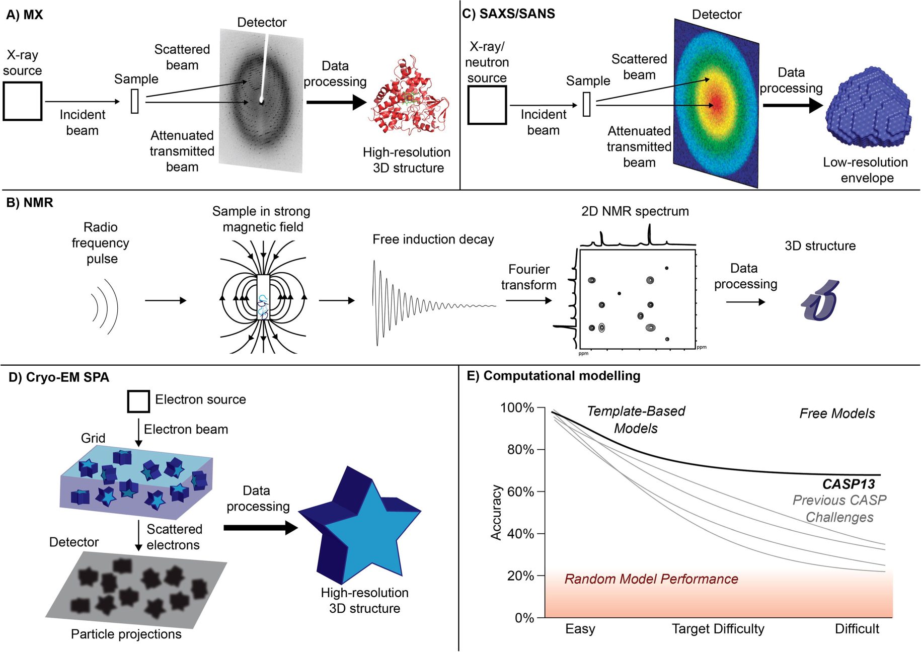
(a–e) One key area of small-angle scattering is the rigid body modeling of macromolecular complexes. State-of-the-art modeling tools can calculate rigid body models by fitting multiple SANS/SAXS diagrams simultaneously. Such a modeling approach involves linear and rotational motions of subunits and domains. Constraints can include symmetry, inter-residue contacts from mutagenesis or chemical shifts, or results from Fourier transform infrared spectroscopy, orientations of subunits from residual dipolar coupling from nuclear magnetic resonance. Reproduced from the work by Ziegler et al. (2021), Computational and Structural Biotechnology Journal, distributed under the terms of the Creative Commons CC-BY license. 87
In bioscience, colloids and macromolecular solution scattering, the co-analysis of X-ray and neutron scattering permits more information than available, alone, due to the differences contrast between the two techniques. A key example is the use of SAXS/SANS measurements for solute–solvent interactions, which are extremely important in bioscience and soft matter in general. These interactions have proven difficult to address with either technique. 88 For example, a NeX measurement on the D-22, X-ray facility at the ILL enabled the analysis of the role of cetyltrimethylammonium bromide in the growth of gold nanorods in situ. 4 The contrast differences between X-ray and neutron scattering removes the ambiguity in data interpretation through co-analysis, which was used in giving a solution to the morphology of PEGylated lipid mixed micelles. 48 Of note is the possibility of integrating these scattering techniques for samples in solution with cryogenic electron microscopy to provide accurate solution structures.
An example of the power of combined scattering techniques is seen in a recent study of the presence of co-existing domains of entangled and disentangled domains in molten, ultrahigh molecular weight polyethylene, 89 the existence of which leads to unusual time-dependent rheology. According to the theory of McLeish, 90 heating of the crystalline polymer below the melting temperature melts the chain ends of the crystalline domains but not the center of the domains, which nonetheless, lose the crystalline packing. When then raised well above the melting temperature, the centers melt, but cannot form a Gaussian chain due to constraints of the elastic Gaussian matrix, which then relaxes to the Gaussian coil equilibrium state, leading to the time-dependent rheology. Here, the sample is a co-crystallized mixture of deuterated high molecular weight polyethylene with protonated low molecular weight polyethylene. The use of SAXS, measuring the time-dependent electron density difference of the two states, and SANS of the entanglement of the deuterated and protonated polymers provided means to show the transition between crystalline and entangled states with different melting procedures and confirmed the theory of McLeish.
Structural changes in protein complexes induced by the interactions between proteins and nucleic acids have shown potential for pathogen binding and might find application in sensing and remediation.
A lesson learned from the recent pandemic is that the knowledge base in science and technology was inadequate to detect, isolate, and identify viral causes of infection and to track variants as the virus evolved. Information derived from protein, nucleic acid, and membrane interactions from genomics, proteomics, and lipidomics is essential for determining in vivo viral infection routes, the spread, and controls needed to mitigate further infections. While the large-scale facilities prioritized measurements germane to this work, access and flexibility are limiting factors. Smaller, more flexible sources that combine analysis by X-rays and neutrons can provide access to enhance and complement measurements at large-scale sources.
In biological membrane research, the contrast variation capabilities of neutron scattering add extra dimensions to the information obtained from X-ray scattering and the ability to match nuclear and electron density profiles to obtain lateral and transverse structural details. Another example is the effect of material properties and surface electrostatics of lipid membranes on the function of biological ion channels. An exciting new development in this area is in the SAS of membranes from whole cells, particularly for photosynthetic thylakoid membranes in algae and whole leaves in the study of responses to stress,91,92 and NeX with imaging provides a new dimension to these studies.
Finally, we stress the unique capabilities of combined X-ray and neutron scattering in probing the dynamics of molecular systems. In-situ measurements over wide ranges of timescales can provide invaluable mechanical and dynamical information on a wide range of biological and material systems. They can be correlated precisely with the detailed structural information obtained from SAS and diffraction.1,2,4,48,49,89,93–98
3.4 Quantum materials
The design, rational synthesis, and thorough characterization of novel quantum materials have made significant scientific and technological progress, essential for the development of advanced technologies. Generally, quantum materials can be described as solids with exotic physical properties arising from the quantum mechanical properties of their constituent electrons. 99
Scientific description of all materials originates from quantum mechanics, describing how electrons interact and atoms bond. There is an increasing interest in material systems where quantum effects can be seen over a broader range of energy and length scales. Examples include superconductors, graphene, topological insulators, Weyl semimetals, quantum spin liquids, and spin ice. 100 Scattering techniques play a central role in revealing the underlying physics of these materials. For example, resulting spin-orbit entangled wave functions cause magnetic interactions that are highly frustrated, even in simple spatial arrangements such as honeycomb lattices, e.g., stoichiometric quantum materials such as RuCl3, Li2IrO3, and Na2IrO3. Resonant X-ray scattering can directly measure the presence of such interactions. 100 Similarly, neutron scattering plays a core role, e.g., to reveal new bosonic quasiparticles at magnetic quantum-critical points. 100
Significant research efforts in quantum materials are related to experimental discovery and characterization, theoretical prediction and interpretation, and potential applications. Discovering novel quantum materials is only possible by implementing advanced experimental techniques, including resonant X-ray and polarized neutron scattering. Along with these techniques, sample environments, e.g., high-pressure cells, are essential tools to modify quantum materials by tuning interatomic distances and, therefore, better understanding the molecular interaction potentials. 101 XRD and neutron studies under ambient and extreme conditions can be essential to determine crystal structures and understand electron dynamics in quantum materials. An important part of such research is elucidating structure–properties relationships, which requires a comprehensive characterization of the quantum materials. Properties measurements include electrical transport measurements (electrical resistivity, Hall effect, electron hydrodynamics), thermal transport properties measurements (Seebeck coefficient, Nernst effect, thermal conductivity), etc.
Examples of quantum materials are superconductors, topological insulators, Dirac Semimetals, Weyl Semimetals, magnetic skyrmion materials, spin liquids, quantum sensors/dots, and those displaying the quantum Hall effect. 102 Potential applications for quantum materials include actively developing fields such as thermoelectric technology, catalysis, photovoltaics, hydrodynamics, and data storage. 103 Moreover, materials with novel exotic electronic properties could revolutionize existing technologies and promote the discovery of entirely new applications, enabling routes to realize state-of-the-art technologies, such as quantum computation. Undoubtedly, such advances will benefit the economy and initiate further research discoveries.
Analyzing the structure of domains is one of the most challenging areas. As the topology of quantum materials is extremely sensitive to crystal and magnetic symmetries, it is imperative to study them at different scales. For example, in systems that form magnetic skyrmions, how do crystal and magnetic structures interplay in a skyrmion, and how do magnetic domains interact? A recent review by Ratcliff et al. identified multiferroics, i.e., materials simultaneously magnetic and ferroelectric, as an essential class of materials requiring X-ray and neutron experiments X-ray and neutron experiments. 104 Using concurrent (polarized) neutrons for the magnetic and (resonant) X-rays for the structural domains allows us to understand the underlying domain physics. This argument is supported by recent experiments of Ueland et al., who studied the origin of electric field-induced magnetization in multiferroic HoMnO3, showing that concurrent X-ray and neutron experiments would be the key to deciphering the underlying domain physics. 105
Concurrent experiments mean benefitting from technique-specific advantages. X-rays are (generally) surface-sensitive, element-specific, and can explicitly probe the chemical and magnetic state. All these examples require energy-resolved experiments like X-ray absorption spectroscopy (XAS), X-ray magnetic circular dichroism (XMCD), and X-ray magnetic linear dichroism (XMLD), for which up-to-date synchrotron radiation is potent. Neutrons are highly penetrating and have an excellent sensitivity to magnetic ordering.
Resonant X-ray reflectivity (RXR) on magnetic films is highly surface-sensitive to energy. Concurrent experiments with more bulk-sensitive polarized neutron reflectometry (PNR) of the same state would be the key to understanding the RXR pattern. Figure 11 illustrates that it is very challenging to unambiguously determine the magnetic vs. structural depth profiles. 105 Determining the magnetization depth profile of thin films and multilayers by polarized neutrons concurrently with structure information from X-ray reflectometry would be precious. The key to answering scientific questions on these thin films and multilayers is to exclude technical questions, such as finding the same spot with the different probes, which is simple for concurrent but challenging for separate experiments. Concurrent experiments would be precious in magneto-ionics, e.g., memristors. In the case of electrical field-controlled magnetic properties, the ion migration can be tracked via PNR, and ion migration and correlated bonding can be tracked via RXR or XAS in fluorescence mode.
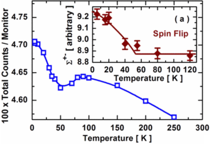
Temperature-dependent intensity and (inset) spin-flip scattering cross section from polarized SANS. Reprinted with permission from Ueland et al. Copyright 2010 American Physical Society. 105
Another area deemed to be essential is single-crystal diffraction. Concurrent techniques could enable the study of magnetic phase transitions, which involve a structural transition due to magnetoelastic coupling. The structural distortion is often very subtle and requires high-resolution synchrotron capabilities, while neutron diffraction can determine the magnetic order. The change of the order parameter could be measured even with a weak neutron source, such as a portable neutron generator that could be installed at a synchrotron. Concurrent experiments eliminate technical challenges that inhibit scientific understanding. Here, the identical thermometry of the sample would be the key to measuring the same transition temperatures and understanding how the (probably coupled) order parameters evolve with temperature, applied electric or magnetic field, and pressure.
Crystalline symmetry is commonly determined through X-ray and neutron diffraction refinement. However, the latter is more advanced when a material contains light elements and is magnetic. The importance of combining neutron and X-ray experiments can already be seen for single-crystal XRD refinement. For example, X-ray analysis of Sr3Ru2O7 shows a tetragonal structure with the I4/mmm space group.106–109 Neutron diffraction refinement on the same Sr3Ru2O7 always results in the orthorhombic structure belonging to the Pban 110 or Bbcb111–114 space group. The reason for this apparent discrepancy traces back to the scattering cross section. In this example, the low sensitivity of XRD to oxygen is the reason. 109 Concurrent experiments are essential because of material imperfections, sample stability over time, and technical imperfections of the instruments and sample environments.
Additional parameters can trigger events, like structural changes, when external manipulations occur. For example, a phase transition can occur if a sample is exposed to an external magnetic or electrical field, pressure, and temperature. From a scientific point of view, it is technically impossible to create precisely the same conditions twice. Hence, slight differences in phase transitions are to be expected. Therefore, concurrent experiments create a unique opportunity to exclude uncertainties.
Due to the neutron's magnetic moment, the neutron is sensitive to unpaired electron spins, thus providing information about material magnetism. Figure 12 displays the contour map of neutron diffraction intensity of EuMnSb2 in the (H K 0) plane and the intensity profile for the area indicated by the dashed rectangular box at 350 K (a–b), 150 K (c–d), and 7 K (e–f), respectively. At 350 K, along the (H −1 0) direction, only two peaks correspond to the crystal lattice. When the temperature is cooled below the magnetic transition temperature of Mn at 346 K, there is an additional peak at (0 −1 0) corresponding to the magnetic ordering of Mn (Figure 12c–d). Two more peaks at (1 −1 0) and (−1 −1 0) corresponding to the magnetic ordering of Eu were observed, further cooling the system below the magnetic transition temperature of Eu at 21 K (Figure 12e–f). Single-crystal neutron diffraction refinement indicates that the Mn sublattice forms the C-type antiferromagnetic structure with moments (4.5 ± 0.6) µB at 7 K pointing along the a-axis (Figure 12g). The Eu sublattice forms the canted A-type antiferromagnetic structure with moments (5.9 ± 0.8) µB at 7 K, lying in the ac plane but pointing (41 ± 1)° away from the a-axis (Figure 12g). Quantitative analysis indicates that the spin–spin correlation length, while anisotropic, has long-range characteristics in all directions for both the Eu and Mn sublattices. The detailed experiments require a high-flux neutron source. However, an in-house NeX setup would be invaluable in preparing and designing the experiments.
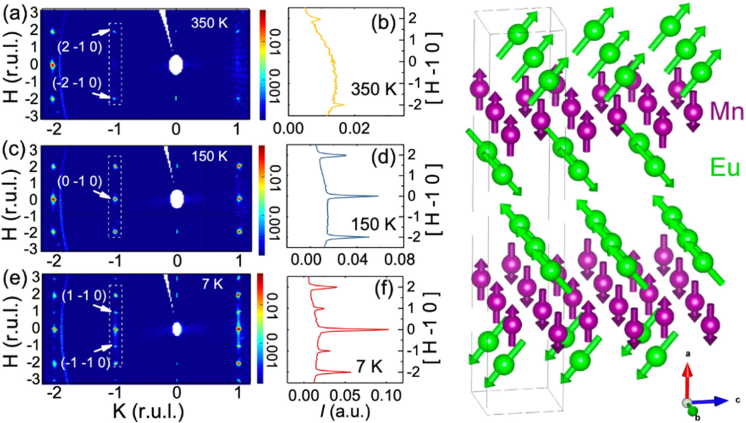
(a)–(f) Contour map of neutron intensity of EuMnSb2 in the (H K 0) plane and the intensity profile for the area indicated by the dashed rectangular box at 350 K (a–b), 150 K (c–d), and 7 K (e–f), respectively. (g) Magnetic structure of Eu sublattice (below 21 K) and Mn sublattice (below 346 K). Reprinted with permission from Gong et al., Copyright 2020 American Physical Society. 115
The materials research community pays close attention to imperfections such as inclusions/vacancies, grain boundaries, and crystal twinning. Figure 13 shows an Sr2(Ru0.99Ti0.01)O4 crystal imaged with grating interferometry, mirroring crystal imperfections. X-ray scattering experiments with a small beam spot can scan a sample and reveal material imperfections.
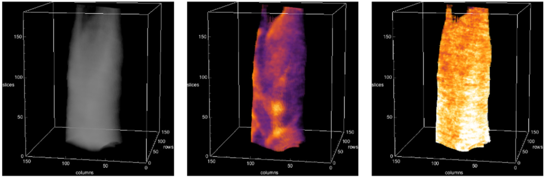
A Sr2(Ru0.99Ti0.01)O4 crystal in absorption (left), dark-field (middle), and differential phase contrast (right). Figure courtesy of Rongying Jin (University of South Carolina).
3.4.1 High-resolution XRD and neutron triple-axis spectroscopy (TAS)
Small changes in the lattice structure (like octahedral tilting in a film) measured by XRD can facilitate changes in the magnetic structure, for example, paramagnetic to antiferromagnetic, which is accessible by TAS. Both must be measured simultaneously to correlate the transition, which is crucial to separate coupled transitions, such as the Mott transition in the material V2O3. 116
3.4.2 Combining TAS with resonant inelastic X-ray spectroscopy (RIXS)
TAS is sensitive to energy exchange with the lattice, so TAS directional information with high Q-resolution, but especially for magnetic excitations, RIXS spectroscopy provides element-specific insights, allowing to determine the active elements and sublattices. Recently, paramagnons and high-temperature superconductivity in a model family of cuprates were studied. Cuprate-based superconductors are known to have the highest critical temperatures, Tc, at ambient temperature. Recent RIXS experiments point to the importance of magnetic fluctuations. 117 Again, avoiding imperfections with concurrent experiments, here TAS and RIXS, would enable a better understanding of paramagnons and the high Tc.
3.4.3 X-ray pump-probe
X-ray pump-probe-type measurements are several orders of magnitude faster than neutrons can achieve. A compact neutron source on a synchrotron beam line is not helpful. On the other hand, having an X-ray beam on a high-flux neutron instrument could be valuable for making simultaneous measurements following the time dependence of magnetic and structural order development as a material evolves below an ordering temperature. Synchrotron measurements are not feasible at low temperatures due to the heating caused by the synchrotron radiation or synchrotron radiation-induced damage. Measuring the time dependence when a periodic perturbation is applied is another area where having simultaneous neutron and X-ray capabilities could provide essential data.
3.5 Chemistry
3.5.1 Electrochemical energy devices
Electrochemical energy storage and conversion devices serve a diverse role as sources of carbon-free electrical power. Lithium-ion batteries are ubiquitous in small mobile electronics and increasingly power light-duty vehicles such as passenger cars and trucks. 118 Hydrogen fuel cells are an emerging platform for heavy-duty vehicles, providing nonpolluting power for semi-tractor-trailer transport. 119 Polymer-based electrolyzers can provide valuable hydrogen using excess electrical power from wind and solar installations. 119 In contrast, flow cell batteries supply power reserves during periods of reduced power generation, for example, at night when co-located with a solar power facility. 120
These systems also share many similarities regarding their essential operation and structural characteristics, see Figure 14. They comprise two porous electrodes, an electrolyte where the conductive ion can migrate, and a membrane separator that electrically isolates the electrodes but permits the ion to pass. They can all be classified as hierarchical structures, as the pore sizes range from the sub-nanometer to 100 µm, electrode particle sizes range from a few nanometers to a few micrometers, electrode thicknesses range from ∼100 nm to 100 µm, separators that are 10's of micrometers thick, and in-plane dimensions from the 10 s of mm to 10 s of cm. Furthermore, the materials used in these devices have a broad range of compositions, from precious metals to transition elements and low atomic weight atoms. As such, observing these devices in-operando rarely, if ever, provides a complete picture of all the phenomena involved. Simultaneously, multimodal characterization techniques are thus essential to develop, as the joint analysis provides significantly more insight than the independent input from each mode.
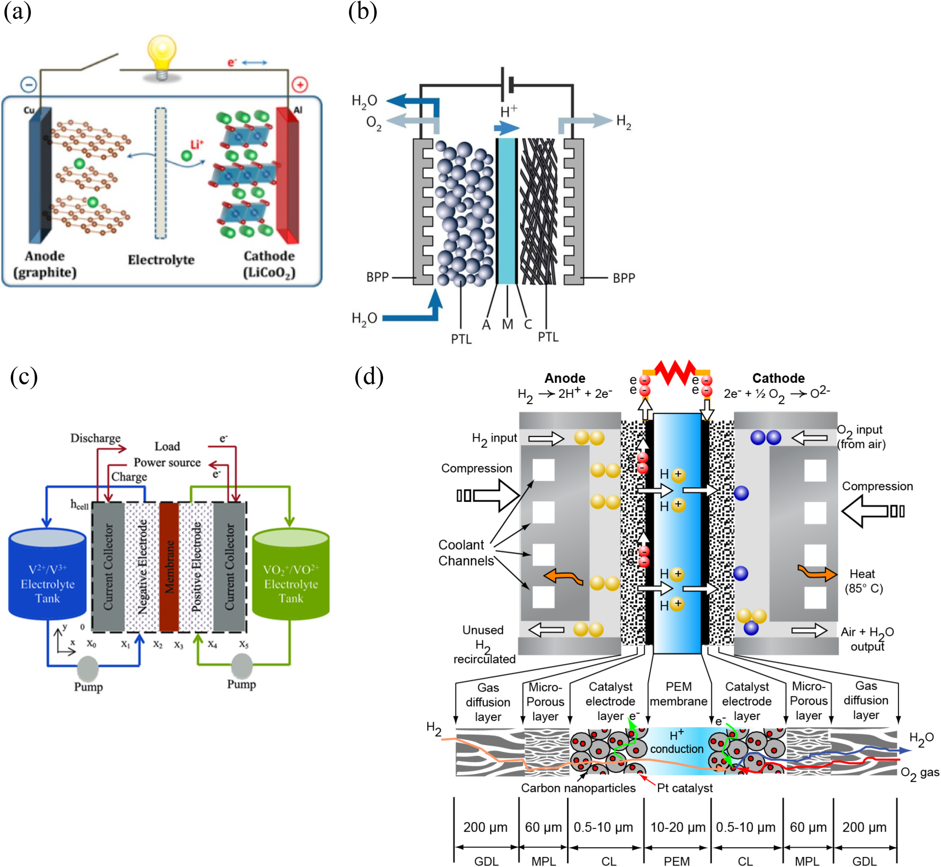
Schematic diagrams of four different electrochemical energy storage and conversion devices. (a) Rechargeable lithium-ion battery. Reprinted with permission from Goodenough et al., Copyright 2013 American Chemical Society. 121 (b) Low-temperature proton exchange membrane (PEM), water electrolyzer. Reprinted from Haas et al., Copyright (2021), under the terms of the Creative Commons Attribution License (CC BY). 122 (c) Flow batteries. Reprinted with permission from Lu et al., Copyright 2020 John Wiley and Sons. 123 (d) PEM Fuel Cell. Adapted with permission from Arif et al., Copyright 2009 Springer. 124
The non-destructive, penetrating radiation of thermal neutrons (energy ≈<25 meV) and hard X-rays (energy ≈>30 keV) are well suited to probe electrochemical energy storage systems in a simultaneous acquisition mode, sometimes referred to as “NeXT.”5,6
A simplifying view is that the X-ray mode can resolve the high-Z structural components with heavier elements. In contrast, the neutron mode observes the electrochemistry via electrolyte distribution and mobile ions in the case of lithium batteries and proton exchange membrane (PEM) systems. The length and time scales are determined mainly by the X-ray and neutron sources used in the experiment. Knowing that the flux of conventional rotating anodes is comparable to that of neutron instruments, a natural choice is a laboratory X-ray instrument at a neutron reactor if space permits such an installation. However, as addressed earlier in this report (see Section 2.1), multicomponent systems require both isotopic labeling, polarization analysis, and X-ray energy variation for a complete sample picture. Such considerations position scientific requirements above technical challenges in concurrent experiments and would enable targeting different aspects of an electrochemical energy storage and conversion device.
3.5.2 Proton exchange membrane fuel cells (PEMFCs)
PEMFCs are being targeted as engines for heavy-duty transportation applications, such as tractor-trailers and auxiliary power units for shipping vessels. It is believed that further development of the technology, cost, durability, and performance, together with the availability of hydrogen refueling infrastructure, will enable PEMFCs to drive light-duty passenger vehicles. The advantages of PEMFCs for transportation include clean operation, as the byproducts include only water, heat, and electricity, and refueling times are similar to gasoline engines. It should be noted that while the discussion below will focus on PEM, many similar investigations into water, electrolyte, and proton transport can be conducted using other fuel cell architectures, including alkaline exchange membrane fuel cells, 125 phosphoric acid fuel cells,126–128 etc.
As shown in Figure 14, PEMFCs comprise four primary components: flow fields, gas diffusion layer (GDL), catalyst layers, and the PEM. Hence, PEMFCs are an excellent example of the need for multiprobe techniques, as explained in Section 2.1. The chemical composition and operation principle show that neutrons and X-rays are required to characterize the sample. For example, the most common PEM is Nafion, a fluoropolymer containing hydrophilic sulfonic acid side chains. When the PEM absorbs water, the proton conduction increases significantly, and thus, ensuring that the PEM is adequately hydrated during PEMFC operation is essential. The protons are conducted through the PEM due to the 1.2 V potential to form water. The anode (hydrogen side) and cathode (oxygen side) catalyst layers are typically composed of carbon support for electrical conductivity; the carbon is “decorated” with catalyst nanoparticles either of pure platinum or a platinum alloy, interspersed with ionomer (typically a Nafion precursor) to enable proton transport from the membrane to the catalyst particle. The anode reaction, H2 → 2H+ + 2e−, is faster than the cathode reaction, O2 + 4H+ + 4e− → 2H2O, and thus requires a lower areal density of catalyst particles. The membrane and catalyst layers are often called the membrane electrode assembly (MEA). The GDL is a carbon fiber cloth or paper that provides mechanical support to the MEA, facilitates homogenizing of the reactant density over the face of the MEA, and promotes passive product water removal. The GDL often contains a microporous layer (MPL) with a high content of polytetrafluorethylene (PTFE) with pore sizes 0.1 µm to a few micrometers, which is in contact with MEA. The larger pores are in contact with the gas flow fields. The flow fields introduce humidified hydrogen (anode) and air (cathode) and remove the product water using the bystander nitrogen from the air or through passive wicking means with channels with cross-sectional dimensions of order millimeter and length of order 20 cm. The flow fields typically have cooling channels on the opposite side to maintain a temperature of 90°C.
We briefly review some in-operando neutron imaging studies of water transport in PEMFCs. The first neutron imaging of a fuel cell measured the water content, which took one day. 129 With this focus, conducting studies at lower-power research reactors, such as the 1 MW Triga Penn State Breazeale Reactor, was feasible. 130 The water content can be estimated from neutron absorption using a simple neutron generator, showing the importance of simple portable neutron generators for concurrent experiments. To obtain some indication about which side of the fuel cell the water in channels was in, General Motors researchers arranged the channels such that the anode side was predominantly vertical. At the same time, the cathode was predominantly horizontal, and significant back diffusion was observed in the anode field. 131 High-speed optical cameras complemented such flow field studies to observe droplet dynamics, which could also be used simultaneously with neutron imaging. 132 These experiments enabled an understanding of the impact of channel cross-sectional geometry and surface treatments on water retention. 133 Work by Hickner et al. showed that the water retention in the fuel cell decreased with increasing current, which indicated that one could not assume a uniform temperature in the fuel cell. Meanwhile, neutron imaging has been established as an essential tool for understanding water transport effects, membrane degradation due to excessive drying, and studying the catalyst layer.134–139
SAS of neutrons and X-rays has been used to understand various aspects of the PEMFC, including in-operando SANS measurements. SAS techniques have given considerable insight into the structure of the dispersion micelles relevant to fuel cells.140–142 More recently, there has been a resurgence in the use of SANS in the role of solvent type in the self-assembly of dispersion micelles and its evolution during the casting procedure, and how this process relates to the structure–property performance of the PEM. The multicomponent nature of PEMFCs complicates the analysis, so a definitive microstructure model is yet to be attainable.143,144
A variety of synchrotron-based X-ray methods provide valuable insight into PEMFC function. Grazing incidence SAXS has been used to measure morphology changes of the thin ionomer film when it is in contact with the carbon support or catalyst particles and to try to correlate these changes with oxygen permeability. 145 SAXS is widely used to investigate changes to catalyst particles induced by aging studies, and XAS yields binding energy and geometry information. 145 STXM or soft X-ray ptychography can provide detailed structural information with tiny samples. 146 Using laboratory-based X-ray tomography systems with about 100 nm resolution, ex-situ studies of catalyst layer degradation have been carried out, and with a coarser resolution of about 1 µm, it is possible to study compression effects on the MPL of the gas diffusion medium.147,148 Tomography at an X-ray synchrotron beamline with a test section that transmits the low-energy X-rays can reveal changes on the 1 s timescale. However, due to the X-ray beam's high intensity and fluorine's K-edge absorption, one must understand how the X-ray beam degrades fuel cell performance. 149 With this consideration, it is possible to image water dynamics in the gas diffusion medium, especially the MPL, under various conditions, including freeze-thaw cycling. 150
The new research using SANS data has been impactful: Developing new solvent dispersion systems for Nafion improved MEA durability beyond benchmark requirements for commercializing automobile applications.10,151–156 Lent support for improved durability through more excellent MEA toughness by increased polymer entanglements, which was shown to lead to more excellent resistance to stress-related damage from voltage cycling, as opposed to crystallinity or loss of electro-catalyst surface area.152,155,157,158 Showed that the formation of homogeneous, reversible gels during evaporation was essential in the formation of entanglements and that there is an inverse correlation between critical gelation concentration and tensile toughness.155,157 Kim et al. demonstrated for the first time that critical gelation followed a distinct series of diols with different chain lengths.
155
The recent work reveals a roadmap for improvements in fuel cell properties and performance through SANS measurements of dispersion morphology. Its evolution is driven by the amphiphilic interactions with different solvents in the solution casting of the MEA.10,155,156,159,160
3.5.2.1
Furthermore, SSC PFSA demonstrates improved oxygen reduction reaction (ORR) hysteresis over that of the LSC ionomer, Nafion.163,165 However, the electrode performance using those ionomers has not been fully optimized in fuel cell electrodes, thus providing impetus to study the solvent dispersion morphology and their solvent cast evolution to films of these ionomers and relating these results to the MEA properties in the same manner as done for Nafion.10,141,155,156,166 While the dispersion morphology of Nafion has been studied extensively, that of SSC ionomers has yet to be.10,140,141,155,156,167 Past and work in progress using SANS shows that the dispersion particle morphologies of SSCs are significantly different from LSC, thus likely affecting the morphology of the MEA.167,168
Refinement of solvent–PFSA interaction parameters and the relationships of SAS measurements of dispersion structure and solvent cast process evolution, SAS with property performance measures to provide engineering rules improved PEM fuel cells.
3.5.2.2
Prospects for combined compact SAS X-ray and neutron sources
The use of neutrons in this work seems optional, as X-ray contrast among the solvents and ionomers is also sufficient to obtain good SAXS data. 98 However, the difference in the origin of the contrast between X-rays and neutrons removes much of the analysis's ambiguity through data co-fitting. The scatter from the dispersions is isotropic, so a neutron line source from Soller collimation with appropriate data correction is feasible. Indeed, the availability of a lab-based source would improve the cycle of dispersion structure measurement and the relationship between properties and performance, as it would shortcut the usual time needed to access a suitable instrument for the scattering measurements—reflectometry and ionomer–catalyst interactions. 169 Interfacial structure at the catalyst–electrolyte interface has long been an essential topic for academic and applied research, as the performance and durability of electrochemical devices significantly rely on the interfacial phenomena.125,170,171 Two sets of recent studies have demonstrated the power of neutron reflectivity (NR) in providing essential information on the interactions of electrolytes with the catalyst surface and its relationship with fuel cell performance.125,169
PFSA ionomers have been included in the electrode catalyst layers to ensure efficient transport pathways from the membrane to the catalyst, significantly reducing the required catalyst loading to achieve high performance. 172 Within the catalyst layer, ionomers form a thin film, which improves ion transport to catalyst sites and plays an impactful role that needs to be fully understood. NR studies have highlighted the complex interface morphology of Nafion. Nafion forms a multilamellar structure with water due to its amphiphilic character, with the charged sulfonate groups in the water layers, as expected.173–175 However, multilayers did not form with Nafion films on Ag or Pt. 173 Such effects can lead to anisotropic ion transport. The amphiphilic character of Nafion in the organization of its layered structure next to Pt, PtO, and glassy carbon was demonstrated in the field of ex-situ NR measurements. 169
In the absence of voltage potential or a low voltage, the catalyst is in the reduced, metallic, hydrophobic state; likewise, glassy carbon is hydrophobic. At higher potential, PtO is formed and is hydrophilic. When Nafion was deposited against Pt, the NR of the sample in a saturated D2O environment revealed a thin hydrophobic layer next to the Pt surface. The electrochemical introduction of PtO in the same sample caused a significant rearrangement of the Nafion hydrophobic and hydrophilic fluorometer sulfonate side chains so that the hydrophilic and hydrophobic domains’ thickness was the same. Films deposited on glassy carbon showed a three-layered Nafion structure. These results gave important insight into the changes that could occur in the ionomer structure near the catalyst–carbon support interface, as the longer-range ionomer structure depends on the interface's properties. Interactions between the catalyst and the electrolyte are paramount for electrochemical device performance. Recently, the cation–hydroxide–water co-adsorption on a Pt surface was addressed using NR to understand the performance degradation of the alkaline membrane fuel cells (AMFCs). 125 The catalyst–alkaline electrolyte interface is unique in that the cation–hydroxide–water can adsorb on the hydrogen oxidation catalyst 176 as opposed to the adsorption of anionic species on the oxygen reduction catalysts in PEMFCs. 177 This attribute makes the AMFCs accessible from anion species poisoning at the cathode, but they suffer from the adverse impact through a cumulative cation–hydroxide–water co-adsorbed layer.133,134 Using a specially designed electrochemical cell to apply voltage potential concurrent with NR, the measurements verified that there was an accumulation of tetramethylammonium deudroxide (TMAOD)–D2O coadsorption that increased in thickness over a time scale similar to the concurrent drop of current density of Pt hydrogen oxidation reaction (HOR). The loss of HOR current is due to the blockage of hydrogen and water (at the anode, due to increased hydrophobicity) on the catalytic surface. 125
3.5.2.3
Current work has demonstrated that the amphiphilic character of the LSC ionomer, Nafion, leads to a layered structure with organization determined by the hydrophilic/hydrophobic character of the ionomer interface with carbon, silica, or catalyst. This affects the water and proton transport channels, implying that altering the decal substrate used in solvent casting could be used in engineering MEAs with better transport properties. This effect leads to the reorganization of the channels through the catalyst oxidation state during the voltage cycle, implying that work must be done in the MEA ORR. Reduction of this work would thus improve the efficiency of PEMFCs. The demonstration of TMAOD–D2O growth at the Pt interface with a concurrent loss of HOR current showed the importance of NR with an NR-capable electrochemical cell to directly show the structure–properties performance of high-performance AMFCs. It illustrated the importance of choosing other AMFC electrolytes to reduce catalytic surface passivation, for example, substituting a bulkier electrolyte, such as tetraethylammonium ion (TEA+), for TMA+. Tetraethylammonium hydroxide is less able to pack at the interface.
3.5.2.4 NR shows that the structure of the ionomer is dependent on the hydrophilic–hydrophobic characteristic of a nearby interface. There is a voltage-dependent rearrangement of Nafion with Pt oxidation. An electrochemical cell has been demonstrated to meet the stringent optical requirements of NR while providing a platform for concurrent electrochemical environments and electrolyte responses. Thus, it is now feasible to study the response of polymer and small molecule electrolytes next to a catalyst interface to voltage cycling, such as the structural bases for ORR hysteresis. These measurements also raise the possibility of pulsed probe and stroboscopic measurements, such as those done on polyvinyl ferrocene films.178,179 NR will also find essential applications with other fuel cell materials, such as high-temperature proton electrolyte membrane fuel cells (HT-PEMFCs), where the high conductivity of phosphate protons is the basis for new technologies for high-energy conversion devices.
180
Prospects for reflectometry at compact sources: neutron specular reflectivity measurements may also be feasible, as this technique uses a line source. This prospect requires further study. X-ray reflectometry is a well-established technique for layered samples in air. Problems arise when the samples need to be in an environment chamber for hydration of fluid interface studies due to the path length in the media. However, the demonstrated success of this geometry in soft X-ray reflectometry suggests that tiny sample cells could be designed to enable reflectometry measurements with lab-based hard X-rays.
181
Neutron spectroscopy on fuel cell polymer electrolytes and model systems. Neutron backscattering (BSNS) and chopper neutron spectroscopy (CNS) have been used to characterize localized water diffusion in the Nafion polymer electrolyte membranes (PEMs). The CNS showed spectra characteristic of Hall-Ross jump diffusion with a jump time of 3 ps.182–187 BSNS showed proton diffusion over longer time scales due to confined and free diffusion.184,186,188,189 BSNS and CNS of the HT-PEMFC polymer electrolyte Polybenzimidazole PEM doped with phosphoric acid showed heterogeneity of proton transport time and length scales by spectra analysis as stretched exponentials due entirely to diffusion under confinement by “fractal” structures.
126
3.5.2.5
3.5.3
An example of the need for this broad range of characterization tools is shown in Figure 15 by the collapse of the cathode catalyst layer subjected to accelerated stress tests to simulate driving conditions. Previous neutron imaging work showed that the corroded catalyst layer increases the overpotential, resulting in more significant thermal gradients and, thus, enhanced membrane dry-out, which can also lead to membrane failure. 190 This unexpected consequence highlights the complexity of realizing robust, high-performing, cost-efficient materials and systems for PEMFCs.
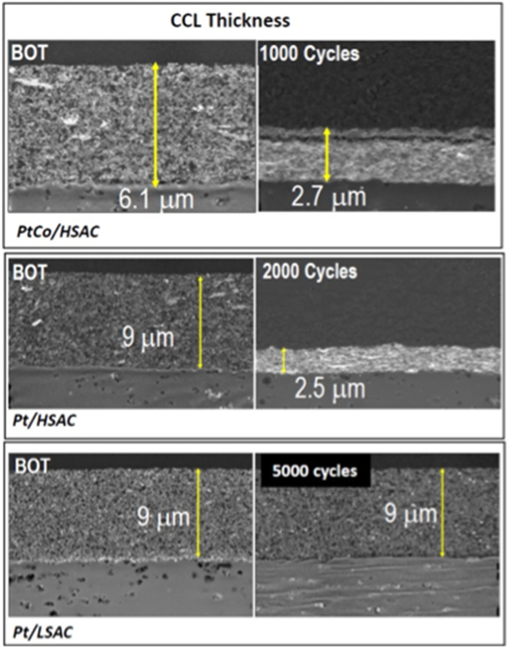
One example of catalyst layer degradation, the loss of porosity and the carbon support limits the operating life of the PEMFC, and the degradation rate sensitively depends on the type of catalyst and the morphology of the carbon support. Figure from Borup et al., “FC-PAD: Fuel Cell Performance and Durability Consortium,” 2018 DOE Hydrogen and Fuel Cells Program Annual Merit Review, Washington, DC, 2018. 191
Figure 15 explains the need for concurrent in-operando experiments on PEMFCs. For example, materials have a hierarchical organization over several orders of magnitude in length scale. While the ability to reach ∼1 µm resolution provides coarse information on the through-plane water gradient, observing the ionomer in the secondary pore space requires a resolution of 100 nm or better. At around 10 nm, changes to the carbon support can be observed. In-operando experiments at these length scales are the domain of SAS experiments. Higher energies provided by synchrotron or inverse Compton effect imaging would be essential to avoid damage to the fluorine components, such as the ionomer in the catalyst layer. More conventional X-ray and neutron imaging systems would still yield valuable insight. However, the potential for the needed 10 nm spatial resolution would no longer be feasible, and the time resolution on the neutron mode would require reliance on sparse angular tomography reconstruction algorithms. With the flux available from a compact neutron source, obtaining the water distribution might be possible.
3.5.4 Corrosion
Corrosion of metals significantly impacts the economy, infrastructure, and conservation of resources. A recent National Association of Corrosion Engineers study estimated the global cost of metallic corrosion to be approximately $2.5 trillion annually. 192 This estimate considers direct and indirect costs associated with materials, labor, equipment, and loss of revenue/productivity. Metallic corrosion also impacts safety and the environment. For example, steel and other metal alloys commonly used for constructing buildings, bridges, pipelines, vehicles, medical implants, and chemical waste containers can develop pits and cracks due to corrosion that can compromise material integrity. Therefore, if corrosion is not controlled, it can lead to significant problems, including medical complications, catastrophic consequences, and environmental contamination.
Corrosion is a natural process where a metal returns to its natural state, and energy is lost. The corrosion rate depends on the metal part's elemental composition, microstructure, and surrounding environment. Corrosion can occur in various forms; however, localized corrosion tends to be particularly problematic as it can occur rapidly and go undetected until the part fails. 193
Although passive metals, such as steel and aluminum alloys, form protective oxide films, the film's integrity can break down in small local areas. This localized corrosion can enable pits of various depths and geometries to form and propagate within the metal. Intergranular corrosion and exfoliation can cause metal components to be removed or flake away. These and other forms of localized corrosion, which can be challenging to detect early on, can cause the metal part to develop cracks and other flaws that lead to premature failure.
As additive manufacturing (AM) of metals shows more potential as an alternative to traditional casting and forging methods for forming metal parts, some unique corrosion concerns have developed due to the microstructural properties of additively manufactured metals. 194 The porosity, roughness, inclusions, and other surface flaws resulting from AM processes can initiate localized corrosion. In addition, anisotropy due to the build direction of AM parts can result in variation in the corrosion behavior of the surfaces parallel and perpendicular to the build direction.
Electrochemical techniques, including impedance spectroscopy, linear polarization, and cyclic potentiodynamic polarization measurements, are valuable for studying the electrochemical characteristics of localized corrosion. 195 The onset and extent of passivation/passivation, pitting initiation and cessation, and corrosion rate can be assessed electrochemically to understand a metal's corrosion behavior and resistance. Although the corrosion behavior of numerous metals is well understood, substantial knowledge gaps need to be filled regarding the corrosion characteristics of additive-manufactured metals and new metal alloys.
Although electrochemical studies provide necessary information regarding corrosion kinetics, the techniques are generally destructive. X-ray and neutron imaging are methods that can not only complement electrochemical corrosion characterization but also enable non-destructive in-situ monitoring. In several recent studies, synchrotron-based X-ray tomography was used for nuclear material characterization, e.g., to reveal early corrosion stages of uranium corrosion within nuclear waste containers.196–198 Figure 16 shows the different stages of the uranium rod following corrosion cycles. As internal corrosion is common in oil and gas pipelines, X-ray tomography and related techniques could facilitate non-destructive in-situ studies in these areas.

The upper halves of the images show a uranium rod cycled 5 times in a corrosive environment resulting in corrosion products shown in the bottom part. Samples A, B, and C are various sections of the uranium rod Reprinted with permission from Paraskevoulakos. Copyright 2019 Elsevier. 196
3.6 Biogeosciences and environmental science
Biogeosciences include soil and rhizosphere processes, plant root imaging in soil, soil chemistry, hydrology, flow, and transport processes.
3.6.1 Geoscience
Recently, the importance of the nanoscale through mesoscale structure of geo-materials has been recognized as a fundamental understanding of materials’ mechanical properties and fluid interactions, critical in a variety of energy-related areas, e.g., nuclear waste management, enhanced oil and gas production, geothermal energy production, CO2 sequestration, and forensics for nuclear non-proliferation.199–205
Porosity is inherently difficult to study since pore sizes can range from nm to mm, which implies the challenge of analyzing broad pore size distributions. Nanopores have been analyzed by transmission electron microscopy (TEM), SANS and USANS, SAXS and USAXS, and microscopy—complemented with other technology, like Brunnauer, Emmet, and Teller (BET) adsorption diagrams (Figure 17). 202 Neutron scattering was essential to measure the transition from the nanoscale to the mesoscale.206–208 Many authors used these techniques, particularly for enhanced oil recovery.209–216
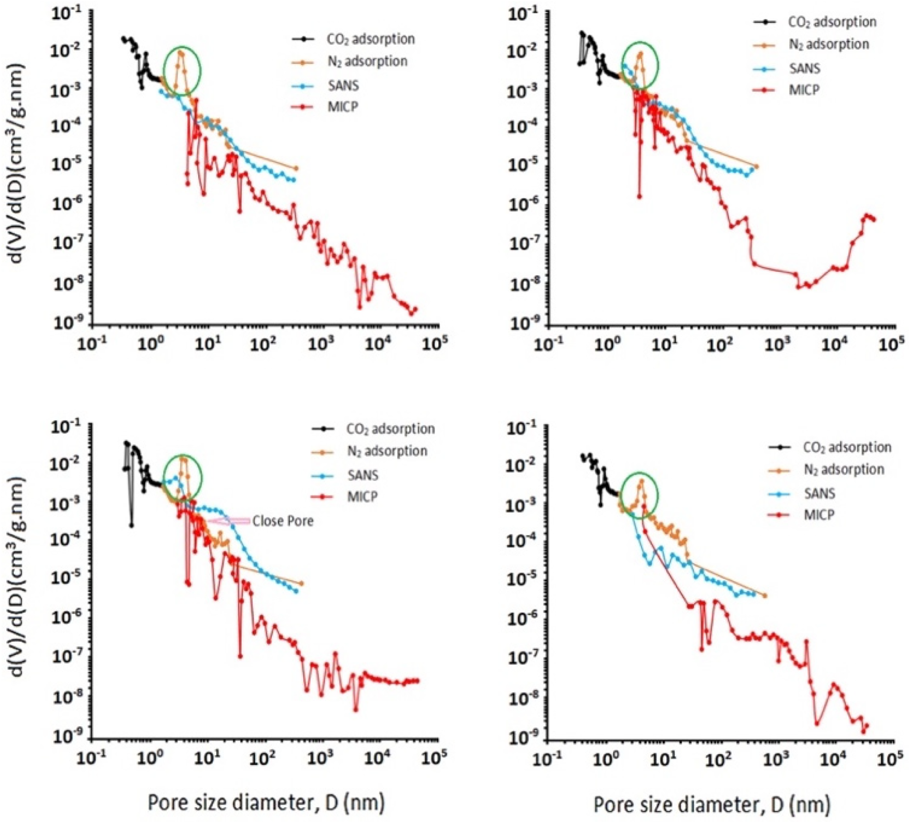
Distribution of pore volume with pore sizes obtained from gas (CO2 and N2) adsorption, mercury injection capillary pressure, and SANS results for four shale samples. The peak at approximately 4 nm for the N2 adsorption data is probably due to an artifact caused by the tensile strength effect. Reprinted with permission from Hosseini et al., Copyright 2021 Elsevier. 217
The high penetrability of neutrons in SANS and USANS measurements is extremely important in providing access to robust sample environments, such as pressure cells, to study supercritical CO2 and enhanced oil recovery under environments emulating field conditions.209–211
Just as significant is the contrast variation/-matching methods using deuterated/protonated mixtures of fluids to determine pore accessibility as a complement to other porosimetry techniques.213,218 The scattering techniques have a distinct advantage over other porosimetry methods, as any fluid of interest (gas or liquid) can be used. 219 Neutron/X-ray scattering has been recently established to characterize porosity from nanoscale to mesoscale.220,221 Since these first demonstrations, significant literature has used these techniques, particularly in enhanced oil recovery.220,222
A point to remember for scattering studies is the inherent anisotropy of geo-materials due to the formation under high pressure and temperature.223,224 Such considerations help drive instrument design, as two-dimensional detectors are the best and easiest means of assessing these characteristics without using special techniques for line sources that are difficult to implement.
One aspect driving compact neutron sources in geoscience is the relatively strong scattering of these materials from the pores and voids, and the considerable, easily detectable reduction of intensity when these features are filled with imbibing fluid. Furthermore, the ability to spend far more time on measurement than available at national facilities is an essential factor. An example is emerging work in low-temperature geological reactions, typically performed at 200°C down to ambient temperature, which are typically slow. Thus, they are inherently difficult to study as most advanced in-situ characterization techniques at user facilities, such as X-ray synchrotrons or neutron facilities, give out beamtime in chunks of 2–3 days. As visible in Figure 18, even after 22 days of reaction time, a rock core of 1.6 cm is not wholly reacted through. 225
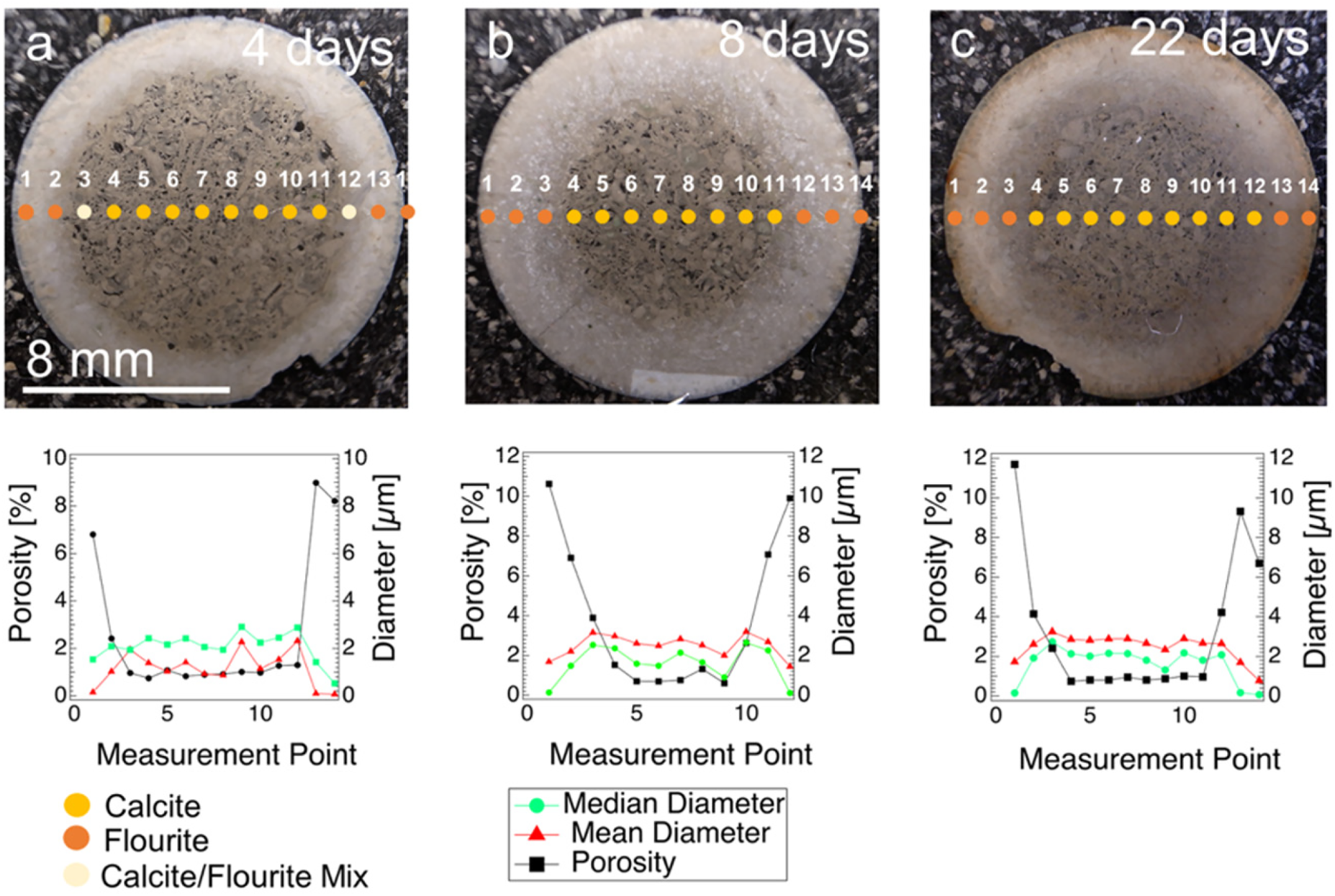
Example of using USAXS for the analysis of porosity changes during replacement reactions. Pictured limestones were reacted with ammonium fluoride, showing increased reaction time: (a) four days reaction time, (b) 8 days reaction time and (c) 22 days reaction time. Diagrams show porosity (%), mean and median diameter (μm) for each (U)SAXS measurement point collected in line scans indicated in images (a)–(c). Wide-angle X-ray scattering was used to identify the phases, and the numbers indicate the positions on the samples. On the outside, it shows fluorite; in the middle, it shows calcite. Between these layers, there is a transition phase consisting of a calcite/fluorite mixture. Reprinted with permission from ACS Earth and Space Chemistry. Copyright 2019 American Chemical Society. 225
A common practice to circumvent this problem is to conduct a time series of experiments, in which parallel experiments are stopped at different times to obtain information about temporal changes in chemical composition or microstructure, e.g., porosity, grain boundary width, and grain sizes.226–229 However, even when using the same starting material, microstructural differences between samples and heterogeneity of natural rocks will always exist. These factors complicate the interpretation of the results.
X-ray sources have essential characteristics that result in emergent methods due to the relatively small beam size and large flux relative to neutrons. Geo-materials are inherently heterogeneous, requiring multiprobe techniques as described in Section 3.1. For example, X-rays can be used to scan the sample grains. The relatively large flux of X-ray sources allows for time-resolved studies of fluid uptake in various geochemical and extraction processes. In many cases, the X-ray scattering length density of imbibed fluids relative to the rock grains with different mineral compositions is sufficient to detect the presence of filled pores. Furthermore, using carbon vapor-deposited diamond windows, a pressure cell can operate over an interesting pressure domain with suitable optical properties that can be designed for X-ray analysis.
The following emergent science areas are examples of areas that will be addressed by concurrent neutrons and X-rays: In-situ chemical and biologically enhanced reactions in mineral extractions and CO2 sequestration. In particular, in-situ concurrent NeX measurements at midscale, compact instruments enable measurements that are not feasible at large-scale facilities due to the mismatch of reaction times and available beam time. Methane sequestration and retention effects due to void and pore size distributions, structure, and interconnections. Enhanced oil recovery methods and the relationship to pore and void structures. Advanced forensics in detecting clandestine nuclear events as part of nuclear non-proliferation. Fluid flow and accessibility of pores and voids in the emergent areas given above, and the relationships to geological structure and properties that impact fluid extraction, insertion, and release that are active over the meter-to-kilometer length scales. The interrelationships of geo-materials' mechanical/viscoelastic properties to nano to mesoscale pore and void structure for predictive models for engineering, seismic data analysis, forecasts, and other applications.
The interrelationships of geomaterials’ mechanical/viscoelastic properties to nano to mesoscale pore and void structure are needed for predictive models for engineering and other applications. Another area of geoscience that could benefit from a combined X-ray/neutron characterization is the geo-mechanical investigation of geomaterials. X-ray tomography has become a reference technique for non-invasive study of micro-mechanical processes in the last decade. 230 Simultaneous vectorial 4D image acquisition (3D+ process + X-rays/neutrons) was used to characterize the water adsorption dynamics in claystone. 231 This allowed us to compare material deformation and water arrival and improved mineral identification by combining neutrons and X-rays (Figure 19).
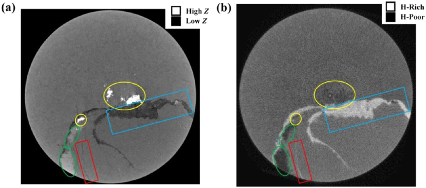
Horizontal slices from (a) X-ray and (b) neutron tomography of a Middle East shale. Regions with higher organic content, indicated by the rectangular boxes, are clearly visible in the neutron images but appear transparent in the X-rays. The ovals highlight areas composed of high-Z minerals (likely pyrite (FeS), with the two upper-right ovals containing anhydrite) which are more prominent in the X-rays. Reprinted with permission from Chiang et al. Copyright 2017 Society of Core Analysts. 232
3.6.2 Meteorites
Scientists are increasingly using a combination of X-ray and neutron imaging techniques to study meteorites, a field ripe for exploration with upcoming asteroid sample return missions like Hayabusa-II and OSIRIS-Rex. This approach offers valuable insights into the early solar system. Pinpointing the location and abundance of hydrogen-containing materials in meteorites can help us understand past processes and identify signs of alteration. While this combined tomography technique is still under development, some initial studies have been conducted on meteorites like the Allende meteorite, a tektite, and the Sutter's Mill chondrite, using either neutron computed tomography alone or the combined X-ray and neutron approach. 233 More recent papers show a combined analysis of meteorite samples. X-ray and neutron tomography can identify non-destructively water-bearing and chlorine-rich materials inside meteorites. 234
3.6.3 Soils
Soil is a multicomponent material with mineral phases, which contain heavier elements and organics (soil organic matter, roots, microbes, biofilms, etc.) and pore space where air and water are held—the latter are built up of lighter elements. The neutron cross section is larger for hydrogen, and the X-ray cross section is larger for heavier elements. Thus, heavier elements are ideal for X-rays, while neutrons are crucial to observe hydrogen.
X-rays are routinely used for XRD 235 and spectroscopy of heavier elements in mineral phases and soft X-rays for light elements, 236 for example, in biogeochemical processes237,238 and imaging of soil and its porous structure and mechanics. 239 Water in soils has been imaged with a microfocus X-ray tube computerized tomography. 240 In contrast, neutrons can image carbon, water, and plant tissue, for example, imaging plant roots in soil. 241 For X-rays, it is generally challenging to distinguish soil pore water from air within the pores, and one can easily distinguish water from air using neutron imaging. A combined X-ray/neutron imaging experiment would see all soil components from the soil matrix with minerals, pore space (filled with air) with X-rays, and pore water, plant roots, and organic matter with neutrons.241,242 Soil hydrology and mapping flow/transport paths within the soil's pore network, following the water, is possible by adding neutron imaging. 243 Water movement in soils is critical for transporting carbon and other nutrients, contaminants, microbial respiration, and carbon sequestration, including climate effects. There is also interest in having some soil samples in a frozen state. A cooling jacket/container would be difficult to penetrate with X-rays, but that would not be a problem for neutrons. Another area of concern in soil research is the formation, distribution, and transport of nanoplastic particles on environmental impact, 244 where NeX would be extremely useful in teasing out scattering contributions from soil constituents, voids, and the nanoparticles. Currently, work using USANS and SANS is only in its initial stages. 245
The ability to perform concurrent X-ray and neutron imaging and spectroscopy using the same instrument is critical to inspect all soil components and gain real-time information on soil processes. In addition, performing such experiments at the home laboratory would allow for long-term access for experiments that last for weeks, experiments that require frequent return of the sample to the instrument, and circumvent potential issues regarding shipping sensitive or hazardous samples.
3.6.4 Plants
X-ray imaging experiments reveal the general morphology of plants. Due to water's good visibility, neutron imaging is an excellent tool to visualize water relocation in plants, including roots and stems.241,243,246,247 Real-time concurrent experiments are essential in living systems in which water flows in channels that may have independent motion. 243 Such experiments are critically important to understand plant nutrient transport and drought tolerance. The measurements help evaluate the effect of draught and recovery on plant morphology. 246 This is especially important for the midwestern United States, where a declining water level threatens farmers.
The ideal techniques for plants at the length scale 1 mm are imaging, including tomography for neutrons as described above, and spectroscopy and imaging (microscopy and tomography) for X-rays. The fate and transport of soil nutrients, fertilizers, and contaminants in the environment are controlled by molecular-level processes, including aqueous complexation, surface complexation to mineral phases, and electron transfer between respiring microorganisms and biogeochemical reactants that occur in the presence of molecular diffusion in moving water. These molecular processes are linked in complex ways that challenge their isolation, quantification, and mechanistic understanding. Studying the structure, chemistry, and nanoscale geometric properties of mineral/water and microbe/mineral interfaces with an array of spectroscopy and imaging methods enabled by a combined X-ray/neutron instrument could provide much-needed insights. Numerous scientific questions need to be addressed. Water and nutrient (e.g., carbon, phosphorus) fluxes inside plants—how does drought affect nutrient uptake, and how does plant phenotype affect drought response? Reducing the ambiguity of organic components or water detection in X-ray measurements by complementary neutron imaging to answer how different soils or sites retain soil carbon and water. How does soil porosity change upon freeze-thaw cycles affect the ability of permafrost soil to resist climate/weather events? Imaging soil or underground tanks with a portable neutron source to complement X-ray-based analysis of soil—how does nuclear waste migrate in the subsurface near legacy nuclear waste tanks? Adding lower-resolution neutron imaging data as complementary to higher-resolution X-ray data—how we can use artificial intelligence/machine learning to combine higher-resolution X-ray imaging information with lower-resolution neutron imaging information to gain more than the sum of the two datasets.
These experiments would require a combination of different techniques to conduct concurrent experiments. Pairing X-ray CT with neutron CT (or just 2D radiography) on the same instrument to ensure that the state of the sample is unchanged (as opposed to needing to move and remount samples between facilities) Pairing XAS with neutron imaging to provide high-resolution X-ray information overlapped with neutron-based information on the location of, e.g., solid–water interfaces. Conducting correlated X-ray imaging studies on the same cell, followed by genetic or proteomic analysis to understand form, function, and mechanisms at work in biological systems. Currently, studies of this type are carried out using chemical fixation and epoxy-based embedding in an anaerobic environment before transport to a remote synchrotron light source. This approach introduces significant artifacts in structure and, possibly, chemistry, and it effectively precludes the use of soft X-ray carbon near-edge spectroscopy to look at the association of different organic functional groups with metals or radionuclides. With a co-localized facility, researchers could prepare frozen-hydrated specimens for electron microscopy and apply them to X-ray.
Aside from the obvious importance of non-invasive X-ray and neutron imaging in assessing the effects of environmental stress on plant functions and development, is the use of non-invasive comparative neutron and X-ray imaging in plant paleontology to probe rare and fragile fossils.248,249
Recent works using SAXS and SANS have demonstrated the effects of environmental stresses on the structure of the light-harvesting antennae of cyanobacteria 250 photosynthetic membrane assembly, the thylakoid membrane, in algae, and in plant leaves.251,252 The state of the thylakoid membrane is assessed by the repeat lamellar structure of the photosynthetic apparatus, which has reasonable contrast with the surrounding aqueous media for both X-rays and neutrons. However, X-rays are not as sensitive to the nuances of the membrane internal structure as neutrons due to the latter's sensitivity to light elements, hydrogen, and deuterium. However, as is the case with other bio and soft matter systems, the differences in contrast mechanism mitigate the ambiguity of scattering data interpretation. X-ray and neutron scattering in cyanobacteria and plants91,92,250,253 studied isolated membranes, leaves, and live organisms. The latter is a particularly interesting innovation, as it opens a new arena of experimental approaches that provide direct evidence of the effects of environment on the same specimen over time without the complications of sample isolation, which of course introduces additional uncertainty in the interpretation of the results.
The general conclusion of the workshop is strong support for concurrent neutron and X-ray instruments. These instruments vary widely, from spanning imaging, scattering, and spectroscopic techniques. The substantial oversubscription of a small number of large-scale facilities that provide access to neutrons has been identified as a significant limitation that impedes scientific progress. The workshop participants envision a new generation of instrumentation that uses novel, relatively compact X-ray and neutron sources for simultaneous X-ray/neutron measurements. University X-ray synchrotrons and neutron sources based on neutron generators, compact accelerators, and eventually lasers will produce X-ray and neutron beams crossing at a sample position. Laser-based neutrons seem to be a compelling perspective, and significant efforts are needed to exceed the capabilities of current facilities.
Nonetheless, the instruments are envisioned to have small footprints, flexible configurations, and additional, more timely access to the research and development and technical communities. Thus, they will provide an essential adjunct to the larger-scale national facilities. Such sources will have fewer shielding requirements than the large-scale national facilities where the emphasis has been on yet more powerful sources. Less heavy shielding will make an open architecture that can be readily reconfigured and accommodate the requirements of different beam optics possible. Small-scale, more flexible sources can be developed and modified to fit programmatic needs at far lower costs than large-scale facilities. Shorter periods for access will be an additional benefit.
Supplemental Material
sj-docx-1-jne-10.1177_10238166251415133 - Supplemental material for The scientific case for concurrent neutron and X-ray scattering and spectroscopy
Supplemental material, sj-docx-1-jne-10.1177_10238166251415133 for The scientific case for concurrent neutron and X-ray scattering and spectroscopy by Rex Hjelm, Juliane Weber, Daniel S Hussey, Michael Bockstaller, Sven C Vogel, Boris Khaykovich, Miriam Siebenburger, Kyungmin Ham, Michael Lerche, Leslie G Butler and Gerald J Schneider in Journal of Neutron Research
Footnotes
Acknowledgements
We gratefully acknowledge support from the U.S. National Science Foundation, CHE-2219790 “MsRI-Planning Workshop: X-ray and Neutron Instrumentation.” Writing by Juliane Weber was supported by the U.S. Department of Energy, Office of Science, Office of Basic Energy Sciences, Geosciences program.
ORCID iDs
Funding
The authors received no financial support for the research, authorship, and/or publication of this article.
Declaration of conflicting interests
The authors declared no potential conflicts of interest with respect to the research, authorship, and/or publication of this article.
Supplemental material
Supplemental material for this article is available online.