
Abstract
Objective
This study aimed to evaluate the clinical efficacy of different rotational alignment techniques for the tibial component in total knee arthroplasty (TKA) using a Medial Pivot (MP) knee system, providing improved strategies for tibial prosthesis positioning.
Methods
A retrospective analysis included 115 patients with end-stage osteoarthritis who underwent MP-TKA at our hospital from January 2022 to June 2025. Patients were grouped based on tibial alignment reference points: the medial one-third of the tibial tubercle (medial 1/3 group) and the midpoint of the tibial tubercle (midpoint group). Intraoperative variables—operation time, incision length, blood loss—were recorded. Postoperative knee function was assessed using the Hospital for Special Surgery (HSS) score, Visual Analogue Scale (VAS), and Western Ontario and McMaster Universities Osteoarthritis Index (WOMAC). Tibial posterior slope angles evaluated prosthesis positioning accuracy, and postoperative complications (infection, anterior knee pain, deep vein thrombosis) were documented.
Results
The average follow-up was 15.87 ± 3.87 months. Baseline and intraoperative data showed no significant differences between groups (p > 0.05). The medial 1/3 group reported one incision infection and four cases of anterior knee pain; the midpoint group had no infections and two cases of anterior knee pain, all resolved with treatment. At 1 and 6 months postoperatively, the midpoint group had significantly higher HSS scores and lower VAS and WOMAC scores compared to the medial 1/3 group (p < 0.05). Tibial posterior slope <1° was more frequent in the midpoint group (75.86% vs 59.65%), indicating more accurate alignment.
Conclusion
Using the tibial tubercle midpoint for tibial component alignment in MP-TKA results in better prosthesis fit, improved knee function, and reduced anterior knee pain compared to the medial one-third reference, offering a preferable clinical approach.
Introduction
Osteoarthritis (OA) is a prevalent chronic degenerative joint disease characterized by progressive loss of articular cartilage, subchondral bone remodeling, and osteophyte formation.1–3 In China, OA affects approximately 300 million people, with a particularly high prevalence among the elderly—62.2% in individuals over 60 years old and up to 80% in those aged 75 and above. Current treatment strategies for early-stage OA include lifestyle modification, oral non-steroidal anti-inflammatory drugs (NSAIDs), intra-articular platelet-rich plasma (PRP) injections, traditional Chinese medicine, and physical therapy.4–6 For patients with end-stage OA, total knee arthroplasty (TKA) remains the most commonly employed and effective surgical intervention. 7
Currently, non-constrained prostheses used in TKA are predominantly of the Posterior-Stabilized (PS) type, which does not preserve the posterior cruciate ligament. Traditional PS femoral components, however, exhibit not only posterior femoral rollback during flexion but also paradoxical anterior translation, deviating from the physiological kinematics of the native knee. Additionally, due to the theoretical “J-curve” design, PS prostheses are prone to mid-flexion instability postoperatively. The central post mechanism also requires increased intercondylar bone resection, potentially compromising postoperative comfort. In contrast, the Medial Pivot (MP) knee system, designed to mimic natural knee biomechanics, offers improved kinematic fidelity and has been associated with higher postoperative patient satisfaction due to its advanced biomimetic design concept.8,9
TKA, the precise rotational alignment of both femoral and tibial components remains a key focus of clinical research. A rotational mismatch exceeding 10° between the femoral and tibial prostheses can lead to abnormal postoperative knee kinematics, adversely affecting clinical outcomes. 10 Moreover, improper prosthesis positioning is associated with anterior knee pain, increased polyethylene wear, and reduced implant longevity.10,11 For femoral component placement, the surgical transepicondylar axis (STEA) is widely recognized as a safe and reliable landmark. This method involves identifying the line connecting the lateral epicondylar prominence and the medial epicondylar sulcus; a perpendicular bisector at the midpoint serves as the reference for implant orientation, enabling satisfactory femoral component positioning. 12 However, tibial component positioning lacks a standardized method, with various techniques proposed such as using the tibial tubercle as a landmark, adaptive technologies, the Akagi line method, and robotic navigation, but no consensus has been reached.13–15 In the early 1990s, Insall et al. introduced the concept of the “Insall line” to establish a reference axis for tibial component rotational alignment. They recommended aligning the tibial prosthesis with the medial one-third of the tibial tubercle and connecting this point to the center of the posterior cruciate ligament tibial insertion, forming the “Insall line”. 16 This line has since become a commonly used anatomical landmark in clinical practice. However, the Insall line method has limitations. For example, Hovell et al. reported that using the medial one-third of the tibial tubercle as a reference resulted in mild rotational misalignment within 5° in approximately 14% of patients postoperatively. 17 Therefore, identifying a more accurate and reliable tibial component alignment landmark remains an urgent clinical need.
To address rotational misalignment of the tibial component, this study utilized the MP prosthesis in TKA procedures. Femoral component positioning was based on STEA, while two different tibial alignment strategies were compared: the classical medial one-third tibial tubercle method (Insall line) and an alternative technique aligning the tibial component center with the midpoint of the tibial tubercle. The study aimed to evaluate the impact of these tibial alignment methods on TKA outcomes, thereby providing theoretical guidance for tibial component positioning in MP knee systems in clinical practice.
Method
General baseline data
This study adopted a retrospective case-control design, including 115 patients with end-stage OA who underwent TKA in the Department of Orthopedics at our hospital between January 2022 and June 2025. All patients received the MP prosthesis manufactured by MicroPort. Based on different tibial component alignment strategies, patients were divided into two groups: the medial one-third of the tibial tubercle group and the tibial tubercle midpoint group.
The medial one-third group included 57 patients (23 males and 34 females) with a mean age of 55.14 ± 2.96 years; 33 underwent left TKA and 24 underwent right TKA. The midpoint group included 58 patients (22 males and 36 females) with a mean age of 57.25 ± 3.19 years; 34 had surgery on the left knee and 24 on the right. There were no statistically significant differences between the two groups in terms of sex, age, or laterality (p > 0.05), indicating comparability between the groups.
Inclusion and exclusion criteria
(1) Inclusion Criteria: ① Patients diagnosed with end-stage OA; ② Patients undergoing primary TKA; ③ Patients with normal cardiopulmonary function and no significant surgical contraindications; ④ All patients provided informed consent and signed the consent form. (2) Exclusion Criteria: ① Patients undergoing revision TKA; ② Patients who received non-MP prostheses; ③ Elderly patients with contraindications to surgery; ④ Patients who did not sign the informed consent form.
Surgical procedure
All surgeries were performed by the same senior orthopedic surgeon and their team to ensure consistency in surgical technique.
Medial one-third tibial tubercle group
A preemptive analgesia protocol was implemented, with patients receiving oral celecoxib (200 mg/day) starting 3 days before surgery. After detailed preoperative counseling with patients and their families, informed consent was obtained. Patients fasted for 8 h and abstained from fluids for 2 h before surgery. General anesthesia combined with nerve block anesthesia was administered.
All patients received a MP prosthesis manufactured by MicroPort. A standard midline skin incision with a parapatellar approach was used to access the knee joint. Femoral component positioning was performed using an intramedullary guide, with STEA used as the reference for rotational alignment. A conventional four-in-one cutting guide was applied for femoral resections.
For the tibial component, an extramedullary alignment system was used. The rotational alignment was based on a line connecting the medial one-third of the tibial tubercle and the center of the PCL insertion (Figure 1(a)). Trial components were inserted to assess flexion and extension stability. After satisfactory joint mechanics were confirmed, the final prosthesis was implanted, and the wound was closed in layers. Schematic illustration of tibial component rotational alignment techniques: (A) Alignment based on the medial one-third of the tibial tubercle (Insall line technique); (B) Alignment based on the midpoint of the tibial tubercle.
Tibial tubercle midpoint group
The anesthesia protocol and surgical approach were the same as those used in the medial one-third group. The key difference lay in the tibial component rotational alignment strategy. In this group, the tibial component was externally rotated so that the center of the tibial baseplate aligned with a perpendicular line passing through the midpoint of the tibial tubercle (Figure 1(b)). All other surgical steps, including femoral alignment using the STEA, bone resections, trial implantation, prosthesis insertion, and layered wound closure, were consistent with the medial one-third group.
Postoperative management
Postoperatively, all patients received routine antibiotic prophylaxis to prevent infection. Pain management was administered with oral celecoxib (Celebrex) at a dose of 200 mg twice daily. Dressings were changed every 2 days to monitor wound healing, and sutures were removed on postoperative day 14.
Knee function rehabilitation began 12 h after surgery, including passive range-of-motion exercises using a continuous passive motion (CPM) machine and guided active quadriceps strengthening exercises. Follow-up evaluations were conducted at 1 and 6 months postoperatively, including outpatient X-ray imaging to assess prosthesis positioning and joint recovery.
Outcome measures
(1) General Data: Baseline characteristics including age, body weight, BMI, and laterality (left or right knee) were collected for both groups. (2) Surgical Data: Intraoperative variables including operative time, incision length, and intraoperative blood loss were recorded. (3) Visual Analogue Scale (VAS) Score: Pain intensity was assessed preoperatively and during follow-up using the VAS, where 0 indicates no pain and 10 indicates the most severe pain. Higher scores reflect greater pain severity. (4) Hospital for Special Surgery (HSS) Score: Knee function was evaluated preoperatively and postoperatively using the HSS score. Higher scores indicate better joint function. (5) Western Ontario and McMaster Universities Osteoarthritis Index (WOMAC) Score: This score was used to assess pain, stiffness, and functional ability. In contrast to the HSS, higher WOMAC scores indicate worse knee function. (6) Radiographic Evaluation: The tibial posterior slope angle was measured and compared between the two groups to assess prosthesis positioning accuracy. (7) Postoperative Complications: The incidence of complications, including incision infection, anterior knee pain, deep vein thrombosis (DVT), prosthesis loosening, and periprosthetic fractures, was recorded.
Statistical analysis
Statistical analyses were performed using SPSS version 19.0. Continuous data were expressed as mean ± standard deviation (x ± s). Comparison of HSS and WOMAC scores between groups at preoperative, 1 week, 1 month, and 3 months postoperative time points was conducted using one-way analysis of variance (ANOVA). Categorical data were analyzed using the chi-square (χ2) test. A p-value less than 0.05 was considered statistically significant.
Results
All enrolled OA patients achieved satisfactory follow-up under the guidance of the medical team, with follow-up durations ranging from 9 to 19 months and an average of 15.87 ± 3.87 months.
Comparison of baseline characteristics between groups
Baseline characteristics and comparison between the two groups.

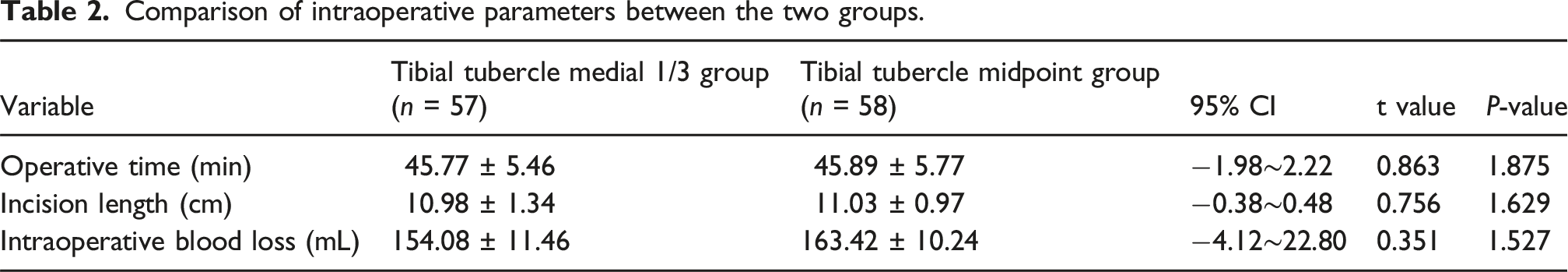
Comparison of intraoperative parameters between the two groups
Comparison of intraoperative parameters between the two groups.
Comparison of postoperative follow-up outcomes between the two groups
During the postoperative recovery period, 1 case of incision infection was reported in the tibial tubercle medial 1/3 group. The patient received timely treatment with vancomycin and early debridement, resulting in effective infection control and good prognosis. No cases of incision infection occurred in the midpoint group. Although the p-values suggested differences, all 95% CIs included 0, indicating limited statistical stability due to the small sample size. These results should therefore be interpreted with caution.
Anterior knee pain was observed in four patients in the medial 1/3 group and in two patients in the midpoint group. All affected patients were treated with regular administration of NSAIDs, such as celecoxib, along with quadriceps strengthening exercises. Symptoms significantly improved within 1 month.
Comparison of postoperative complications between the two groups.

Comparison of clinical scores between the two groups
There were no significant differences in the three preoperative scores (p > 0.05, 95% CIs included 0). The results showed that both the medial 1/3 and midpoint tibial alignment groups experienced significant postoperative improvement. Specifically, WOMAC and VAS scores decreased significantly, while HSS scores increased, all with statistical significance (p < 0.05, 95% CIs did not include 0).
Comparison of HSS, VAS, and WOMAC scores between the two groups.

Radiographic comparison results
This study selected the comparison of the posterior tibial slope angle as an evaluation criterion for the placement position of the tibial component. The results showed that in the group with the tibial tubercle midpoint placement, 44 patients (75.86%) exhibited a posterior tibial slope angle of less than 1°, 10 patients (17.24%) had an angle between 1° and 2°, 3 patients (5.17%) between 2° and 3°, and 1 patient (1.73%) between 3° and 4°. In the group with the tibial tubercle medial one-third placement, 34 patients (59.65%) had a posterior tibial slope angle less than 1°, 11 patients (19.30%) between 1° and 2°, 9 patients (15.79%) between 2° and 3°, and 3 patients (5.26%) between 3° and 4°.
Discussion
In total knee arthroplasty (TKA), the rotational alignment of the tibial component is considered one of the critical factors influencing postoperative functional outcomes, patient satisfaction, and prosthesis survival rates.10,14,18,19 Malrotation of the tibial component is widely recognized as a primary cause of complications such as anterior knee pain, patellar instability, premature polyethylene wear, and prosthesis loosening.10,14,20 Previous studies have demonstrated that a rotational mismatch exceeding 10° between the tibial and femoral components significantly reduces postoperative knee function scores and leads to abnormal gait patterns. 21 Furthermore, during high-flexion activities, rotational malalignment of the tibial prosthesis markedly increases stress on the polyethylene insert, with internal rotation of 15° elevating stress levels to 160%, thereby exacerbating wear and increasing the risk of insert failure. 10
This study analyzed 115 osteoarthritis patients who underwent TKA using the MP prosthesis at our institution, comparing two technical approaches for tibial component placement based on the midpoint versus the medial one-third of the tibial tubercle as reference points. The findings indicate that patients in the group utilizing the tibial tubercle midpoint for tibial component rotational alignment demonstrated superior outcomes in HSS scores, VAS scores, WOMAC indices, and lower complication rates compared to those in the medial one-third reference group.
The increasing prevalence of osteoarthritis has placed greater emphasis on optimizing surgical techniques and selecting biomimetic prosthetic designs in TKA to achieve pain-free outcomes and a “forgotten knee.” MP prostheses allow 0–120° of motion with the medial compartment serving as the rotational center, thereby more closely reproducing native knee kinematics. This design provides superior stability while reducing the need for soft tissue release and bone resection. In addition, the absence of a post–cam mechanism minimizes surgical trauma and may be advantageous for future revision procedures.
Common techniques for rotational alignment of the tibial component during TKA include anatomical landmark-based methods such as the Insall line, Akagi line, anterior tibial cortex curve method, and the posterior tibial condylar axis refs 15,18. However, there is no universally accepted gold standard, and the reliability and reproducibility of these intraoperative alignment techniques vary significantly.19,22 Traditionally, many surgeons have favored using the Insall line as a reference for prosthesis alignment. Recent studies, however, indicate that this method may predispose the tibial component to external rotation, which can increase shear forces at the patellofemoral interface, potentially leading to postoperative pain and functional impairment.23–25 This may explain why the Insall line group in this study exhibited a higher incidence of anterior knee pain postoperatively, as well as why their postoperative WOMAC and VAS scores were higher compared to the tibial tubercle midpoint group. The Akagi line method uses the line connecting the center of PCL attachment to the medial border of the tibial tubercle (i.e., the tibial insertion of the patellar tendon) as a ref 26. However, there remains considerable debate regarding the accuracy of the Akagi line for tibial component alignment. Johncy et al. reported that the Akagi line shows the least variability relative to STEA. 27 Similarly, studies by Shota M and Mo S also support the Akagi line as a reliable landmark for tibial prosthesis placement.19,22 Conversely, Mehmet Barış E et al. found that using the Akagi line may result in malrotation of the tibial component in patients with poor femoral condyle alignment, and based on this, they proposed modifications to improve accuracy. 28 Regarding the anterior tibial cortex curve (ATCC) method, Andrea B. et al. Considered it a more reliable and easily identifiable landmark for accurate tibial component alignment compared to the Akagi or Insall lines. Their study reported that 89% of patients aligned using the ATCC exhibited rotational errors within ±3°. 29 However, Mohammad Shahnawaz K. et al. Found that the rotational alignment of the tibial component in osteoarthritic patients varies with the degree of varus deformity; when the varus angle is less than 10°, the ATCC serves as the most precise and suitable landmark for tibial rotation in TKA, but its accuracy decreases as the varus angle increases. 30 Furthermore, Guomin L. et al. Demonstrated that the posterior tibial condylar axis appears to be a reliable and even more accurate reference than STEA for tibial component placement in varus OA knees. These findings suggest that, in clinical practice, surgeons should be proficient with multiple alignment and positioning references to enable flexible and accurate prosthesis placement. 24
Secondly, dynamic kinematic techniques, such as the Range of Motion (ROM) method, rely primarily on intraoperative observation of the knee joint’s movement trajectory during flexion and extension to determine the natural rotational alignment of the tibial component. This approach emphasizes the “natural” congruence of prosthetic components under dynamic conditions and theoretically minimizes ligament tension imbalance and prosthesis wear.18,22 However, Tatsunori K. et al. highlighted that the ROM method is highly subjective, exhibits significant inter-individual variability, and has poor reproducibility—especially in knees with varus deformity or cases requiring extensive soft tissue release, where notable external rotational deviations often occur. 18 The technique is also influenced by factors such as surgeon experience, soft tissue laxity, and capsular tightness. Their study further reported that approximately 18% of patients showed rotational discrepancies exceeding 10° when comparing ROM-based alignment to the Akagi line. 18 To address the limitations of the traditional ROM method, a hybrid technique termed ROM-A (ROM combined with navigation-assisted correction) has been proposed. 31 This method initially establishes a preliminary prosthesis orientation using ROM, followed by fine-tuning of the rotational angle with computer navigation to avoid abnormal rotational positioning. The postoperative rate of rotational malalignment with ROM-A was only 3%, outperforming the 11.7% rate observed with the conventional ROM method alone. These results demonstrate the advantages of integrating dynamic assessment with navigation guidance, representing a promising future direction for improving tibial component alignment.
To improve the accuracy of rotational alignment and reduce human error, various image-guided navigation systems have been increasingly applied in recent years, including CT-based navigation, imageless navigation, and patient-specific 3D instrumentation (PSI) systems.22,32–34 Navigation technology has been shown to significantly reduce postoperative rotational outliers, particularly in preventing tibial internal rotation exceeding 9°. However, skepticism regarding the reliability of navigation systems remains. Lennart S. et al. argued that no single technique can consistently achieve stable tibial rotational alignment, and navigation systems based on anatomical landmark recognition are not necessarily more reliable than conventional instrumentation. 35 This underscores the need for further large-scale studies and extended validation to establish definitive standards for tibial component positioning. Additionally, Apisit P. et al. proposed a novel tibial component alignment technique termed “self-aligned,” which intraoperatively allows automatic adjustment of the tibial component’s rotational orientation by controlling the contact interface with the femoral component to achieve maximal congruence. 34
This technique offers advantages of operational simplicity and strong dynamic adaptability, potentially reducing intraoperative rotational errors. However, it remains in the early stages of development and requires further validation through large-scale studies to assess its stability and long-term outcomes. With the advancement of digital medicine and personalized treatment concepts, some studies have begun to integrate multiple techniques into a so-called “comprehensive alignment strategy.” For example, Dong-Hoon L. et al. proposed a femoro-tibial synchronization technique, emphasizing that the femoral and tibial osteotomy surfaces should be adjusted synchronously during surgery to maintain mechanical axis consistency.25,29 Kohei K. et al. highlighted the importance of preoperative CT evaluation of femorotibial rotational relationships in osteoarthritis patients, enabling preoperative planning to adjust osteotomy angles based on the predicted native rotational alignment. 36 Nuttawut C. also reported that the use of PSI enhances rotational accuracy; compared to conventional cutting instruments, customized cutting blocks improve tibial component rotational alignment. 37 These integrative approaches collectively emphasize that rotational alignment should not rely solely on a single anatomical landmark but instead incorporate preoperative structural assessment, intraoperative kinematic data, prosthesis design features, navigation assistance, and soft tissue conditions to develop a more individualized and precise alignment strategy.
In this study, four osteoarthritis patients in the medial one-third of the tibial tubercle group experienced anterior knee pain, compared to two patients in the tibial tubercle midpoint group. Several factors may explain this difference. Firstly, the MP prosthesis replicates the natural patellofemoral anatomy by deepening the trochlear groove, elevating the lateral edge, and removing metal that could cause impingement on the femoral side, thereby reducing patellofemoral joint tension. During surgery, patellofemoral tracking is more easily controlled in the tibial tubercle midpoint group through selective release of the patellofemoral ligament, which helps reduce the incidence of anterior knee pain. Secondly, inflammation of the infrapatellar fat pad may also contribute; selecting the medial one-third of the tibial tubercle as the reference point can increase friction on the fat pad, exacerbating synovial irritation during lower limb movement and thereby causing anterior knee pain. Additionally, comparisons of postoperative outcomes using HSS and WOMAC scores revealed that the tibial tubercle midpoint group had significantly higher HSS scores at both 1 month and 6 months post-surgery compared to the medial one-third group (p < 0.05). Conversely, WOMAC and VAS scores were significantly lower in the midpoint group (p < 0.05). These findings indicate that choosing the tibial tubercle midpoint as the reference point for tibial component positioning in TKA can significantly improve postoperative knee function.
In addition to evaluating knee joint function, we also used the posterior tibial slope angle as an assessment metric for tibial component positioning. In the medial pivot knee system, the polyethylene insert does not have a built-in posterior slope, and the tibial cutting guide offers options for 0° or 3° posterior slope osteotomies. In this study, we found that the tibial tubercle midpoint group achieved better balance of flexion and extension gaps, with only one patient exhibiting a posterior slope greater than 3°. The posterior tibial slope in the midpoint group was significantly superior to that in the medial one-third tibial tubercle group. These results indicate that using the tibial tubercle midpoint as a reference point can lead to improved postoperative outcomes.
Although this study suggests that using the tibial tubercle midpoint as the reference point for tibial component placement in medial pivot knee systems yields better postoperative functional outcomes, several limitations remain. First, as a retrospective case-control study, it is subject to selection and information biases, limiting the ability to establish causal relationships. Second, the relatively small sample size and the low incidence of certain complications may affect the statistical power. Third, the mean follow-up duration of 15.87 months is insufficient to comprehensively evaluate the long-term stability of the prosthesis and its extended clinical efficacy. Fourth, the study did not assess patient-reported satisfaction or quality of life, nor did it specify whether postoperative rehabilitation protocols were standardized, potentially impacting the comprehensiveness of outcome evaluation. Fifth, the results of this study are limited to MP prostheses, and further investigations are needed to determine whether the midpoint of the tibial tubercle is equally effective across other knee prosthetic designs. Therefore, further validation through multicenter, large-scale prospective studies is warranted to confirm these findings.
Conclusion
In summary, during total knee arthroplasty (TKA) with the medial pivot (MP) prosthesis, selecting the tibial tubercle midpoint as the reference point for tibial component placement can achieve better conformity between the tibial plateau and femoral component, improved patellofemoral tracking, enhanced postoperative knee function, and a reduced incidence of anterior knee pain.
Footnotes
Ethics Considerations
This study was reviewed and approved by the Ethics Committee of Shanxian Central Hospital (Approval No. 20250118).
Funding
The authors received no financial support for the research, authorship, and/or publication of this article.
Declaration of conflicting interests
The authors declared no potential conflicts of interest with respect to the research, authorship, and/or publication of this article.