
Abstract
The primary hurdle in the treatment of cancer is acquisition of resistance by the tumor cells toward multiple drugs and selectively targeting the cancer stem cells. This problem was overcome by the chemotherapeutic property of recently discovered drug salinomycin. Exact mechanism of action of salinomycin is not yet known, but there are multiple pathways by which salinomycin inhibits tumor growth. Salinomycin decreases the expression of adenosine triphosphate–binding cassette transporter in multidrug resistance cells and interferes with Akt signaling pathway, Wnt/β-catenin, Hedgehog, and Notch pathways of cancer progression. Salinomycin selectively targets cancer stem cells. The potential of salinomycin to eliminate both cancer stem cells and therapy-resistant cancer cells may characterize the compound as a novel and an efficient chemotherapeutic drug.
Introduction
Salinomycin was isolated from the Streptomyces albus species of bacterium and produced by tank fermentation technology. 1 This monocarboxylic polyether ionophore is a 751-Da antibiotic (Figure 1) and is weakly acidic in nature. Salinomycin is a membrane ionophore, which shows affinity for alkali ions, preferentially for potassium ions. 2 Salinomycin eases the ion flux through cytoplasmic membrane and the mitochondrial membrane. It works as a mobile carrier and discharges K+ rapidly.1,3 For a long time, salinomycin has been used as an anticoccidial agent in the poultry industries. Coccidiosis is a single-cell parasitic disease of the intestinal tract, which leads to huge economic loss. Salinomycin improves mortality, weight gain, and lesion score at different concentrations. Low dose of salinomycin was as effective as higher dose of monensin, another polyether ionophore, in controlling coccidiosis. 4 Salinomycin also has antimalarial property. Plasmodium berghei–infected rats showed complete destruction of parasite when 20 mg/kg of salinomycin was given subcutaneously or 80 mg/kg of salinomycin was given orally. In fact, low dose of salinomycin was able to destroy asexual stage of P. berghei. 5 In sheep, rumen fungal count was diminished after salinomycin treatment, but restored when the treatment was halted. 6

Structure of salinomycin.
Recently, Gupta et al. unveiled that salinomycin selectively kills breast cancer stem cells (CSCs). Salinomycin activity toward CSCs is 100 folds higher than the comparatively conventional chemotherapeutic drug paclitaxel. 7 Previously, Zhou et al. 8 and Antoszczak and Huczynski 9 discussed about anticancer aspects of salinomycin and their selective targeting to cancer stem cells. P-glycoprotein (P-gp) is a class of adenosine triphosphate (ATP)-binding cassette (ABC) transporter that facilitates movement of drug out of cells making them multidrug resistant. Salinomycin is a potent inhibitor of P-gp, which makes cells sensitive to chemotherapeutic drugs. 10 Salinomycin exhibits antibacterial activity, especially against Gram-positive bacteria, including various antibiotic-resistant species of Streptomyces. Antifungal, antiparasitic, antiviral, and anti-inflammatory activities have also been reported for this molecule. 11 Various studies demonstrated the anticancer potential of salinomycin in different cancers. In this review, we have discussed the challenges in cancer treatment and the mechanisms by which salinomycin overcomes them.
Challenges in cancer treatment
Multidrug resistance
Multidrug resistance (MDR) is a phenomenon by which cancer cells escape the cytotoxic effect of various chemotherapeutic agents. 12 MDR in cancer cells is a major problem and sensitizing them toward chemotherapeutic drugs is a challenging task. 13 MDR is a strategy exhibited by diverse type of cancers involving cellular and non-cellular mechanisms for resistance to survive. 14 Major mechanisms of MDR in cancer cells are energy-dependent efflux of drug, driven by P-gp pump.12,13 In humans, P-gp is encoded by MDR1 gene and is one of the members of ABC family which is an ATP-dependent transporters. 15 According to Goldstein, MDR1 RNA level was elevated in more than 400 human cancers like colon, kidney, liver, pancreas, and adrenal gland, as well as in chronic myelogenous leukemia and non-small-cell lung carcinoma with neuroendocrine properties. However, MDR1 expression was occasionally elevated in other untreated cancers, like neuroblastoma, acute lymphocytic leukemia in adults, acute non-lymphocytic leukemia in adults, and indolent non-Hodgkin’s lymphoma. 16 There are other subfamilies of ABC transporters which confer drug resistance, such as breast cancer resistance protein (BCRP) and MDR proteins (MRPs). BCRP is a G-subfamily member of ABC transporter that plays an important role in efflux of anticancer drugs like methotrexate, mitoxantrone, indolocarbazole topoisomerase I inhibitors, camptothecin, flavopiridol, and quinazoline ErbB1 inhibitors, whereas MRP transports anionic drugs across membrane.17,18
Cells acquire resistance by the following three major mechanisms: (1) water-soluble drugs like cisplatin and folate antagonists are less absorbed by cells. (2) Variety of changes in cells like increased DNA repair, changes in the cell-cycle protein, and reduced level of apoptosis. (3) Efflux of hydrophobic drug via ABC transporter. In humans, ABC transporter is encoded by 48 genes. 19 In breast cancer, ABC transporters play vital role in making it multidrug resistant. High-level expression of ABC transporter decreases chemotherapeutic drug response in cancer cell lines. 20 Kickhoefer et al. 13 found involvement of vault protein in MDR of cancer cells. According to Nygren et al., 21 MDR can be generated because of the elevated intracellular calcium ion level in small-cell lung cancer cells. In some MDR cell lines, transporter associated with antigen processing (TAP) is more expressed. It was found that TAP is overexpressed in parallel with major histocompatibility complex class I (MHC I), which helps in antigen presentation on endoplasmic reticulum membrane. Increased expression levels of TAP and MHC I confer resistance to cells. 22
Cancer stem cells and its role in cancer
Cancer stem cells are “stem-like” cells within a tumor that “possess the capacity of self-renewal and can produce the heterogeneous lineages of cancer cells that complete the tumor.” 23 These cells facilitate tumor maintenance and are also referred to as tumorigenic cells or tumor-initiating cells. 24 First, these cells develop resistance to chemotherapy and radiation due to their ability to regenerate, accumulate mutations, and differentiate into chemoresistant cells and second because of their quiescent behavior which protects them from cytotoxic therapy that targets rapidly dividing cells. Therefore, CSCs are primarily responsible for relapse and poor survival in various cancers. CSCs can evade the chemopreventive strategies for cancer treatment, and hence, it is necessary to identify drugs that selectively target this population25,26 (Figure 2).
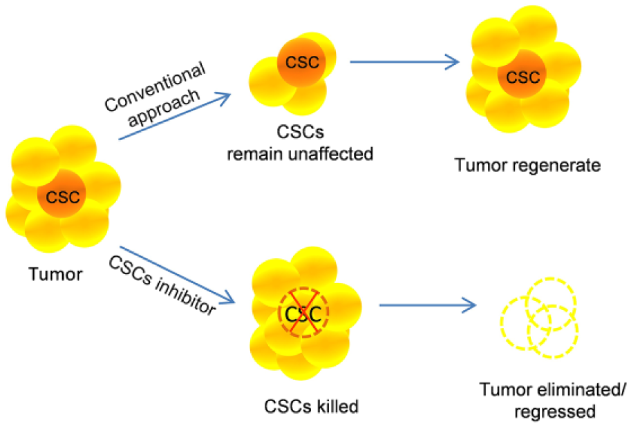
Schematic representation of CSC targeting in chemotherapeutic approach and its importance.
Cell surface markers (immunophenotype) help in phenotypically distinguishing stem cells from their differentiated progeny. Different patterns of cell surface markers are expressed in CSCs of different tumor types. Various putative surface biomarkers include CD44, CD24, LGR5, and CD44v6. ALDH1A1 and ALDH1A3 are functional biomarkers. These cells show elevated levels of stem cell–associated transcription factors like Nanog, Oct4, Sox2, Hes1, and Klf4. 27 CD34+ and CD38− leukemia cells, which overexpress ALDH1 (aldehyde dehydrogenase 1), show resistance toward cyclophosphamide. 28 Colorectal cancer stem cells express CD133 and CD44 cell surface biomarkers; high expression of these biomarkers is associated with metastasis and worse prognosis. 29 Pancreatic cancer stem cells expressing CD44+/CD24+/ESA+, CD133+ and ALDH1 are the most common cancer stem cell markers. 30
Similar signaling pathways that occur in normal stem cells regulate the self-renewal in CSCs. Wnt, Hedgehog, NF-κB, Notch, and PI3K/AKT/mTOR are the major pathways that play a pivotal role in CSC self-renewal and lineage fate regulation.23,31 Suppression of GSK3β activity was shown to be critical for maintenance of murine pluripotent stem cells. 32 Wnt signaling inhibition by Axin decreases hematopoietic stem cell (HSC) proliferation. Stimulation of sonic hedgehog (Shh) pathway in human HSC in vitro culture (CD34+Lin−CD38−) exhibits increased self-renewal response. Role of Notch activation in self-renewal of HSCs was confirmed by Reya et al. 33 through Jagged-1 ligand culture that constantly expresses increased amount of primitive progenitor activity in vitro and in vivo. Shh is also linked to transcription factor NF-κB signaling. NF-κB activates overexpression of Shh in pancreatic cancer and accelerates pancreatic cancer cell proliferation. 34 Loss of tumor-suppressor gene phosphatase and tensin homolog (PTEN), which regulates PI3K signaling, has been shown to mediate AKT activation and increase stemness properties of CSC populations in prostate cancer. 35 Furthermore, crosstalk between PI3K/AKT and other pro-survival as well as mitogenic pathways has been shown to drive cancer growth. 36
Salinomycin as an anticancer agent
Antitumor property of salinomycin was revealed by Gupta et al. after high-throughput screening of 16,000 compound libraries. Salinomycin selectively targets breast CSCs and inhibits tumor proliferation. 7 Salinomycin has shown anticancer property, specifically targets the CSCs of multiple types of cancer, and sensitizes MDR human cancer cells. 8
Exciting effects of salinomycin on cancer
Gupta et al. showed that salinomycin inhibits growth of human breast cancer and decreased metastasis in non-obese diabetic (NOD)/severe combined immunodeficient (SCID) mice. 7 Salinomycin caused apoptosis in human CD4+ T-cell of acute T-cell leukemia, MDR human MES-SA/Dx5 uterine sarcoma cells which express P-gp. 2
Salinomycin mediates its anticancer effect by various mechanisms in different cancer types. It reduced the activity of ABC transporters in leukemia stem cells, inhibited Wnt signaling cascade in colorectal cancer cells, 11 inhibited growth and migration of prostate cancer cells, 37 and led to reactive oxygen species (ROS) accumulation in androgen-dependent and independent prostate cancer cells. 38 It reduced cisplatin-resistant mesenchymal-like subpopulation of squamous cell carcinoma cells expressing high or low levels of E-cadherin in vitro; 39 expression of apoptotic and anti-apoptotic genes is dysregulated in cisplatin-resistant SW620 colorectal cancer cells. 27 It also induced apoptosis in human hepatocellular carcinoma cells 40 and autophagy response in prostate and breast cancer cells. 41
Salinomycin caused apoptosis in human nasopharyngeal carcinoma cells CNE-1, CNE-2, and CNE-2/DDP via caspase activation and destabilization of mitochondrial membrane potential. 42 Time- and concentration-dependent cytotoxic effect of salinomycin was exhibited in LNM35 and A549 lung cancer cell lines 43 and human cholangiocarcinoma cells in vitro. 44 Lower salinomycin concentration reduced proliferation in high-density cell populations of Hs578T and MCF-7 breast cancer cells. 45 Salinomycin also acted on OVCAR-3 human ovarian cancer cells through caspase-mediated apoptosis without harming normal cells. 46 Recently, salinomycin demonstrated anti-angiogenic activity by VEGFR2 signaling pathway in gastric cancer xenografts. 47
How salinomycin works?
Mechanism of action of salinomycin as chemotherapeutic drug is not well understood. Various mechanisms of action and molecular targets of salinomycin have been summarized in this review (Figure 3; Table 1).

Salinomycin interferes with cell-cycle regulation in cancer cells by inhibiting Skp-2 activity.
In vitro and in vivo studies elucidating molecular targets of salinomycin.

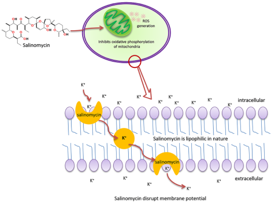
Ionophoric action of salinomycin
Salinomycin shows cytotoxic effect by disrupting the balance of sodium and potassium ions across the cell membrane and the mitochondrial membrane 11 (Figure 4). Rapid efflux of K+ from cell membrane and mitochondrial membrane led to apoptosis of cells. 61 In rat liver, salinomycin caused efflux of alkali cations, especially K+ from mitochondrial membrane. Salinomycin inhibited mitochondrial oxidative phosphorylation without affecting the substrate-level phosphorylation. 3
Overview of multiple targets of salinomycin and its mechanism of action through acting on cancer cells and CSCs.
Selective targeting of CSCs by salinomycin
In a study, a chemical library screened by Gupta et al., 7 salinomycin was shown to decrease the percentage of CD44high/CD24low breast cancer stem cells by 20 folds. Salinomycin has been shown to kill CSCs in different types of human cancers. The breast CSC population was reduced by eliminating ALDH1+ and SOX2-positive cells. Salinomycin inhibited Oct4, Nanog, Sox2 expressing lung CSCs, and CD44+ expressing VCaP- and LNCaP-derived prostate CSCs. Salinomycin also killed CD133+ colorectal and pancreatic CSCs. Lower expression of E-cadherin makes cells drug resistant and promotes epithelial-to-mesenchymal transition (EMT). Salinomycin eliminated squamous cell carcinoma CSCs population by inhibiting low E-cadherin expressing cell proliferation. Interestingly, salinomycin inhibited tumorosphere formation in breast, lung, gastric, osteosarcoma, colorectal, and pancreatic CSCs. 23
Sensitization of MDR cells by salinomycin
Salinomycin inhibited P-gp, type of ABC transporter which is expressed in most of the cancers and involved in efflux of chemotherapeutic drugs. 62 Human promyeloblastic leukemia KG-1a cells, which express ABC transporters, such as P-gp, BCRP, and MRP8, made them resistant toward many chemotherapeutic drugs. Polyether ionophore antibiotic salinomycin sensitized KG-1a cells for apoptosis. 56 Riccioni et al. 10 reported that salinomycin also inhibited MRP gp170. Fuchs showed that salinomycin triggers apoptotic cascade, independent of the cell-cycle arrest, p53, and proteasome. Salinomycin induced apoptosis even in those cells which acquire resistance to the apoptosis by higher expression of ABC transporters, anti-apoptotic protein Bcl-2, or 26S proteasomes. 2
Molecular pathways targeted by salinomycin
Salinomycin targeted prostate cancer cells by reducing activities of NF-κB pathway and ALDH. It induced oxidative stress and finally reduced CD44 cell population. 37 Salinomycin induced oxidative stress in PC-3 prostate cancer and declined pro-caspase 3 activity. However, it increased Bax/Bcl-2 ratio and PARP-1 cleavage. 38 Salinomycin also activated mitogen-activated protein (MAP) kinase pathway through ROS generation by involvement of Jun N-terminal kinase (JNK) pathway. 63 Salinomycin killed cisplatin-resistant colorectal cancer cells via ROS-mediated apoptosis. 27 It induced apoptosis in NCI/ADR-RES, DXR, and OVCAR-8 ovarian cancer cells through downregulation of S-phase kinase-associated protein-2 (Skp-2) and Stat3 inactivation. Skp-2 overexpressed in most of the cancer stem cells that target tumor-suppressor genes. Skp, cullin, and F-box together form SCF complex; the SCF complex and anaphase-promoting complex (APC) are major E3 ubiquitin ligases that regulate cyclin and cdk levels. However, negative cell-cycle regulators Cip and Kip inhibit cdk, leading to cell-cycle arrest. Skp-2 ubiquitinized Kip that induced cell-cycle progression. Salinomycin downregulated Skp-2, ultimately cell proliferation was stopped and apoptosis was induced 54 (Figure 5). Salinomycin inhibited proliferation of CD44+ Oct4+ gastric CSC population growth by targeting Wnt signaling proteins. 58 Salinomycin also decreased CD44(+)/CD24(−) breast CSC population through modulation of hedgehog signaling pathway in MCF-7 cells and MCF-7 mammosphere cells.48,64 However, in MDA-MB-231 triple-negative breast cancer cells, CSC population was inhibited by STAT3 down-regulation. 49

Schematic diagram representing salinomycin-induced ROS generation inside the cell which blocked oxidative phosphorylation in mitochondria; salinomycin acts as ionophore and causes mitochondrial and cytoplasmic K+ ions efflux which led to cell death.
DNA damage and autophagy induction by salinomycin
Salinomycin increased tumor-suppressor protein p53 and DNA damaging protein pH2AX and decreased cyclin D1 level, which led to cell-cycle arrest and high DNA damage. Comet assay and immunocytochemical staining of pH2AX protein confer salinomycin-mediated DNA damage. In Hs578T and MDA-MB231 cells, salinomycin reduced cyclin D1 expression, consequently leading to cell death. 50
In salinomycin-treated MCF-7 cells, autophagy pathways of cell death via vacuole formation have been observed. Salinomycin treatment to MCF-7 cells generates multiple vacuoles, which is confirmed in other cell lines like SW480, SW620, and RKO to check monodansyl cadaverine uptake, which is a specific autophagic marker. Salinomycin also led to ROS generation in above-mentioned cells. ROS like O2N and H2O2 are responsible for autophagic response. Autophagy was also dependent on endoplasmic reticulum via the elevation in the level of DNA-damage-inducible transcript 3 (DDIT3) and activating transcription factor 4 (ATF4) after salinomycin treatment in non-small-cell lung carcinoma. Endoplasmic reticulum regulates the autophagy through ATF4, DDIT3, AKT1, and mechanistic target of rapamycin (MTOR) pathways which are modulated by salinomycin. 51 Salinomycin worked effectively in glucose starvation and serum starvation conditions; since there is nutrient and oxygen deprivation in tumor, it worked efficiently to kill autophagic cancer cells. 60
Anti-angiogenic activity of salinomycin
Fibronectin promoted elongation of micro-vessels during angiogenesis in vitro. 65 Francis et al. 66 showed that fibronectin is required in early blood vessel development. Salinomycin reduced the level of fibronectin expression in human endometrial cancer (Hec-1-SP) cells. 59 In chronic lymphocytic leukemia cells with constitutive Wnt activation, nanomolar concentrations of salinomycin downregulated the expression of Wnt target gene, such as fibronectin. 57 VEGFR2 signaling plays crucial role in angiogenesis and tumor progression. Salinomycin inhibited VEGFR2 phosphorylation and STAT3 activation in Human Umbilical Vein Endothelial Cells (HUVEC). Therefore, salinomycin hampered VEGF-induced angiogenesis in gastric cancer. 47
Effect of salinomycin on EMT phenomenon
EMT allows cells to detach from the tissue and gain motility by losing its basoapical polarity. A series of transcriptional and post-translational events cause epithelial cells to acquire mesenchymal characteristics. 67 This process enables them to invade and form macroscopic metastases in different organs. Re-expression of E-cadherin causes cells to regain epithelial phenotype, a phenomenon called mesenchymal-to-epithelial transition (MET). The EMT-MET-related differentiation status is identified by the differential expression of markers of extracellular (fibronectin and matrix metalloproteinases) and cellular (vimentin and E-cadherin) localization. 68 Several studies conducted in vitro as well as in vivo suggest that salinomycin inhibits migratory and invasive potential of cancer cells.59,69–73
Salinomycin was able to significantly reduce the metastasis and invasion abilities of bladder cancer cell line T24. Tumor tissues from rats inoculated with T24 cells showed high expression of E-cadherin and lower vimentin expression level in the salinomycin-treated group. 69 Salinomycin inhibited migratory and invasive potential of side population cells in human and rat endometrial cells Hec-1 and RK12V, respectively. 59 Another study reported that salinomycin was able to inhibit transforming growth factor-β1 (TGF-β1)-induced EMT phenotypic transition in human breast cancer cell line MCF-7 and activated key signaling molecules involved in Smad (p-Smad2/3 and Snail1) and non-Smad (β-catenin and p-p38 MAPK) signals which cooperatively regulate the induction of EMT. 73
Salinomycin works synergistically with conventional chemotherapeutic drugs to inhibit invasion and migration of cancer cells. Transcription factor ZEB1, known for promoting metastasis in cancers, is highly expressed in primary mantle cell lymphoma (MCL) orchestrated by active Wnt signaling. Salinomycin blocked Wnt signaling and downregulated ZEB1, thereby increasing the sensitivity of MCL cells to the cytotoxic effect of gemcitabine, cytarabine, and doxorubicin. 70 In combination with metformin, salinomycin was able to block TGFβ-induced EMT and inhibit EMT-induced cell migration in the two non-small-cell lung cancer cell lines A549 and HCC4006. 71 Salinomycin inhibited doxorubicin-induced EMT by activation of FOXO3a which disrupted the interaction of β-catenin and TCF and suppressed its downstream targets like ZEB1, CyclinD1, and c-Myc, involved in doxorubicin-induced EMT in hepatocellular carcinoma cells. 72
Epigenetic modifications induced by salinomycin
Epigenetics governs normal cell maintenance and gene expression in mammalian cells. It is the study of alterations in gene expression without affecting prime DNA sequence. Deleterious epigenetic changes lead to malignant cellular transformation. Epigenetic regulation in cancer progression includes DNA methylation, covalent histone modifications, nucleosome positioning, and noncoding microRNAs which post-transcriptionally regulate gene expression.74,75 High degree of histone hyperacetylation is associated with increased cell death and senescence. 76 Acetylation profiling of histone H3 and H4 in MDA-MB-231 cells showed a gradual increase in their acetylation status upon salinomycin treatment. 77
CSC-associated phenotypes including drug resistance are modulated by polycomb group (PcG) protein-mediated dynamic epigenetic alternations in histone methylation landscapes. TRAIL (ligand for DR4/DR5) resistance in colon CSCs may be attributed to the catalytic component of polycomb repressive complex 2, enhancer of zeste homolog 2 (EZH2)-mediated H3K27me3 in promoter region, thereby epigenetically repressing the expression of DR4/DR5. Salinomycin treatment increased the DR4/DR5 expression with a simultaneous decrease in EZH2 localization in the nucleus and hence reduced EZH2 recruitment and histone methylation near promoters, thereby restoring their TRAIL sensitivity. 78
Dysregulation of microRNAs is involved in many aspects of cancer progression including EMT, invasion, and stem cell properties. Microarray analysis of global microRNA expression in a putative head and neck squamous cell carcinoma stem cell culture JLO-1 treated with increasing doses of salinomycin revealed a set of microRNAs that were consistently upregulated or downregulated, among these microRNAs were miR-328 and miR-199a-3p, both with known roles in promoting drug sensitivity.79–81 Expression of miR-203, associated with the inhibition of EMT, was downregulated. 82 This suggests that the changes in microRNA expression may be mediated by salinomycin.
Effects of salinomycin derivative
Salinomycin reacts with many divalent metal ions to form new derivative which shows enhanced bioactivities such as antifungal, antibacterial, herbicidal, and anti-inflammatory. Sodium salinomycin (SalNa) formed novel derivative with Co(II), Cu(II), Ni(II), or Zn(II). Antibacterial activity improved especially against Gram-positive bacteria Bacillus cereus. These derivatives showed dose-dependent cell toxicity in human leukemia cell lines. Complexes with Co(II) and Cu(II) showed more prominent cytotoxic effect. 83
Huczynski et al. screened the anticancer activity of different salinomycin amide substitutes like n-butylamine, phenylamine, 4′-aminobenzo-15-crown-5, 3-morpholine-propylamine, benzylamine, tryptamine, and 3,6,9-trioxadecylamine, benzotriazole ester of salinomycin. Antiproliferative activities of all these compounds were evaluated against MDR phenotype of colon cancer cells LoVo and leukemia cells HL-60. Many derivatives of salinomycin showed less toxicity and stronger anticancer activity against doxorubicin-resistant colon cancer subline LoVo/DX than unmodified salinomycin. 11
The cell count of MDA-MB-231 breast cancer cells with CD44 expression was decreased when treated with hyaluronic acid–coated salinomycin nanoparticles indicating their effectiveness toward cancer stem cells. 84 The glioblastoma polysorbate 80–coated poly(lactic-co-glycolic acid)-encapsulated salinomycin nanoparticle worked effectively with enhanced cell death. 85
Advantage of salinomycin over other anticancer agents
A chemotherapeutic drug should show cytotoxic effect on cancer cells, but at the same time, growth of normal cells must remain unaffected. There are recent reports published on salinomycin preferentially acting on tumor cells over non-tumorigenic cells. Salinomycin induced cell-cycle arrest and decreased expression of anti-apoptotic genes in MCF-7 cells, thereby causing apoptotic cell death of tumor cells, whereas non-tumor cells were able to evade this toxic effect. 86
Salinomycin induced apoptosis in human T-cells of leukemia, but Fuchs et al. 2 reported that normal human T-lymphocytes were not harmed. Salinomycin is more potent in inhibiting P-gp than verapamil, which is a known drug for inhibiting ABC transporter. 62 Recently, Huczynski et al. 11 studied salinomycin-mediated induction of apoptosis in tumor cells of different tissue origin and its ability to overcome MDR phenomenon.
High-density cancer cell populations were reduced by salinomycin at lower dose upon long time exposure. This dose helped in detachment of cells, and these detached cells undergo apoptosis in breast cancer cells like MCF-7 and Hs578T. The advantage of using lower dose with longer exposure is that salinomycin toxicity can be reduced and multidrug-resistant tumors can be conquered. 45
Salinomycin toxicity
Although salinomycin has great chemotherapeutic effect over large number of cancer cells, it has shown to be toxic in vivo. Some researches and case studies have been presented here.
Weight of mice in the salinomycin-treated group greatly reduced as compared to control group. Salinomycin toxicity to male reproductive system was revealed by Ojo et al. Weight of testis, seminal vesicle, and epididymis was reduced irreversibly. Lot of structural changes occurred in interstitium of the testis and seminiferous tubules. Oxidative stress was generated in testicular and epididymal tissues, and significant decrease in enzymes lactate dehydrogenase (LDH), superoxide dismutase (SOD), catalase (CAT), and reduced glutathione (GSH) was noticed. 87
Neuronal cells damaged by salinomycin treatment came in light by histopathologic analysis. Severe vacuolation occurred in dorsal root ganglia of neurons in horses. Horses developed ataxia, muscular atrophy, and persistent weakness. 88 Cardiac arrhythmia, tachycardia, recumbency, sudoresis, groaning, incoordination, and paddling movements of limbs were observed in horse after monensin poisoning; along with myoglobinuria, elevated serum creatine phosphokinase level was observed. 89 Once hybrid turkey hens accidently ingested sodium salinomycin in feed, which resulted in death of 34% of turkey hens. 90 Dorsal root ganglia and Schwann cells were also affected by salinomycin. Toxicity is due to elevated cytosolic Na+ concentrations, ultimately leading to increase in cytosolic Ca2+ level by means of Na+/Ca2+ exchangers in the both plasma membrane and mitochondria. Higher Ca2+ level induced caspase-dependent apoptosis. 91
In pig, toxicity was seen after consumption of food containing 720 and 441 ppm of salinomycin. Pigs behaved abnormally, some were unwilling to stand, and elevated rectal temperatures were recorded, which finally led to death. 92 Shortness of breath, dizziness, nausea, leg weakness, photophobia, and increased blood pressure are the symptoms of salinomycin toxicity in humans.
Amelioration of toxicity and synergistic effects
Cancer stem cells express certain markers specific to them. HER2high and HER2low mammosphere reveal markers like OCT4, SOX2, and NANOG. Salinomycin selectively targeted these CSCs by decreasing SOX2 expressing cell population. However, combinatorial treatment of salinomycin with anti-HER2 monoclonal antibody trastuzumab showed enhanced cell death, which could not be achieved by salinomycin or trastuzumab alone. This combination treatment was also effective on MCF-7, MDA-MB-231, and BT-474 cell lines. 93
Salinomycin combination with doxorubicin enhanced caspase activation and pause the cells in sub-G1 phase. Greater transcription level of p21, p53, PUMA, and NF-κB activity was achieved in co-treatment. Chemosensitivity of sarcoma cell line increased upon combined treatment of salinomycin and doxorubicin even at minute concentrations, so the toxic effect of salinomycin is reduced and relative cell growth rate decreased from 1.5 to 3 fold in the sarcoma cell lines SW872, A204 and HT-1080. 94 Combinatorial treatment of salinomycin and doxorubicin shows variability in their cytotoxic effect in different cell lines. This treatment showed significant reduction of cell number in MES-SA/Dx5 and MCF-7 cell lines, while HepG2 and MDA-MB231 showed a lower inhibition rate. 50
Salinomycin and gemcitabine eradicated pancreatic cancer cells. However, poly(ethylene glycol)-b-poly(lactic acid) micelles mediated salinomycin delivery in gemcitabine-resistant pancreatic cancer cells with increased efficacy.95,96 Salinomycin significantly inhibited cell proliferation of imatinib-resistant KitlowCd44+ Cd34+ interstitial cells of Cajal stem cells. Treatment of salinomycin along with imatinib retarded cell division effectively than when these drugs were used alone. 97 Trace amount of salinomycin sensitized tumor cells when treated with antimitotic drugs like docetaxel, paclitaxel, colchicine, and vinblastine. Combination reduced salinomycin toxicity and increased apoptosis. 45 Salinomycin with cisplatin or paclitaxel revealed strong synergism for antitumor activity in head and neck squamous cell carcinoma stem cells. 53 MCF-7 xenografts and salinomycin-loaded PEG-b-PCL polymeric micelles combined with octreotide-modified paclitaxel-loaded PEG-b-PCL polymeric micelles showed stronger antitumor activity. 98
Lapatinib is an anticancer drug used in the treatment of breast cancer. Lapatinib or salinomycin combination therapy with adriamycin for breast cancer was capable of eliminating breast tumor–initiating cells by 50 fold. These two drugs significantly reduced mammosphere formation and proportion of ALDH1+ cells by 10–100 folds in CD44+/CD24− breast tumor–initiating cells isolated from MCF-7 cells, primary cells, and epirubicin-resistant SKBR3 cells. Adriamycin and salinomycin together increased cell death by 32%–77% in breast tumor–initiating cells. 99 Activation of Akt led to cancer progression by the PI3K-mediated pathway, which can be impeded by the PI3K inhibitors like LY294002 and wortmannin. Combined treatment of salinomycin with PI3K inhibitors led to increased apoptosis as compared to single treatment. 100 Aydin developed a novel approach to deliver salinomycin in tumor cells. He synthesized Herceptin (HER)-immobilized nanoparticle made up of poly(lactic-co-glycolic acid) encapsulated with salinomycin to target breast cancer cells. Its small size and Herceptin immobilization efficiency made it suitable for uptake by MCF-7 cells. 101
Tumor requires vascularization (angiogenesis) for productive growth and metastasis. Overexpression of HER2 in human tumor cells is closely associated with increased expression of vascular endothelial growth factor (VEGF) and angiogenesis. 102 HER2 signaling upregulated angiogenesis at different levels and different mechanisms. 103 Combinatorial treatment of mammospheres with trastuzumab and salinomycin efficiently targets HER2-positive cancer cells and CSCs. 93 In gefitinib-resistant colorectal cancer, salinomycin synergies induce apoptosis through caspase-dependent and independent pathways and are involved in loss of membrane potential of lysosome and mitochondria. 104 Salinomycin was also found useful in treatment of T-cell acute lymphoblastic leukemia Jurkat cells by treating along with vincristine. 55 Histone deacetylase inhibitor and salinomycin together worked on CSCs of triple-negative breast cancer in NOD/severe combined immunodeficiency gamma mice model. This combination induced apoptosis, cell-cycle arrest, and modulation of EMT in breast CSCs. 105
Conclusion
Polyether ionophore antibiotic salinomycin has been used as a coccidiostat agent in the poultry industry for a long period, but now it has emerged as an effective anti-proliferative agent. There are very few drugs available in market which target CSCs. Salinomycin is known for selective killing CSCs and sensitizing tumor cells at a very low concentration. It has exhibited toxicity over a broad range of cancers. Salinomycin sensitized tumor cells for many chemotherapeutic drugs by inhibiting many MDR genes. Selective targeting and evasion of MDR phenomenon make salinomycin a promising anticancer drug. Clinical pilot studies conducted by Naujokat and Steinhart 23 revealed that salinomycin induced partial clinical regression of heavily pretreated and therapy-resistant cancers, particularly in combination with novel tumor-targeted drugs. Further clinical studies need to be conducted for evaluating the safety and effectiveness of this drug. Approaches for drug delivery, dose optimization, and amelioration of its systemic toxicity should be deciphered.
Footnotes
Acknowledgements
CSIR-CDRI communication Number for this article is 9452.
Declaration of conflicting interests
The author(s) declared no potential conflicts of interest with respect to the research, authorship, and/or publication of this article.
Funding
The author(s) received no financial support for the research, authorship, and/or publication of this article.