
Abstract
Background
Osteoarthritis (OA) is the most common form of arthritis affecting the elderly and obese population, and requires long-term medical management. Inflammatory pathways, possibly initiated by tissue damage and stress are involved in the pathogenesis of OA. Citrus medica Linn., known for its anti-inflammatory and analgesic properties, has been the focus of management of OA in recent years.
Purpose
To explore the therapeutic potential of Citrus medica gel in Wistar Albino rat OA model with standard Aceclofenac gel.
Materials and Methods
An experimental interventional animal study was conducted in which OA was induced in 24 male Wistar Albino rats (12-week-old) using monosodium iodoacetate. The animals were observed for 1 week for signs of joint inflammation. They were then divided into four groups. Group A (standard) received Aceclofenac gel twice daily (BD) for 4 weeks. Group B received C. medica gel (0.2 g, 0.33% w/w) once daily (OD). Group C received C. medica gel (0.2 g, 0.33% w/w) BD, and Group D was kept as a disease control throughout the study. The results were compared between groups using one-way analysis of variance (ANOVA).
Results
The double-dose C. medica gel regimen displayed a significant and earlier reduction in ankle joint circumference (ANOVA, p value < .001). Pain relief was observed, as evidenced by a favorable reversal of the arthritic index and score compared to the standard treatment.
Conclusion
This study highlights the potential of C. medica gel in reducing inflammation and pain associated with OA. These findings warrant further investigation through clinical trials.
Keywords
Introduction
Osteoarthritis (OA) is the most common form of arthritis worldwide. Classically, it presents with joint pain and loss of function; however, the clinical presentation of OA is highly variable. It can range from an asymptomatic incidental finding to a severely debilitating and permanently disabling disorder. OA affects the entire joint, sparing no tissue. Its pathogenesis involves the interplay of multiple risk factors, mechanical stress, and abnormal joint mechanics. This combination leads to the release of pro-inflammatory cytokines and proteases that ultimately mediate joint destruction. However, the complete pathway leading to the progressive destruction of joint tissues remains incompletely understood. 1
Morphological changes observed in OA include cartilage erosion as well as synovial inflammation. Current research attributes these changes to a complex network of biochemical factors. One such factor is tumor necrosis factor-alpha (TNF-α). Human articular chondrocytes from OA cartilage have been shown to express a significantly higher number of p55 TNF-α receptors, which could make OA cartilage particularly susceptible to degeneration. Specific cytokines also have anti-inflammatory properties. Three such cytokines—interleukins (IL-4, IL-10, and IL-13) have been identified from previous studies as modulators of inflammatory processes. However, their efficacy depends significantly on the target cell type. IL-4, when tested in vitro, in OA tissues, has been shown to suppress the synthesis of both TNF-α and IL-1β. Naturally occurring anti-inflammatory cytokines such as IL-10 inhibit the synthesis of pro-inflammatory mediators like IL-1 and TNF-α, making them potential targets for therapy in OA. Inflammatory signaling pathways in OA, including those mediated by nuclear factor kappa B (NF-κB), are likely triggered by products of tissue damage and stress via cell surface pattern recognition receptors (PRRs).2–4
Citrus medica Linn., commonly known as C. medica in English and “Narthangai” in Tamil, is a fruit that is globose, ovoid, or oblong, often mammillated at the apex. Various parts of Narthangai are widely used in the Indian traditional system of medicine. Ripe fruits are known for their anti-scorbutic, stomachic, cardiac tonic, stimulant, sedative, and analgesic properties. They are traditionally used to treat dyspepsia, bilious vomiting, cold, fever, palpitations, sore throat, cough, asthma, hiccups, and earaches. The root is known to be analgesic and anti-spasmodic, and it is used for conditions such as diarrhea, piles, and constipation. Seeds are considered anti-helmintic, sedative, and cardiotonic and are also used for palpitations. Fruit extracts have demonstrated potent antioxidant activity. Both the leaves and juice of C. medica are used by the people of South-Eastern Nigeria for febrile illnesses. In local Indian customs, many people use fresh C. medica juice as a topical application for joint pain relief.5–7
To date, there is no literature exploring the effects of C. medica gel on in vivo arthritic animal models. This study aims to evaluate the anti-inflammatory and analgesic effects of C. medica gel using a whole animal model.
Aims and Objectives
To evaluate the therapeutic potential (anti-inflammatory and analgesic) of the topical application of C. medica gel in monoiodoacetate (MIA)-induced OA in Wistar Albino rat models.
To compare the efficacy of C. medica gel with Aceclofenac gel in MIA-induced OA in Wistar Albino arthritic rat models.
Materials and Methods
This experimental study was conducted in the Department of Pharmacology, Medical College, Coimbatore, Tamil Nadu, India, in August 2023, in accordance with institutional animal ethics norms. Approval was obtained from the Institutional Animal Ethics Committee (444/IAEC/2022).
Plant Authentication Certificate
The C. medica fruit specimens were collected from Coimbatore (Tamil Nadu, India). The plant authentication certificate was obtained from the Botanical Survey of India (BSI), Coimbatore, with certificate number: BSI/SRC/5/23/2022/Tech./181.
Preparation of C. medica Powder
20 mL of C. medica juice was extracted from 30 g of C. medica fruit. This solution was kept in a hot air oven at 60°C for 3–4 days, and 100 mg of C. medica powder was obtained. Qualitative analysis using Salkowski and Liebermann–Burchardt’s tests was performed on C. medica powder to determine its anti-inflammatory potential.
Phytochemical Analysis C. medica Powder—Test for Sterols
Salkowski Test
1 mL of the C. medica powder sample, diluted in chloroform (10 mg/mL), was added to concentrated sulfuric acid (H2SO4), which yielded a red-colored precipitate. 8
Leibermann–Burchard Test
Two drops of acetic anhydride and 1 mL of concentrated H2SO4 were added along the sides of the test tube to 1 mL of C. medica powder sample. A reddish ring was formed at the junction of the two solution layers. 8
Spectrophotometric Analysis of C. medica Powder
Quantitative analysis of C. medica powder was done using spectrophotometric analysis. 20 mL C. medica powder was used to prepare five different concentrations of methanolic extracts (1, 2, 3, 4, and 5 mL). For comparison, methylprednisolone was prepared in five concentrations (100, 200, 300, 400, and 500 µg). Each concentration was mixed with 2 mL of sulfuric acid (H2SO4, 4N), 2 mL of ferric chloride (0.5%), and 0.5 mL of potassium ferricyanide (0.5%). This mixture was heated in a water bath and maintained at 70°C for 30 min. Absorbance was measured at 780 nm using a SmartSpec spectrophotometer (Bio-Rad). 9 The results were compared with standard corticosteroids.
Fourier Transform Infrared Spectroscopy (FTIR) of C. medica Powder
C. medica powder was subjected to FTIR (Shimadzu), and its structure was determined. The results were compared with methylprednisolone. 10
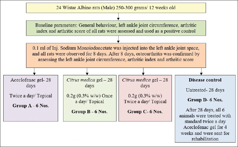
In Vitro Permeability Test of C. medica Powder
Topical penetration of the C. medica powder was subsequently evaluated using an in vitro topical permeability test as described by a standard protocol.11–15
The samples were analyzed using a spectrophotometer (Bio-Rad) at 780 nm at the end of 30 min, 1, 1.5, 2, 2.5, and 3 h.
Preparation of C. medica Gel
C. medica gel was prepared using a standard procedure. 16 0.2 g of C. medica powder was mixed with 1.5 g of Carbopol 934 in 60 mL of demineralized water. The solution was stirred manually and continuously with a magnetic stirrer for 1 h; this sample was considered Solution 1. 0.005 g of disodium edetate and 1.33 mL of triethanolamine were dissolved in 10 mL of demineralized water and were stirred for 30 min; this sample was considered Solution 2. 4.83 mL of propylene glycol and 12 mL of distilled water (Solution 3) were added drop by drop to Solution 2, with continuous stirring for 10 min; this sample was considered Solution 4. The required quantity of Solution 4 was added to Solution 1 until the desired gel consistency was achieved. 60 g of final C. medica gel was obtained. The procedure was done at room temperature.
Analysis of C. medica Gel
C. medica gel was analyzed to determine its strength and spreadability.
Calculation of C. medica Gel Dosage Required for Application in Each Rat
0.2 g of C. medica gel was applied to the left ankle joint of each rat.
17
The gel was produced twice. A total of 0.4 g of C. medica powder was used to produce 120 g of C. medica gel, sufficient for daily topical application in the left ankle joint of 12 rats (Groups B and C) over 28 days.
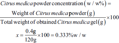
Formula A. Showing formula to calculate the dose of C. medica gel in w/w.
Thus, 0.2 g (0.33% w/w) of C. medica gel was used per application in each rat (Formula A). Based on the literature, higher concentrations (5% w/w) of topical C. medica extract may exert acute anti-inflammatory effects within a single day. We formulated a lower concentration of C. medica gel (0.33% w/w) for chronic application. This concentration was chosen to evaluate the cumulative therapeutic effect while minimizing the risk of local adverse effects. This standardized dose was used for all topical applications in the study and was applied to the left ankle joint.
Animals
Twenty-four male Wistar Albino rats weighing 250–300 g (12-week-old) were obtained from the PSG Central Animal House. Rats were housed in individually ventilated cages with corncob bedding from the Central Animal House.
All the animals were maintained under controlled temperature (20°C–25°C), relative humidity (60% ± 10%), and a light/dark cycle (12 h light:12 h dark; lights were on between 7
Exclusion Criteria
(a) Female rats: Female Wistar rats were excluded from the study to avoid variability due to hormonal fluctuations and the risk of pregnancy, which could influence inflammatory responses in the arthritis model. (b) Abnormal body weight: Rats weighing less than 250 g or more than 300 g at the start of the study were excluded to maintain consistency in physiological baseline parameters. (c) Pre-existing health conditions: Animals exhibiting signs of systemic illness (e.g., diarrhea, skin lesions, or eye discharge) before arthritis induction were excluded. (d) Pre-existing joint abnormalities: Rats showing signs of limb deformity or joint swelling before arthritis induction were excluded to avoid confounding with induced arthritis symptoms.
Animals were managed according to standard institutional ethical norms to reduce suffering. Each animal was handled for 5 min every day for three consecutive days to become familiar with the researcher and to minimize stress during procedures.
Initially, all animals were assessed for baseline parameters, general behavior, right and left ankle joint circumference using vernier calipers, arthritic index, and arthritic score, which served as the positive control.
Induction of Arthritis
Under strict aseptic precautions, all animals were given general anesthesia intraperitoneally using ketamine (Ketaset, 100 mg/mL; Zoetis) (91 mg/kg) and xylazine (Xzin, 20 mg/mL; Nicosia) (9.1 mg/kg) using a 31G needle, as per standard reference. 18 All animals were sedated within 15–20 min. Breathing movements were observed throughout the procedure. The response to pain sensation was checked by touching the right ankle joint with a 31G needle, and the left ankle joint space was identified. After confirmation of complete anesthesia, under aseptic precautions, 0.1 mL of sodium MIA (Sigma–Aldrich, 57858-5G-F) was injected intra-articularly into the left ankle joint of all rats using an insulin syringe and a 31G needle.19–23 Bleeding from the injection site and breathing movements were observed. All animals regained consciousness and started moving by around 1.5–2 h. All rats were monitored for 8 days.
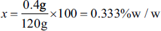
Animal Design
All animals were randomly divided into four groups, with each animal in a separate cage to avoid internal conflicts and cofounders. General behavior, left ankle joint circumference, arthritic index, and arthritic score were assessed. Group D was kept as the disease control (Figure 1). Animals were housed as per the institutional ethical norms.
Flowchart Showing the Design of the Animal Study.
General Behavior
General behavior of all animals was noted before, during, and after treatment. Mobility, phonation, gait, reaction, and mood were observed.
Left Ankle Joint Circumference
The left ankle circumference was measured using vernier calipers at the end of the 8th day and at the end of each week for 4 weeks in all four groups.
Arthritic Index
Arthritic index was calculated using the formula (Formula B), which indicates the relative left-ankle joint circumference. 24
Arthritic Index (%)

Formula B. Formula for arthritic index, day X indicates the day of measurement.
Arthritic Score
The arthritic score was assessed for each rat. 0 = rat walks and runs normally, 1 = rat walks and runs with difficulty, 2 = rat shows limping without retraction of the hind paw, 3 = rat shows limping with retraction of the hind paw (does not touch the hind paw on the floor), and 4 = rat crawls or lies down only.24, 25
At the end of the study, the disease control groups were treated with standard Aceclofenac gel for 4 weeks and were returned to the Central Animal House for rehabilitation.
Statistical Analysis
All groups were compared with one-way analysis of variance (ANOVA). Statistical significance was set at p < .05. All statistical analyzes were performed using the Statistical Package for Social Sciences (SPSS) software version 24.0.
Results
C. medica powder sample underwent a series of qualitative and quantitative tests to determine its anti-inflammatory potential.
Phytochemical Analysis of C. medica Powder
The C. medica powder sample showed the presence of a reddish precipitate. This shows the presence of sterols and a corticosteroid-like component in C. medica.
Spectrophotometric Analysis of C. medica Powder
Spectrophotometric analysis at 780 nm showed that the graph of C. medica powder was similar to that of methylprednisolone. This confirms the presence of a steroid-like component in C. medica, ensuring its anti-inflammatory property.
FTIR Analysis of C. medica Powder
The FTIR graph of C. medica showed the presence of functional groups consistent with a corticosteroid-like nucleus.
In Vitro Permeability Testing of C. medica Powder
Samples from the beaker at 1 and 1.5 h of diffusion demonstrated complete diffusion of C. medica powder, confirming in vitro permeability and topical penetration through the skin (Figure 2).
Graph Showing Spectrophotometric Readings of In Vitro Permeability of Citrus medica Powder Taken at 780 nm over 3 h, Demonstrating the Complete Diffusion of C. medica Powder Within 1.5 h.
Analysis of Gel
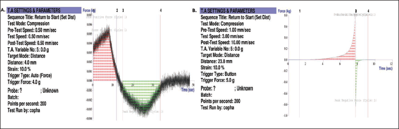
The prepared gel underwent mechanical evaluations to assess its suitability for topical application. The gel demonstrated a peak positive force of 1.387 kg, with a displacement of 0.063 mm and a force at the target of 0.005 kg. These values correspond to bloom strength within the lower range of bloom-strength gels, indicating a relatively soft texture. This is consistent with the requirements of International Organization for Standardization (ISO) 9665 standards for topical formulations and confirms the gel’s appropriateness for topical application (Figure 3A). The gel exhibited a firmness of 686.59 g, a spread distance of 7.66 mm, and a work of shear of 0.863 g s, with a force at the target of 0.702 kg. These parameters indicate that the gel maintains adequate firmness to hold its shape while still spreading easily under light pressure. The low shear work confirms the ease of application. At the same time, the moderate resistance indicated by the area under the curve and the peak force reflects optimal mechanical behavior for topical pharmaceutical gels. Overall, these findings support the gel’s favorable consistency, spreadability, and user compliance for topical application (Figure 3B).
(A) Figure Showing the Strength of Citrus medica Gel. (B) Figure Showing the Spreadability of C. medica Gel.
In Vivo Analysis of C. medica Gel on Arthritic Models
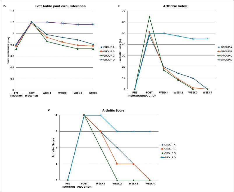
The general behavior of the rats was observed. After inducing arthritis, all the rats were aggressive, limping, lethargic, whining, and had a reduced appetite. By the fourth week, all the rats became calm, active, and alert, showing no signs of whining, and they had a good appetite. The general behavior of rats in Group C returned to their usual behavior much earlier than that of the other three groups (by the second week post-treatment). Group C exhibited the most significant reduction in swelling, erythema, and arthritic (pain) scores, with early resolution of limping and joint stiffness observed by the end of the first week (p value: .004), followed by complete recovery by the second week (p value < .001). Group B showed similar clinical improvements by the end of the third week, while Group A demonstrated delayed recovery, with noticeable improvement only by the fourth week. Group D (disease control) showed no resolution of swelling or erythema until the end of 28 days (Figure 4A–4C). Later, it was subjected to standard Aceclofenac gel treatment for 4 weeks. No signs of skin irritation or adverse effects were noted throughout the study period. Data from the disease control and treatment groups were analyzed by one-way ANOVA in the SPSS software version 24.0. Statistical analysis using one-way ANOVA confirmed that the differences among the groups were statistically significant (p value: .004) in Group C by the end of first week, which subsequently showed significant improvement in the further weeks (p value < .001) thus, demonstrating non-inferior therapeutic efficacy compared to the once daily (OD) application of C. medica gel (Group B) and standard Aceclofenac treatment group (Group A).
(A) The Graph Depicts Changes in Left Ankle Joint Circumference Compared to Baseline Values Recorded Before Arthritis Induction. The Results Were Also Compared with the Disease Control Group (Group D). (B) The Graph Shows the Changes in the Arthritic Index. (C) The Graph Illustrates That the Maximum Arthritic Score Recorded for Each Rat was Four, and This Score Subsequently Decreased Over the Following Weeks in Each Test Group.
Discussion
OA is a chronic degenerative joint disease that primarily affects the elderly population, causing pain, swelling, and reduced mobility.1–5 Current pharmacological interventions mainly focus on symptomatic relief and are often associated with systemic side effects. As a result, there is growing interest in exploring alternative and complementary therapies derived from natural sources with anti-inflammatory potential. C. medica melon (C. medica) has been traditionally used by local populations as a topical remedy for joint pain. However, there has been a lack of scientific validation for this practice.
The findings from this study demonstrated a significant reduction in joint swelling, the arthritic index (relative ankle joint circumference), and arthritis scores (pain score) in the twice-a-day C. medica melon gel-treated group (Group C) when compared to the standard group (Group A), which was treated with twice-a-day Aceclofenac gel, and the disease control group (Group D). Additionally, Group C also exhibited improvements in general behavior and ankle joint circumference in the early recovery phase of treatment (Figure 4A). In comparison, Group B showed similar clinical improvements only by the end of the third week, probably due to its OD application (Figure 4A–4C). Group A, the Aceclofenac gel-treated group, demonstrated delayed recovery, with noticeable improvement only by the fourth week, indicating a later recovery compared to the twice daily (BD) topical application of C. medica gel in OA. No signs of skin irritation or adverse effects were noted throughout the study period. Group D showed no improvement in signs of joint swelling, arthritic index (relative ankle joint circumference), or arthritic scores (pain score). It was later subjected to rehabilitation using standard Aceclofenac gel for 4 weeks to aid recovery.
Previous phytochemical analyzes of C. medica species have reported the presence of flavonoids, phenolic acids, and carotenoids, which are known for their antioxidant, anti-inflammatory, analgesic, and many other properties as well. These outcomes suggest that the C. medica melon possesses biologically active compounds capable of exerting therapeutic effects on inflamed joints, similar to the previously published anti-inflammatory effects of C. medica. In this study, the early improvement seen in the C. medica gel-treated group (Group C) may be attributed to the modulation of pro-inflammatory mediators and oxidative stress pathways due to the presence of flavonoids, carotenoids, and steroid-like nucleus in C. medica compared to the Aceclofenac gel-treated group (Group A), which is known for its anti-inflammatory and analgesic properties. The reduction in the arthritic pain index in Group C at the earliest could be attributed to the additional analgesic property of C. medica, as proven by previous studies.5–7
In our study, the BD application of the 0.33% w/w C. medica gel showed a significant reduction in inflammation within 2 weeks. OD application achieved comparable effects by 3 weeks, supporting its use as an effective topical treatment for OA. The topical application of C. medica melon may reduce the inflammatory burden locally, thereby minimizing systemic side effects in the long-term management of OA. The easy availability of C. medica melon also makes it a potentially valuable candidate for further development as a topical formulation for OA, especially in resource-limited settings. The observed behavioral and morphological improvements support the traditional use of C. medica melon and lay the foundation for future preclinical and clinical investigations.
Conclusion
In the present study, topical application of C. medica melon (C. medica) gel BD demonstrated earlier anti-inflammatory activity compared to OD application and standard BD Aceclofenac gel. These findings suggest that C. medica gel applied BD may serve as an effective and safe topical therapeutic option for the management of OA on a long-term basis. Further clinical studies are warranted to validate these results in human populations.
Limitations
The limitations of the present study include the use of basic parameters such as joint circumference, arthritic score, arthritic index, and general behavior to assess anti-inflammatory effects. Advanced investigations such as histopathological analysis, biochemical markers of inflammation, and imaging modalities were not performed, which may have provided a more detailed evaluation of disease modification. The therapeutic role of C. medica was assessed and compared with a standard non-steroidal anti-inflammatory drugs (NSAID) gel; future studies can also explore its effects in combination with standard therapies to determine any synergistic benefit. The efficacy of further concentrations of C. medica can be tested in upcoming studies.
Footnotes
Abbreviations
Acknowledgments
The authors would like to thank the Department of Pharmacology and the Central Animal House, PSG Institute of Medical Sciences and Research, for their support and for providing the necessary facilities to conduct the study.
Authors Contribution
Snehalakshmi Kavacheri Subramaniam: Conceptualization, data curation, formal analysis, funding acquisition, investigation, methodology, project administration, resources, supervision, validation, visualization, writing—original draft, writing—review & editing.
Umamaheswari Anbarasu: Data curation, formal analysis, investigation, methodology, supervision, validation, visualization, writing—review & editing.
Bhuvaneswari Krishnamurthy: Conceptualization, formal analysis, supervision, investigation, methodology, validation, visualization.
Manashaa Gnanaprakasam Poonguzhali: Data curation, project administration, visualization.
Declaration of Conflict of Interests
The authors declared no potential conflicts of interest with respect to the research, authorship, and/or publication of this article.
Ethical Approval
Approval was obtained from the Institutional Animal Ethics Committee (444/IAEC/2022).
Funding
The authors received no financial support for the research, authorship, and/or publication of this article.
Informed Consent
Not applicable.