
Abstract
Introduction:
Whole-body magnetic resonance imaging (WB-MRI) is increasingly used in asymptomatic individuals as part of health checkups due to its comprehensive nature and lack of radiation. It frequently detects incidental findings (IFs), the significance of which can vary from benign to potentially significant conditions. The study aims to evaluate the role of WB-MRI in detecting IFs in asymptomatic individuals and to assess the clinical relevance of these findings.
Materials and Methods:
A retrospective observational study was conducted on 62 asymptomatic individuals who underwent WB-MRI from November 2023 to April 2024. Findings were categorised by organ system and clinical relevance. Descriptive statistics were performed using Statistical Product and Service Solutions version 28.0 (SPSS v28.0).
Results:
Among 62 asymptomatic individuals, IFs were identified in multiple organ systems, most commonly in the spine (46.8%), abdomen (41.9%), musculoskeletal system (32.3%) and genitourinary system (24.2%). The majority of IFs were clinically significant: 33.9% required follow-up, 41.9% needed further investigation and 14.5% were potentially significant. Only 9.7% were benign.
Conclusion:
Our study found that WB-MRI effectively identified a wide range of IFs in asymptomatic individuals, many of which warrant clinical attention. Its role in preventive diagnostics is valuable, though appropriate pathways for follow-up and management are essential.
Introduction
Whole-body magnetic resonance imaging (WB-MRI) is a non-invasive, radiation-free imaging modality increasingly used for health screening. Its ability to provide high-resolution imaging across multiple systems has made it a valuable tool in detecting clinically silent abnormalities, known as incidental findings (IFs). These findings, though often asymptomatic, can range from benign lesions to potentially serious diseases, emphasising the importance of determining their clinical relevance.[1]
The increasing trend toward preventive health screening has led to the development and utilisation of advanced imaging modalities such as WB-MRI. Unlike other imaging techniques, WB-MRI provides a comprehensive assessment without ionising radiation, making it ideal for screening purposes.[2,3] The ability to detect IFs across multiple systems in a single scan has significant implications for early diagnosis and preventive management.[4] This study explores the role of WB-MRI in the detection of IFs in a healthy population and investigates how often these findings require medical attention.
Materials and Methods
This is a retrospective observational study conducted at a tertiary care hospital, Apollo One Hospitals, Greams Road, Chennai, over a period of six months, from November 2023 to April 2024. We have a total of 62 asymptomatic adults, aged 19–75 years, in this study period. Healthy individuals undergoing WB-MRI for a routine health checkup were included in the study. People with known malignancies or chronic illnesses were excluded.
All patients fulfilling the inclusion criteria were subjected to the study. WB-MRI sequences were performed on a 1.5 T (Philips BlueSeal helium-free MR operations magnet [BLUE SEAL]). The sequences included were axial and coronal sections of T2-weighted imaging, axial and coronal sections of Short Tau Inversion Recovery (STIR) and axial sections of Diffusion-Weighted Imaging (DWI).
IFs in WB-MRI were recorded in different organ systems. The IFs were classified as benign, for which no follow-up is required; indeterminate, which requires imaging follow-up; potentially significant, which requires further investigation; and significant, which requires urgent referral to a specialist.
Summary statistics were presented with mean (SD) and frequency (percentage) for the continuous and categorical factors, respectively. The descriptive statistics were done using SPSS (IBM, 28.0).
Results
Demographics
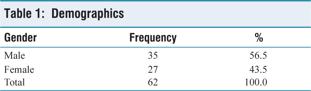
In our study, we found that out of 62 people, the mean age was 54.3 years, with a standard deviation was ± 11.4 years. The range is from 19 to 75 years. The males were 35 (56.5%) and the females were 27 (43.5%). The males were slightly higher than the females [Table 1].
Demographics
Incidental Findings by Organ System
Spine
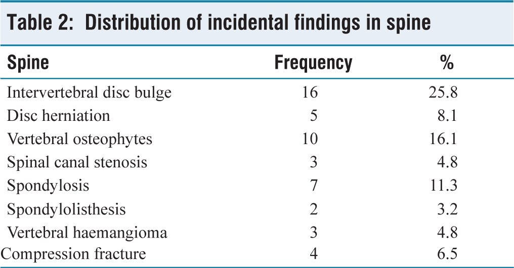
We found that the most common IFs were frequently in the spine (46.8%). Of which, 25.8% had disc bulge, 16.1% had osteophytes and 11.3% had spondylosis [Table 2].
Distribution of incidental findings in spine
Abdomen
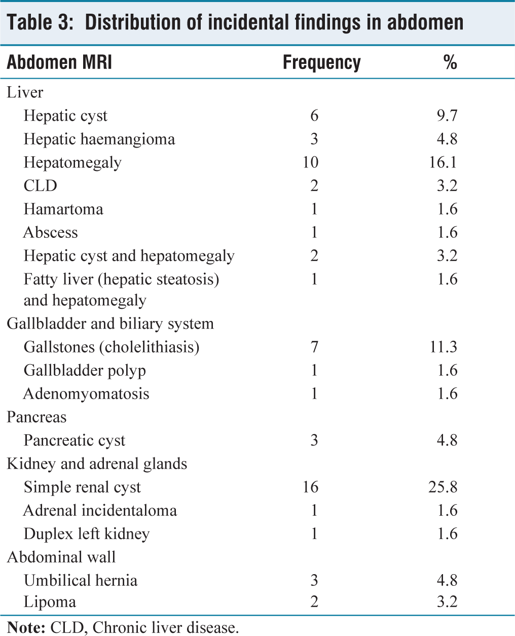
The second most common IFs were located in the abdomen (41.9%). In the abdomen, hepatomegaly was present in 16.1% of cases and renal cysts were found in 25.8% [Table 3].
Distribution of incidental findings in abdomen
Musculoskeletal System
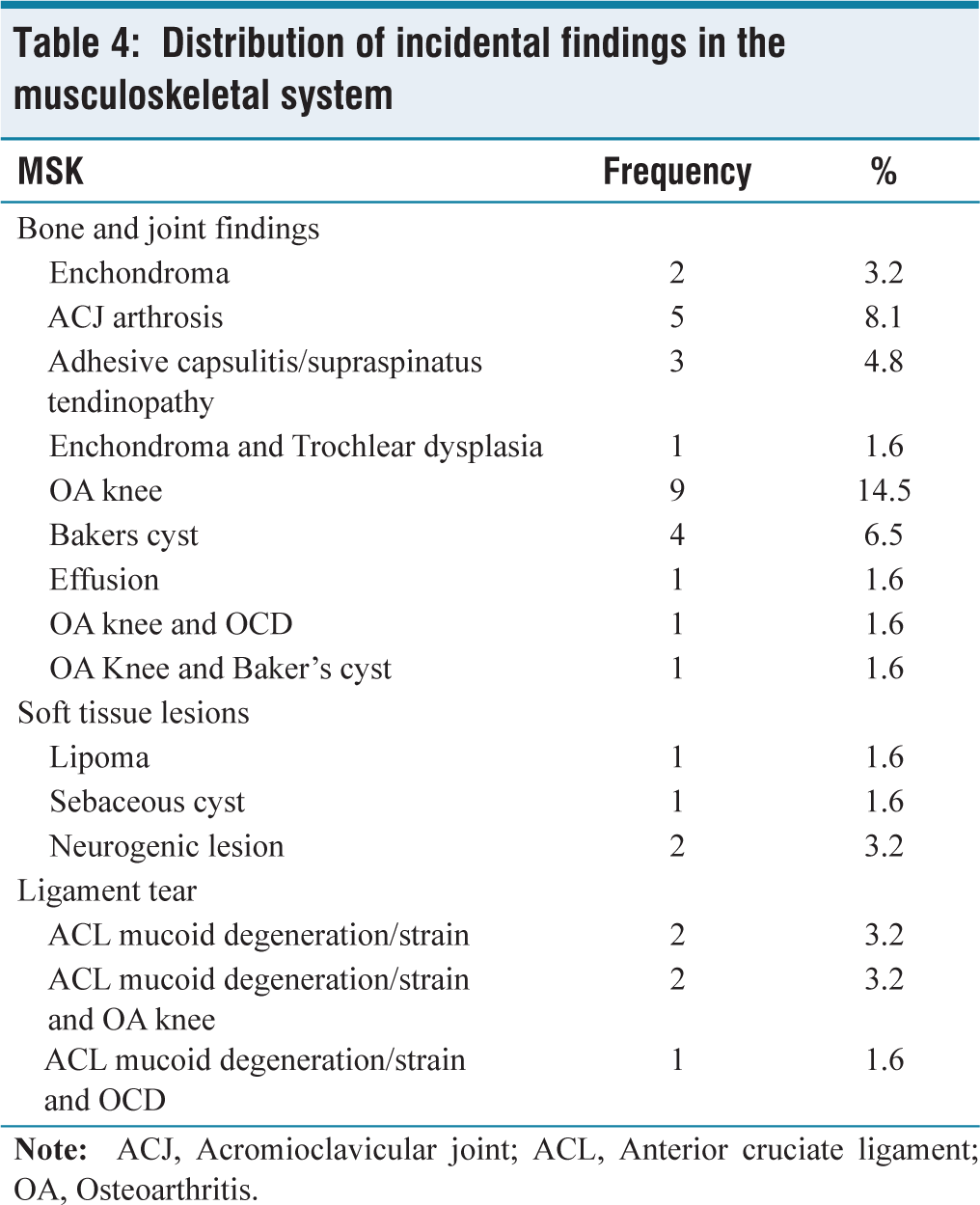
The third most common IFs occurred in the musculoskeletal system (32.3%). In musculoskeletal system (MSK), osteoarthritis of the knee (14.5%) and acromioclavicular arthrosis (8.1%) were prevalent [Table 4].
Distribution of incidental findings in the musculoskeletal system
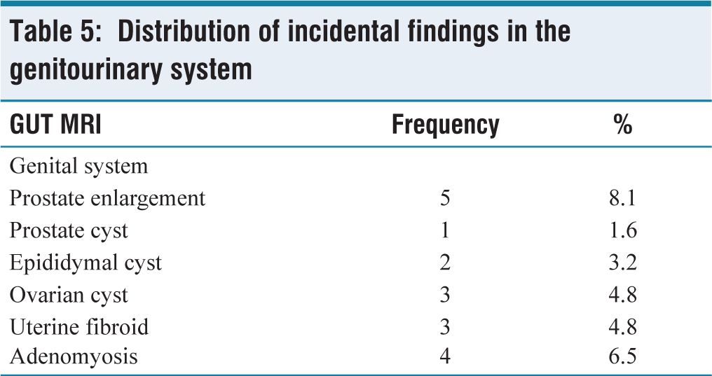
Genitourinary System
The fourth most common IFs were found in the genitourinary system (24.2%). Of which, prostate hypertrophy (8.1%) and adenomyosis (6.1%) were frequently detected [Table 5].
Distribution of incidental findings in the genitourinary system
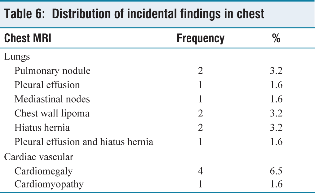
Chest
The fifth most common IFs were observed in the chest (12.9%). Specifically, cardiomegaly accounted for 6.5%, and hiatus hernia accounted for 3.2% [Table 6].
Distribution of incidental findings in chest
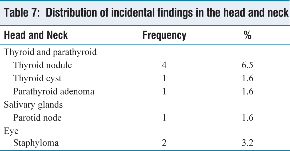
Head and Neck
The sixth most common IFs were found in the head and neck (9.7%), with thyroid nodules 6.5% and staphyloma 3.2% [Table 7].
Distribution of incidental findings in the head and neck
Clinical Relevance of Whole-body MRI Incidental Findings
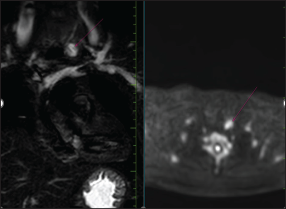
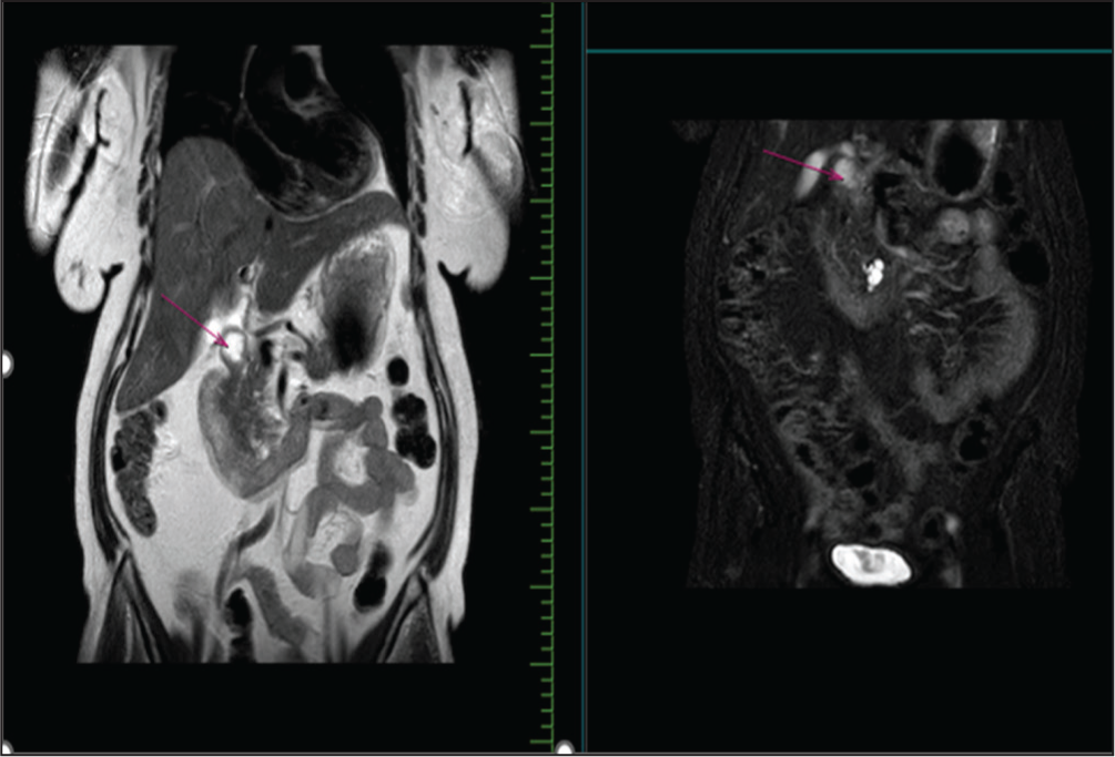
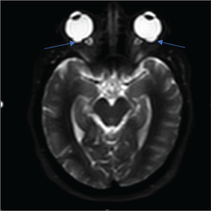
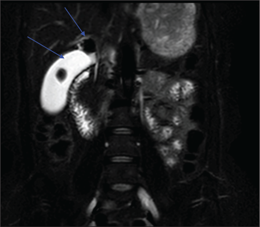
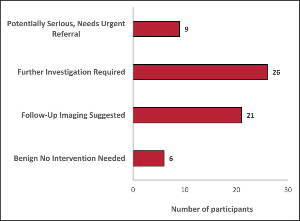
Out of 62 people, 9 (14.5%) had significant findings such as adrenal incidentaloma, parathyroid adenoma [Figure 1] and hepatic abscess, which need urgent referral and other interventions. Twenty-six (41.9%) had potentially significant findings such as compression fracture, spondylolisthesis, neurogenic lesion, chronic liver disease, branch duct IPMN [Figure 2], prostate hypertrophy, posterior staphyloma [Figure 3] and thyroid nodule that require further investigations. Twenty-one (33.9%) had indeterminate findings such as lipoma, hiatus hernia, enchondroma, hamartoma, lung nodule, cholelithiasis [Figure 4] and spinal canal stenosis that require follow-up imaging. Six (9.7%) had benign findings such as hepatic and renal cysts and cardiomegaly which require no follow-up [Figure 5].
Coronal T2 (Right) and STIR (Left) of abdomen showed multiseptated cystic lesion in the uncinated process of pancreas with suspicious communication to a side branch (pink arrow), a potentially significant finding; MRCP is suggested. On MRCP a branch-duct intraductal papillary mucinous neoplasm was found out
Axial STIR coronal images of the head and neck region showed bilateral posterior staphyloma, a potentially significant finding that requires further investigation
STIR coronal section of abdomen showed two well-defined STIR hypointense filling defects (blue arrows) in the body and neck of the gallbladder, cholelithiasis. The finding is indeterminate and requires follow-up imaging
Clinical relevance of whole-body MRI incidental findings
Discussion
This study demonstrated the utility of WB-MRI in screening asymptomatic individuals, revealing a significant number of IFs, with more than 90% requiring some form of follow-up or intervention. The spine and abdomen were the most frequently affected regions. While WB-MRI improves early detection, it also introduces challenges such as overdiagnosis, patient anxiety and the need for additional diagnostic workup. These findings support the integration of WB-MRI into preventive screening, but clinical guidelines must be in place to manage IFs appropriately.
IFs were identified in 62 asymptomatic individuals, with the most common in the spine (46.8%), abdomen (41.9%), musculoskeletal system (32.3%) and genitourinary system (24.2%). Hegenscheid et al. (2012), in a study of 2,500 adults, 36.2% had potentially relevant IFs, with the abdominal organs (6.8%), urinary tract (6.8%) and skeletal system (6.0%) being most affected. Ulus et al. (2016), among 118 subjects, 70% had benign lesions, with renal cysts, liver haemangiomas and uterine leiomyomas being common. Our study reported a higher prevalence of IFs, particularly in the spine and musculoskeletal system, which may be attributed to differences in imaging protocols or population demographics.
Findings were categorised as benign (9.7%), requiring follow-up (33.9%), needing further investigation (41.9%) and potentially serious (14.5%). Hegenscheid et al. (2012), out of 1,330 IFs, 0.4% required immediate referral, 36.4% were benign and 57.7% were unclear. Ulus et al. (2016) detected two malignant and one precancerous lesions among the 118 subjects. Our study indicates a higher proportion of findings necessitating further investigation or being potentially serious, suggesting a more proactive approach in categorising and managing IFs.
Our study found that the spine (46.8%) and the abdomen (41.9%) had the highest incidence of IFs. Hegenscheid et al. (2012) found that the abdominal organs and urinary tract were most commonly affected.[5] Ulus et al. (2016) found that renal cysts and liver haemangiomas were among the most frequent findings.[6] While abdominal findings were common across studies, your study reports a notably higher incidence in the spine, which may reflect differences in imaging techniques or population characteristics.
Our study found the utility of WB-MRI in detecting clinically significant IFs in asymptomatic individuals, highlighting the importance of structured follow-up pathways to manage these findings effectively.
Conclusion
In conclusion, we highlight the role of WB-MRI serves as an effective non-invasive screening tool for detecting clinically relevant IFs in asymptomatic individuals. A structured follow-up pathway is crucial to balance the benefits of early detection against the risks of overdiagnosis and unnecessary intervention.
Footnotes
Acknowledgements
To the Research Department, Apollo Main Hospital, Chennai.
Declaration of conflicting interests
The authors declared no potential conflicts of interest with respect to the research, authorship and/or publication of this article.
Funding
The authors received no financial support for the research, authorship and/or publication of this article.
Institutional ethical committee approval number
Approval was obtained from the Institutional Ethical Committee on 18 March 2025.
Informed consent
Informed consent has been obtained from the patients who met the eligible criteria.
Credit author statement
ND Participated in conceptualisation, methodology, data collection, data analysis, literature search and manuscript preparation.
SPS, AS and KN were involved in conceptualisation, literature search, validation, supervision and manuscript revision.
All the authors have reviewed and approved the manuscript.
Data availability
The datasets used and/or analysed during the current study are available from the corresponding author upon request.
Use of artificial intelligence
Nil.