
Abstract
Background
Cerasus serrulata has an excellent antioxidative, antibacterial, and anti-alopecia effect, but its active ingredients and the underlying mechanism are currently unclear, in addition to the fact that the active products vary with the extraction methods.
Aim
Therefore, this study aims to clarify the active components and antioxidative property of C. serrulata extracted by two different methods (low-temperature vacuum extraction and ethanol extraction) and explore its potential biofunction.
Materials and Methods
First, the compositions in extracts were characterized by gas chromatographic mass spectrometry (GC-MS) and liquid chromatography-mass spectrography (LC-MS). Then, the antioxidative activity of both extracts was evaluated using free radical scavenging assay (ABTS) and ORAC. Next, the antibacterial activity of both extracts was measured via a bacteriostatic circle experiment. The anti-alopecia effects of both extracts were studied using a mouse model of alopecia. The hematoxylin and eosin (H&E) staining was used to evaluate hair follicles; vascular endothelial-derived growth factor (VEGF) and interleukin-6 (IL-6) expression in mouse skin were evaluated by immunohistochemistry, while β-catenin expression in tissues was used by immunofluorescence. The expression of FGFR and insulin-like growth factor 1 (IGF-1) in the skin was determined to evaluate using real-time quantitative polymerase chain reaction (RT-qPCR). The levels of DHT and 5α-reductase in the experimental mice were determined by ELISA.
Results
The GC-MS analysis results showed that the extracts were rich in alkanes and aromatic compounds, specifically benzaldehyde, which was the highest component in the cold vacuum extraction, while pipecolic acid was the main component in the ethanol extract. This indicates that C. serrulata has components that exhibit strong inhibitory ability against the growth of five common bacterial strains, expressing excellent free radical scavenging ability. In addition, the active ingredients in C. serrulata were found to significantly deregulate the protein levels in the skin of androgenetic alopecia (AGA) mice, with the levels of IL-6. They were also able to significantly inhibit VEGF reduction. The β-catenin was being significantly upregulated in skin tissue. In addition, FGFR and IGF-1 gene expression also increased. The 5α-reductase and dihydrotestosterone (DHT) can be reduced in the C. serrulata treatment.
Conclusion
Our data indicate that C. serrulata extract could be beneficial supplements of natural antioxidative, antibacterial, and anti-alopecia agents.
Introduction
Androgenetic alopecia (AGA) is the most common form of alopecia, which is characterized by a progressive decrease in the non-vellus, diameter, length, and pigmentation of the hair (Adil & Godwin, 2017). The World Health Organization (WHO) reports that AGA is one of the top 10 esthetic problems, affecting approximately 50% of women and 80% of men during their whole life. Furthermore, AGA results from the effects of the testosterone metabolite dihydrotestosterone (DHT) on androgen-sensitive hair follicles. The medical treatment of AGA includes topical minoxidil, antiandrogen agents, and 5α-reductase inhibitors. In fact, only oral finasteride and topical minoxidil have been authorized by the US Food and Drug Administration and the European Medicines Agency. The causes and underlying mechanisms of AGA are complex, and it may be caused by a variety of factors but mainly by genetics and endocrine disorders (Kelly et al., 2016; Lolli et al., 2017). Many researchers prefer to search for plants’ extract containing natural ingredients instead of artificially synthesized ones because plant extract is usually considered as safer and more eco-friendly. Meanwhile, research on alopecia has increased to elucidate its mechanisms for further good application (Leem et al., 2018).
Cerasus serrulata is an ornamental and medicinal plant belonging to the genus Cerasus in the Rosaceae family (Yi et al., 2020). It has reported that C. serrulata extract such as phenolic acids, flavonoids, alkaloids, polysaccharides, and essential oils depend on different extraction methods that exhibit a variety of biological activities, including excellent antioxidative ability, broad-spectrum antibacterial function, antiviral property, anti-inflammatory, and anti-alopecia effect (Mansoori et al., 2021; Peixoto et al., 2020; Zhang et al., 2014). The mouse model of alopecia induced by acetone injection has indicated that the Wnt/β-catenin signaling pathway may be the key to solving hair growth abnormalities, which can be used to screen target drugs and elucidate potential mechanisms of action (Gentile & Garcovich, 2019). Furthermore, research has suggested that the peroxidation stress response of the cortex, free radical clearing, and hair growth metabolism are closely related. Therefore, there is a need to study the relationship between the antioxidant capacity of C. serrulata and the Wnt/β-catenin pathway regulation.
In this study, two extracts of C. serrulata were obtained using different methods: C. serrulata ethanol extract (CSEE) and C. serrulata cold vacuum extract (CSCE). Next, the active ingredients in these extracts were determined by gas chromatographic mass spectrometry (GC-MS) and liquid chromatography-mass spectrography (LC-MS), followed by the evaluation of the antibacterial activity and anti-free radical scavenging ability. Then, CSCE and CSEE were administered in an experimental mouse alopecia model to determine the potential anti-alopecia effects of these extracts using H&E staining, immunohistochemistry, immunofluorescence, and ELISA. Thus, this study aims to elucidate the effects of C. serrulata on alopecia and provide a reference for the development of novel therapeutic options for the treatment of this common hair disorder.
Materials and Methods
Materials and Chemicals
The extract was stored in the Institute of Nature Medicine and Green Chemistry (Guangdong University of Technology, Guangzhou, China) as a voucher specimen (no. ZLY-20210403-006). The plant materials were authenticated by Prof. Nian Liu (Zhongkai University of Agriculture and Engineering, Guangzhou, China), according to the morphological description presented in the China Species. Refined peanut oil was purchased from Luhua Group Co., Ltd., Shandong Province. Testosterone propionate was purchased from the Ningbo Second Hormone Factory, China. Minoxidil topical solutions were purchased from the Wansheng Pharmaceutical Co., Ltd. DHT and 5α-reductase were obtained from the Jiangsu Sumeike Biological Technology Co., Ltd. library.
Extraction Methods
Ethanol Extraction
Briefly, 50 g of C. serrulata powder, 100 mL of anhydrous ethanol and 20ml of deionized water as a solvent were added to a Soxhlet and allowed to stand for 3 h. After ethanol and deionized water are removed, spray dry under vacuum at 40°C to achieve constant weight, brown solid extracts were obtained and stored for further use. The corresponding yield was calculated based on the weight of the extract.
Cold Vacuum Extraction
Briefly, 50 g of C. serrulata flower petals were placed in low-temperature vacuum extraction equipment under the following conditions: extraction temperature, 40°C–50°C; extraction pressure, 80–100 kPa; and extraction time, 6 h. The resultant solution was filtered through a membrane with a 0.22-micron pore size to obtain the CSCE.
Component Analysis of C. serrulata
The studies of CSCE were performed on a DSQ-II Ultra (Thermo Electron, UAS) GC-MS equipment with a DB-5MS capillary column (0.25 mm × 30 m, 0.25-µm film thickness) (Zhang et al., 2018). The GC temperature program was first held at 40°C for 1 min, then increased at a rate of 5°C/min from 40°C to 280°C, and kept at 280°C for 5 min. The injector temperature was 250°C, the sample injection volume was 1 µL, the carrier gas was helium, the flow rate was 1.0 mL/min, and the split ratio was 100:1. Following were the mass spectrometry conditions: electron energy, 70 eV; electron pressure before columniation, 70 kPa; and ion source temperature, 230℃. Using the Kovats method (Huang et al., 2022), retention indices (RIs) were computed for each chemical in comparison to n-alkane standards (C6–C40). By comparing RIs and matching the recorded mass spectra of each chemical with the published literature and the NIST Chemistry digital book, the contents were identified.
The separation and identification of CSEE was performed by ultra-high performance LC-MS (Ultimate 3000 LC; Thermo Fisher Scientific) equipped with a C18 column (Zorbax Eclipse C18, 1.8 µm × 2.1 × 100 mm; Agilent), combined with the annotation and classification of mass spectrometry database information. The separation conditions were as follows: column temperature, 30°C; flow rate, 0.3 mL/min; mobile phase A, water + 0.1% formic acid; mobile phase B, pure acetonitrile; injection volume, 2 µL; and active autokinetic nozzle temperature, 4°C.
Determination of the Antioxidant Activity
The ABTS free radical reserve formulation was comprised of 20 mL of 7 mmol/L ABTS and 352 L of 140 nmol/L potassium persulfate, which was allowed to stand overnight at 25°C under dark conditions (Wołosiak et al., 2021). The solution was diluted into a working solution using anhydrous ethanol with an absorbance of 0.7 + 0.02 at 30°C and wavelength of 734 nm, to which 0.1 mL of the extracted solution and 5 mL of the ABTS working solution were added. After mixing evenly and allowing to stand for 10 min under dark conditions at room temperature (Thermo Fisher Scientific, USA), the absorbance of 0.1 mL sample and 5 mL ethanol mixed uniformly was measured at 734 nm (A0), using ABTS solution as the blank (Ar):
ABTS + free radical clearance rate (%) = [1 ‒ (At ‒ A0)/Ar] × 100,
where At is the absorbance of the sample and Ar is the absorption value of control.
Oxygen Radical Absorption Capacity
To determine the oxygen radical absorption capacity of the extracts, azo compounds were used as free radical sources, fluorescein sodium was used as an indicator, and water-soluble Vitamin E (Trolox) was used as a reference substance for antioxygen analysis (Gunawardena et al., 2019). Oxygen free radicals oxidize the fluorescent probes via hydrogen transfer, quenching the fluorescent probes overtime (Kameya et al., 2014). Antioxidants can prevent the oxidation of the fluorescence probe and delay fluorescence quenching until the antioxidant activity in the sample is exhausted.
Bacteriostatic Zone Test
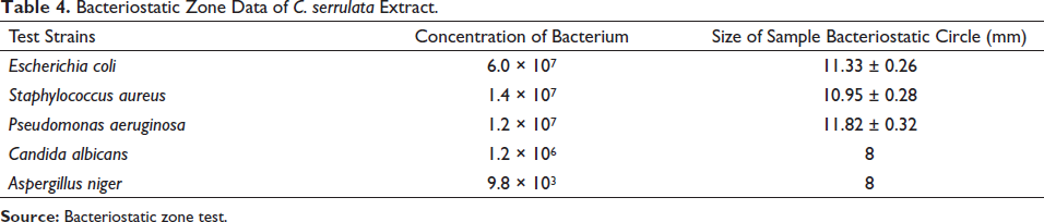
The sterilized AGAR medium was heated until it was completely melted. The medium was then poured into Petri dishes, with 15 mL in each plate (lower layer), and allowed to set. The melted PDA medium was cooled to about 50°C and mixed with test bacteria. Then, 5 mL of the medium mixed with bacteria was added to the solidified medium to be solidified (upper layer). Each sample was tested in triplicates for each strain (one negative control and one positive control). Briefly, the Oxford cup was placed on the surface of the plate with the aperture facing upwards, onto which the culture medium with the test bacteria was poured. After coagulation, the Oxford cup was removed and 100 µL of CSCE and 250 mg/mL of CSEE solution were added. Sterile distilled water was used as the negative control, while silver ion bacteriostatic agent was used as the positive control. After the sample solution was fully diffused and dried and transferred to culture. The bacteria and fungi were cultured at 37°C for 24 and 36 h, respectively. The diameter of the inhibition zone was measured using vernier calipers, and the resulting measurements were averaged. The diameter of the Oxford cup used in the test was 8 mm, while the diameter of bacteriostatic zone was >10 mm, which was estimated to exhibit antibacterial activity in vitro. The bacteria inhibition zone diameter of ≤10 mm was judged to have no bacteriostatic effect (Jing et al., 2021).
Animals
C57BL/6LJ mice (n = 70) (male, 5 weeks old) (weight, 22–24 g) were purchased from the Guangdong Medical Laboratory Animal Research Center. The mice were randomly divided into seven groups (n = 10, with five mice in each group fed for 1 week): control, testosterone (TES), minoxidil, CSCE-H, 5-fold diluted CSCE-L, 10% CSEE-H, and 1% CSEE-L. Except for the control group, all mice were injected with testosterone propionate 5 mg/kg on their back (Chen et al., 2017). After 4 weeks of successful modeling, the mice were administered with the corresponding samples once a day for 8 weeks.
Hematoxylin and Eosin Staining of Hair Follicles
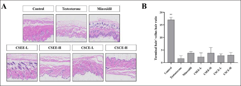
An H&E staining kit (Solarbio, Beijing, China) was used to evaluate the hair follicles. At the end of week 8, samples of skin from the back of the alopecia model mice were obtained from each group (1 × 1 cm paraffin sections) and stained with hematoxylin and eosin (H&E). The paraffin sections were placed in xylene (20 min), alcohol (10 min), and distilled water (5 min) for dewaxing treatment, sequentially, followed by staining with hematoxylin (3–8 min). After staining, the sections were observed microscopically and analyzed in Image-Pro Plus to calculate the ratio of stained hair follicles to unstained hair follicles (Shin et al., 2022).
Immunohistochemical Staining
The mouse skin (2 × 0.8 cm) was rinsed to remove residual blood, dried, and sliced for dewaxing before placing it in a repair chamber comprised of citric acid antigen repair buffer (pH 6.0) in a microwave oven for antigen repair. After heating and cooling, the samples were washed with PBS (pH 7.4) (3 × 5 min) and 3% hydrogen peroxide solution was added, and they were incubated under dark conditions for 25 min. Next, the samples were washed again with PBS at room temperature and 3% BSA was added, and they were allowed to stand for 30 min at room temperature. The closure was removed and primary antibodies were added (4°C), followed by washing with PBS. Then, the second antibody was added, and the sections were incubated at room temperature under dark conditions for 50 min. After washing once more with PBS, the slices were dehydrated and observed microscopically (Nikon, Japan) to measure the vascular endothelial-derived growth factor (VEGF) and interleukin-6 (IL-6) levels in the mouse skin samples (Kim et al., 2019).
Immunofluorescence Assays
Next, the sections were placed in xylene, anhydrous ethanol, 85% alcohol, 75% alcohol, and distilled water for washing (5 min). Paraffin sections were repaired with serial antigens. After drying, the slices were incubated in spontaneous fluorescent quenching agent for 5 min, washed with running water for 10 min, incubated with BSA for 30 min, and treated with one and two resistances. After washing with PBS, DAPI dye was added to the sections, and they were incubated at room temperature for 10 min. Finally, the sections were washed with PBS before observing β-catenin expression level with an inverted fluorescence microscope (Nikon).
Real-Time Quantitative Polymerase Chain Reaction
RNA was extracted from the sample tissues using RNA extraction reagents (Khantham et al., 2021). The RNA concentration and purity were evaluated using Nanodrop 2000 in a reverse recording reaction system using 20 µL (MB-2917A, MB-6079A, Jiangsu Meibiao Biotechnology Co., Ltd.) as recommended. This was mixed and centrifuged before analyzing using the following reverse recording program: 25°C for 5 min, 42°C for 30 min, and 85°C for 5 s. The RNA extract was amplified for 10 min: 40 cycles at 90°C (from 60°C to 95°C), followed by heating by 0.3°C per 15 s.
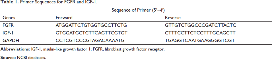
The reaction consisted of 5× reaction buffer (4 µL), oligo (dT)18 primer (100 µM) (0.5 µL), random hexamer primer (100 µM) (0.5 µL), Servicebio RT enzyme mix (1 µL), total RNA × 0.1 ng–5 µg/10 pg–0.5 µg, and free water add to 20 µL (Waltham, MA, USA, Thermo Scientific). Primers used for real-time quantitative polymerase chain reaction (RT-qPCR) are listed in Table 1. GAPDH was used as the internal control and the expression of each target gene was normalized by GAPDH level.
Primer Sequences for FGFR and IGF-1.
Statistical Analysis
All data were analyzed statistically using GraphPad.8.0.2. Significant differences were calculated by single-factor analysis of variance.
Results and Discussion
Components of C. serrulata Extract
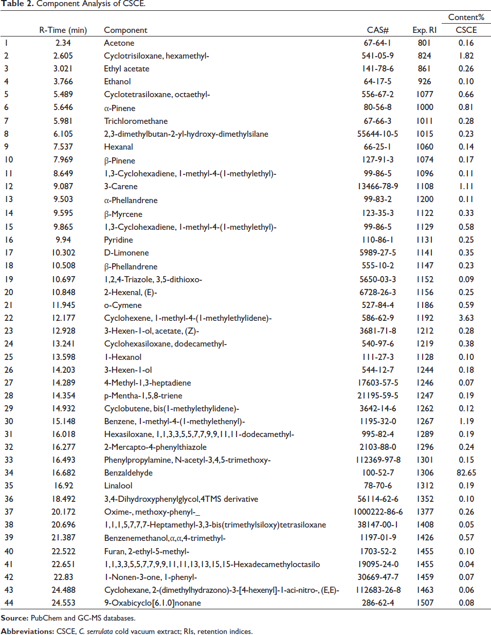
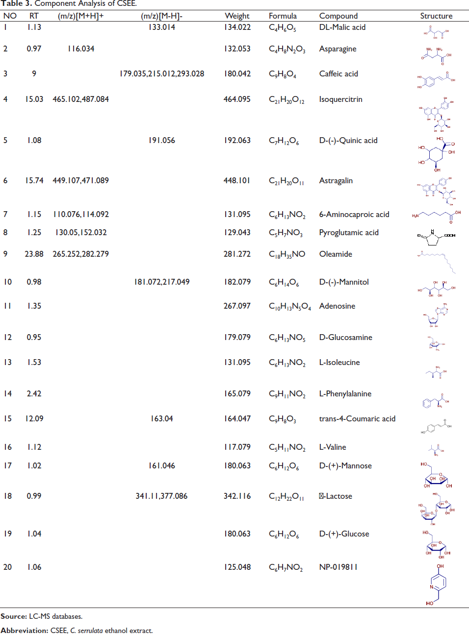
As shown in Tables 2 and 3, benzaldehyde (82.65%) was the highest component in CSCE and was significantly more abundant than the other components (Patsner & Harti, 2020). This suggests that benzoin plays a major role in the results obtained for the hair and skin of the experimental mice. Other aromatic compounds included hexamethyl-cyclotrisiloxane (1.82%), 3-carene (1.11%), 1-methyl-4-(1-methylethylidene)-cyclohexene (3.63%), and 1-methyl-4-(1-methylethenyl)-benzene (1.19%). As shown in Table 3, the main compounds in the CSEE were DL-malic acid, asparagine caffeic acid, isoquercitrin, and D-(-)-quinic acid. Studies have shown that aromatic compounds can promote the decomposition of oil in the skin, which can alleviate the occurrence of seborrheic alopecia to an extent (Szollosi et al., 2016). These compounds, most of which have a slightly acidic pH, are ideal for the scalp environment, leaving the hair soft and smooth. C. serrulata is found widely across the globe (Liu et al., 2019). In addition to the excellent ornamental value of this flower, its potential as a sustainable resource of plant compounds is gradually being developed. According to previous studies, C. serrulata has an excellent antioxidant capacity and free radical scavenging ability (Damar et al., 2012). In fact, some oxidative stress response signaling pathways have recently been identified in human experiments in relation to hair loss, with the considerable progress being made. Herein, extracts obtained using low-temperature vacuum extraction methods exhibited the highest content and activity of aromatic compounds and are thereby likely to achieve the best results for preventing alopecia.
Component Analysis of CSCE.
Component Analysis of CSEE.
Anti-free Radical Scavenging Ability
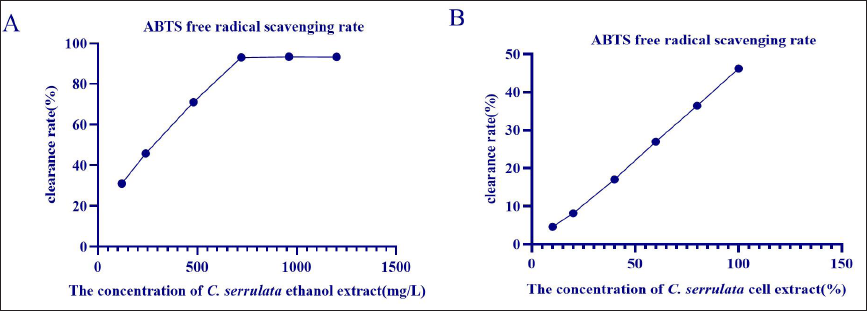
ABTS is a medium used to measure laccase activity in compost (Ruksiriwanich et al., 2022). The rate at which laccase oxidizes ABTS determines the levels of laccase activity. As shown in Figure 1, C. serrulata showed a good clearing ability for ABTS free radicals. Moreover, the free radical scavenging ability of CSEE was found to increase with concentration, reaching the highest scavenging rate concentration at 720 mg/L before stabilizing gradually. The maximum clearance rate reached over 90%. The maximum scavenging rate for ABTS radicals by CSCE was proportional to the content; however, the maximum free radical scavenging rate was found to be lower than 50%. In summary, CSEE exhibited stronger ABTS radical absorbance capacity. According to the free radical theory, the accumulation of free radicals is detrimental, not only interfering with the metabolism of normal cells, but also triggering chain reactions, which can lead to the development of different diseases. The results presented here indicate that CSEE had a stronger ability to clear ABTS, with the effective content of the CSEE being higher than that of the CSCE.
Antibacterial Activity of C. serrulata Extract
As shown in Table 4, CSCE has a bacteriostatic effect on five types of bacteria flora. Among these, the antibacterial effect on Pseudomonas aeruginosa was the most marked. Pseudomonas aeruginosa is common in humans and is harmless in small amounts. However, this fungus also has the ability to penetrate blood, adversely affecting the throat, the small and large intestine, and the heart valves. Furthermore, Pseudomonas aeruginosa infection in the skin can lead to dermatitis, baldness, and itching (Singh et al., 2019). The main components containing weak acidity in C. serrulata extract may inhibit the growth of Pseudomonas aeruginosa in the back skin of the experimental mice, thus inhibiting the damage to dermal hair follicles by bacteria (Moosavi et al., 2020). CSCE had the highest antibacterial effect against Pseudomonas aeruginosa.
Bacteriostatic Zone Data of C. serrulata Extract.
Evaluation of Terminal and Vellus Hair
The control group had a higher density of hair follicles, with almost all terminal hair in the field of vision, and only a small amount of vellus hair. In the TES group, the total number of hair follicles decreased, while the number of vellus hairs increased significantly. In the minoxidil group, many terminal hairs and few trichomes were observed in the visual field. In the other experimental groups, CSCE-H showed the highest ratio of terminal hair to setae, with a high density of hair follicles, almost all terminal hair in the visual field, and less vellus hair. These results indicate that the effect of the four types of C. serrulata extract was better than TES, while CSCE-H showed the best effect, which was better than the effects observed in the minoxidil group. Therefore, in terms of alopecia prevention, the extracts could mitigate hair loss caused by TES injection to a certain extent.
Immunohistochemistry for the Detection of VEGF and IL-6 in Skin
VEGF plays a key role during tissue repair (Liu et al., 2022), which is of great significance for maintaining skin homeostasis. As shown in Figure 3B, the VEGF levels in the control and minoxidil groups were significantly higher than those in the TES group. The experimental groups all promoted the increase of VEGF level, among them, CSEE-H, CSCE-L, and CSCE-H significantly increased the level of VEGF (**p < 0.01). IL-6 is a proinflammatory cytokine that is paradoxically associated with skin healing and inflammation. IL-6 expression was evaluated and found to be more highly expressed in the TES group than in the control group (Lee et al., 2013). When compared to the TES group, the other experimental groups showed a significant therapeutic effect, wherein IL-6 expression was effectively decreased. CSCE was found to overtly decrease IL-6 expression, while no significant difference was observed between the CSEE-L and the TES groups. Among these, CSCE-H was found to have the best therapeutic effect.
These results highlight the close association between VEGF and IL-6 expression in alopecia. Thus, the extracts used in the experimental mouse model could be used to regulate these four growth factors to achieve an anti-alopecia effect, wherein CSEE-H showed a better performance than minoxidil.
Immunofluorescence
It is well-known that the one of the key factors in stimulating hair growth is the stimulation of Wnt signaling in dermal cells, wherein β-catenin is an important element (Gentile et al., 2019; Nakamura et al., 2018). As shown in Figure 4, the content of β-catenin in the TES group was found to be significantly decreased (p < 0.01) when compared with that in the control group. After treatment, in comparison to that of the TES group, the treatment effect of CSCE-L and CSCE-H significantly improved the expression of β-catenin (p < 0.01), wherein CSCE-L showed the best treatment effects.
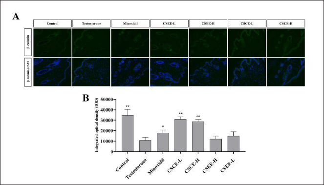
These results highlight the correlation between the Wnt signaling pathway and the alopecia, further confirming that the extracts tested could prevent hair loss by upregulating in this pathway. Furthermore, this correlation was found to be negative in relation to the concentration of the CSCE within a certain range.
FGFR and IGF-1 Expression
FGFR is mainly expressed in the inner root sheath (Kawano et al., 2005), which can control the direction of hair follicle growth. Insulin-like growth factor (IGF) is known to promote hair growth. IGF-1 is secreted by the dermal papilla and is one of the main growth regulators of epithelial cells produced by the paracrine and autocrine systems. As a strong mitogen, IGF-1 stimulates the growth of hair follicle epithelial cells.
As shown in Figure 5, FGFR and IGF-1 expression in the TES group was found to be markedly lower than in the control group. By contrast, the expression of FGFR and IGF-1 in other treatment groups was found to be higher than in the TES group, wherein expression in the group treated with CSCE-H was markedly higher than that in the TES and minoxidil groups, indicating that CSCE-H exhibited the best therapeutic effects.
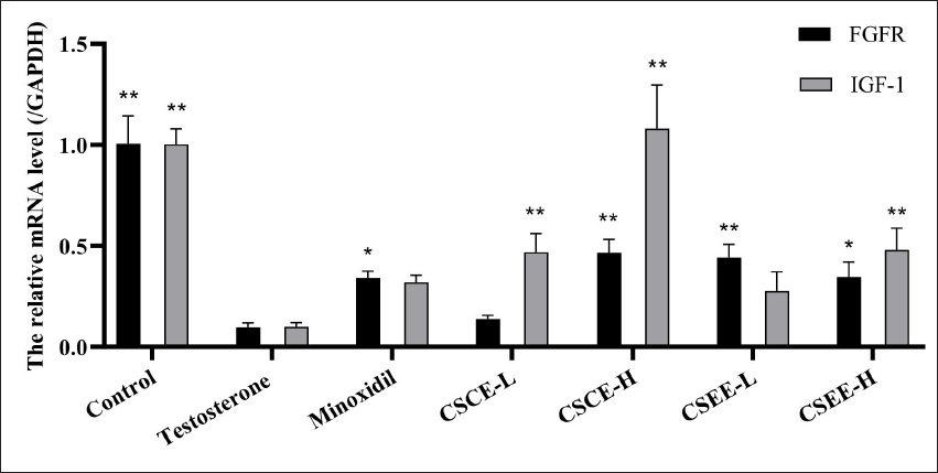
The comparison between the results of the experimental groups suggests that IGF-1 and FGFR produced by paracrine and autocrine can be influenced by experimental extracts, with these effects potentially either directly or indirectly related to alopecia (Zhang et al., 2019). These findings indicate that the extract tested can interfere with hair loss caused by androgens through this pathway. Furthermore, this association was positively correlated with the concentration of the CSCE within a certain range.
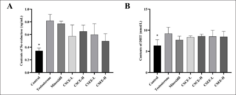
DHT and 5α-reductase Levels
5α-reductase can catalyze the conversion of testosterone to DHT (Audet-Walsh et al., 2017). The increased levels of DHT activity facilitate binding to androgen receptors, leading to stimulation of hair follicles to initiate the degenerative phase at a faster pace, ultimately resulting in alopecia. Therefore, if 5α-reductase and DHT can be reduced in vivo, the development of seborrheic alopecia could be inhibited to an extent.
As shown in Figure 6, in the experimental mice, the expression of DHT in the TES group was found to increase significantly when compared to that in the control group (Figure 6B), while the expression of DHT in the treatment groups and TEST group decreased markedly, although not significantly. The expression of DHT in the C. serrulata group was found to be higher than that in the positive group, while the expression of DHT in the CSCE-H group was higher than in other C. serrulata treatment groups (Zgonc Škulj et al., 2020). These results indicate that CSCE-H exhibited the best therapeutic effect among the four treatment groups.
Discussion
The concentration of active components was found to be significantly higher in those extracts obtained using low-temperature vacuum extraction than in ethanol extraction. Furthermore, the active components between the two extraction samples were found to differ. The GC-MS and LC-MS results indicated that benzaldehyde (82.65%) had the highest content in CSCE, at much higher levels than any of the other main components. Therefore, benzoin was speculated to play a major role in the alopecia mouse model. By contrast, in CSEE, the highest concentration was observed for 1-[1-benzyl-4 -(4-benzyl-1-piperazinyl)-6-methyl-2-thioxo-1,2-dihydro-5-pyrimidinyl]ethanone, which is also an aromatic compound.
Subsequent antioxidant and bacteriostatic experiments using the two extracts showed that the ethanol extract exhibited a better free radical scavenging ability. However, the cold vacuum extract showed a better antibacterial effect on five common bacteria. According to the results of H&E staining, the group with the highest ratio of the terminal to vellus hair was the control group. The expression of VEGF was the highest in the control group, wherein the content of IL-6 was the lowest.
According to immunofluorescence analysis, the highest optical density of catenin was observed in the control group. Furthermore, according to the qPCR results, the highest expression of FGFR was observed in the control group, while the highest expression of IGF-1 was observed in the CSCE (stock solution). Finally, using ELISA, the levels of 5α-reductase and DHT were found to be the lowest in the control group.
Conclusion
In this study, the composition of C. serrulata extract obtained using two different extraction methods was evaluated, and their corresponding antioxidant and bacteriostatic abilities were determined and compared. To this end, the function and mechanisms underlying the potential anti-alopecia effects of C. serrulata were studied in an experimental mouse model of alopecia. Herein, the correlation between C. serrulata and the Wnt pathway was evaluated by targeting proteins and factors known to be related to alopecia. As a result, both the C. serrulata cold vacuum and ethanol extracts were found to regulate the expression of FGFR-1, VEGF, and IL-6 proteins in the pathway, thereby affecting the Wnt/β-catenin pathway, thus preventing alopecia. Thus, this study provides theoretical support for the research and development of a treatment for alopecia wherein C. serrulata is the primary raw material.
Footnotes
Summary
The main components and biological activity of C. serrulata extracts were studied by two different methods (ethanol extraction and cold vacuum extraction).
To study the correlation between the expression of VEGF, IL-6, 5α-reductase, and Wnt/β-catenin signaling pathway during hair growth metabolism, and the anti-baldness efficacy evaluation and mechanism study of C. serrulata extract.
Abbreviations
AGA: androgenetic alopecia; ABTS: 2,2′-Azino-bis(3-ethylbenzothiazoline-6-sulfonic acid) diammonium salt; ORAC: oxygen radical absorption capacity; CSCE: C. serrulata cold vacuum extract; CSEE: C. serrulata ethanol extract; DHT: dihydrotestosterone; ELISA: enzyme-linked immunosorbent assay; PBS: phosphate buffered saline; BSA: bovine serum albumin; TES: testosterone; FGFR: fibroblast growth factor receptor; GC-MS: gas chromatography-mass spectrometry; IGF-1: insulin-like growth factor 1; IL-6: interleukin-6; LC-MS: liquid chromatography-mass spectrography; RT-qPCR: real-time quantitative polymerase chain reaction; VEGF: vascular endothelial-derived growth factor; HE: hematoxylin and eosin.
Acknowledgments
We acknowledge the Innovation and Entrepreneurship Leading Team Project of Panyu District and the Guangdong Provincial Key Laboratory of Plant Resources Biorefinery, China, for providing support.
Declaration of Conflicting Interests
The authors declared no potential conflicts of interest with respect to the research, authorship, and/or publication of this article.
Funding
This research was carried out with the Innovation and Entrepreneurship Leading Team Project of Panyu District (Grant No. 2019-R01-6) and the Guangdong Provincial Key Laboratory of Plant Resources Biorefinery (No. 2021GDKLPRB02).
Statement of Informed Consent and Ethical Approval
All animals used in this study were reviewed and approved by the Ethics Committee on Animal Use of Guangdong University of Technology (Guangzhou, China).