
Abstract
Background
Osteoporosis is a progressive bone disease, which is characterized by high bone resorption, and low bone density and mass. Osteoporosis elevates the risk of bone fragility and fracture, which leads to significant morbidity and poor life quality.
Objectives
The current study focuses to explore the anti-osteoporosis activity of cynaropicrin against the ovariectomized (OVX)-induced osteoporosis in rats.
Materials and Methods
The OVX method was performed on the female rats to induce osteoporosis and then treated with 10 and 20 mg/kg of cynaropicrin, respectively, for 16 weeks. The uterus and femur tissues were excised from rats after the sacrification. The biomechanical properties of femur bones were analyzed to detect the stiffness, maximum load, young modulus, and energy absorption. The levels of acid phosphatase (ACP), bone-gla-protein (BGP), and estradiol (E2) in the serum were analyzed. The status of TNF-α, IL-6, IL-1β, osteoprotegerin (OPG), and receptor activator of nuclear factor-κB ligand (RANKL) was analyzed in the serum samples of experimental rats. The femur tissues were examined by histopathological analysis.
Results
The cynaropicrin treatment effectively increased the uterine and femoral weight in the OVX rats. The stiffness, maximum load, young modulus, energy, and femoral length were effectively increased by the cynaropicrin treatment. Cynaropicrin decreased the levels of BGP and ACP and increased the E2 level. The Cr, Ca, and P levels were appreciably increased and TNF-α, IL-6 and IL-1β levels were decreased by cynaropicrin on the OVX rats. The cynaropicrin treatment also increased the OPG and reduced the RANKL in the OVX rats. The histological findings revealed that cynaropicrin improved the femur trabecular bone microarchitecture in the OVX rats.
Conclusion
Our findings revealed that cynaropicrin increased the femoral weight and length, restored bone turnover markers and mineral profiles, and decreased inflammatory markers.
Introduction
Osteoporosis is a progressive bone disease, which is characterized by excessive bone resorption, low bone density and mass, and trabecular micro-architectural damage. Osteoporosis elevates the risk of bone fracture, which leads to considerable morbidity and poor life quality. 1 At present, osteoporosis is one of the severe threats to human health. 2 Osteoporosis mainly affects aged people worldwide. 3 Particularly, women are more vulnerable to osteoporosis risks followed by menopause due to accelerated bone turnover.4−5
The development of osteoporosis may be due to the involvement of several factors, comprising inflammation and oxidative stress, which interrupt the bone remodeling process through increased resorption.6–8 Pro-inflammatory cytokines such as TNF-α, IL-6, and IL-1β play pivotal functions in the development of osteoporosis. The TNF-α triggers pre-osteoblasts to further worsen the inflammatory condition to activate the receptor activator of nuclear factor-κB ligand (RANKL) receptors. These mechanisms enhance osteoclast differentiation and bone resorption. 9 The increased osteoclast production and resorption are believed to be an imperative pathological change in osteoporosis. RANKL is a cytokine, which is vital for osteoclastogenesis. It is expressed by the osteoblasts as a membrane-bound cytokine. Osteoprotegerin (OPG) is a soluble RANKL decoy receptor mainly generated by the osteoblasts, which inhibits the osteoclast formation and resorption by preventing the RANKL–RANK interaction.10–11
The initial diagnosis of osteoporosis may be helpful to prevent or evade the functional damages of osteoporosis. 12 However, the complete primary mechanisms of osteoporosis development have not been completely reported yet. Furthermore, the medications for osteoporosis treatment have several restrictions due to their side effects. The existing osteoporosis treatments mainly depend on preventing the osteoclasts with anti-resorptive and/or improving the activities of osteoblasts. 13 Even though well-reported beneficial properties on bone health, these treatments have often experienced several side effects, including stomachache, vomiting, nausea, and high risks of developing cancers. 14 The major purpose of osteoporosis treatment is to discover new therapeutic candidates with fewer adverse effects along with the dual mechanism, which either prevents osteoclast differentiation and/or improves the activity of the osteoblasts. Although in-depth studies of discovering novel drugs with these dual effects on osteoblasts and osteoclasts, no potential agents have been discovered. 15
Cynaropicrin is a sesquiterpene lactone majorly found on the Cynara cardunculus (artichoke) plant. 16 Cynaropicrin has reported to exhibit several pharmacological actions, such as anti-photo ageing, 17 anti-hepatic, 18 anti-inflammatory, 19 anti-parasitic, 20 and anti-tumour,21–22 properties. Earlier reports also highlighted that cynaropicrin has immunoregulatory and anti-inflammatory, 23 anti-melan, 24 anti-colorectal cancer, 25 and reno-protective 26 properties. However, the beneficial properties of cynaropicrin against osteoporosis are not studied yet. Hence, the present work focuses to explore the anti-osteoporosis activity of cynaropicrin on the ovariectomized (OVX) rats.
Materials and Methods
Chemicals and Reagents
Cynaropicrin (≥90%), ketamine, and journal chemicals, which are used in this work, were procured from Sigma-Aldrich, USA. To estimate the biochemical parameters, the ELISA assay kits were purchased from Thermofisher and MyBioSource, USA. All reagents were used in analytical grade.
Experimental Rats
The healthy Sprague Dawley rats (10−14 weeks aged female breeds) weighing 230−245 g were selected for this study and maintained in polypropylene cages with utmost care. All the experiments were performed in accordance with the animal ethical approval committee. The animal house ambiance was maintained with adequate light sources and air-conditioning. The rats were permitted freely to access the standard pellet diet with pure drinking water throughout the study. All rats were acclimatized for a week in a laboratory before conducting the experiments.
Ovariectomy Procedure
The ovariectomy procedure was performed on experimental rats under ketamine anesthesia by making incisions on the dorsolateral using surgical scissors. The ovariectomy was executed based on the previous technique described. 27 The skin and dorsal muscles of the rats were incised, and fat tissues surrounding the ovary were taken out. The connection of the uterine and fallopian tube was clamped and cut to excise the ovary. Followed by the excision of the ovary, the suture was made to close the surgical wound.
Experimental Groups
Followed by the successful surgical recovery, all the rats were distributed into four groups with six in each (n = 6). Group I was sham-operated and fed with a regular diet and saline solution. Group II was OVX rats and sustained for 3 weeks followed by the ovariectomy procedure. Group III and IV were OVX-rats treated with 10 and 20 mg/kg of cynaropicrin, respectively, for 16 weeks. The body weight of each rat from all the groups was measured respectively. Followed by the completion of the experiments, the rats were anesthetized and sacrificed. Then, blood (by retro-orbital plexus) and tissues were gathered for additional assays.
Measurement of Organ Weights
The uterus and femur (left) tissues were excised from the rats, weighed precisely and after dehydrating at 65°C, the dry weight was measured. The organ coefficients were estimated as per the earlier technique by ref. 28
Biomechanical Analysis of Femur
The biomechanical study of the femurs was performed on the MTS-858 Mini Bionix apparatus. All the procedures were carried out as per the protocols described by the manufacturer. The femur diameter and length were determined by testing the caliper. The stiffness, maximum load, young modulus, and energy absorption were determined by measuring the load-deformation curve.
Quantification of ACP, BGP, and E2
The status of acid phosphatase (ACP), bone-gla-protein (BGP), and E2 in the control and treated rats was analyzed using commercially purchased assay kits according to the guidelines recommended by the manufacturer (MyBioSource, USA).
Determination of Bone Mineral Density (BMD)
The level of BMD in the femur was analyzed using dual-energy X-ray absorptiometry (DEXA, HOLOGIC discovery WA, USA). The experiments were done according to the protocols described by the manufacturer.
Micro CT Analysis of Trabecular Bone
The microarchitecture of the trabecular of the femoral metaphysic was assessed with SIEMENS Inveon PET.SPECT.CT, Germany. The collected bone was processed with 70% ethanol and sliced into the 21 µm thickness. Then, the trabecular thickness, number, separation, and trabecular area were determined.
Quantification of Inflammatory Biomarkers
The levels of TNF-α, IL-6, and IL-1β in the control and treated rats were investigated using ELISA assay kits by the guidelines recommended by the manufacturer (Thermofisher Scientific, USA).
Determination of OPG and RANKL
The status of OPG and RANKL in the serum samples of control and treated rats was examined with the aid of commercially purchased assay kits according to protocols described by the manufacturer (MyBioSource, USA).
Histopathological Analysis
The collected femur bone tissues were processed with formalin and fixed in paraffin wax. Then, paraffinized tissues were cut into slices using a microtome and then stained with hematoxylin and eosin (H&E). Finally, the tissues were analyzed using a microscope to detect the histopathological alterations at 40× magnification. 28
Statistical Analysis
The values are depicted as a mean ± SD of triplicates values. The outcomes were analyzed statistically using one-way ANOVA and DMRT with the help of GraphPad Prism software (GraphPad Software, Inc., San Diego, USA). The significant level was fixed at p < 0.05.
Results
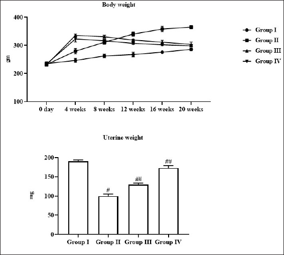
Effect of Cynaropicrin on the Uterine and Bodyweight of Experimental Rats
Figure 1 reveals the effect of cynaropicrin on the alterations in uterine and body weight of the control and treated rats. The OVX rats exhibited a remarkable elevation in body weight and a reduction in uterine weight. Nonetheless, 10 and 20 mg/kg of cynaropicrin treatment effectively decreased the body weight and improved the uterine weight in the OVX rats (Figure 1).
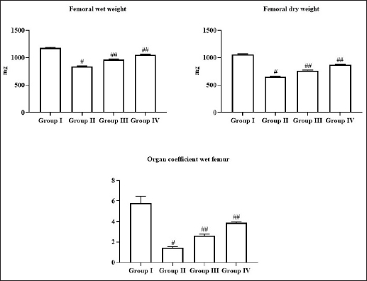
Effect of Cynaropicrin on the Changes in Femoral Weight in the Experimental Rats
The impacts of cynaropicrin treatment on the changes in the femoral wet and dry weights and organ coefficient are presented in Figure 2. The OVX rats revealed a drastic reduction in the femoral wet and dry weights and organ coefficients. Interestingly, these alterations were effectively regulated by the 10 and 20 mg/kg of cynaropicrin treatment (Figure 2).
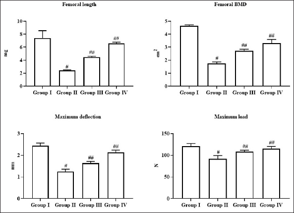
Effect of Cynaropicrin on the Femoral Length, BMD, Maximum Deflection and Load in the Experimental Rats
Figure 3 demonstrates the impacts of cynaropicrin on the BMD, femoral length, maximum deflection, and load in the control and experimental rats. The remarkably decreased levels of femoral length, femoral BMD, maximum load, and deflection were observed in the OVX rats when compared with the control. Meanwhile, the administration of 10 and 20mg/kg of cynaropicrin appreciably improved the femoral length, femoral BMD, maximum load, and deflection in the OVX rats (Figure 3).
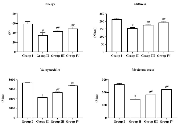
Effect of Cynaropicrin on the Biomechanical Characteristics in the Experimental Rats
The effects of cynaropicrin treatment on biomechanical characteristics such as energy, stiffness, young modulus, and maximum stress were evaluated and findings were revealed in Figure 4. The OVX rats have exhibited a remarkable reduction in energy, stiffness, young modulus, and maximum stress. Interestingly, these alterations were appreciably attenuated by 10 and 20mg/kg of cynaropicrin treatment (Figure 4).
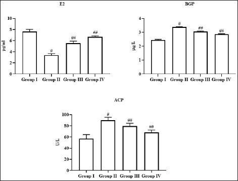
Effect of Cynaropicrin on the Status of Estradiol (E2), BGP, and ACP in the Experimental Rats
The status of E2, bone-gla-protein (BGP), and acid phosphatase (ACP) in the control and treated rats are represented in Figure 5. As shown in Figure 5, the status of E2 was found decreased meanwhile BGP and ACP levels were augmented in the serum of OVX rats. However, the level of E2 was increased and BGP and ACP status were remarkably reduced in 10 and 20 mg/kg of cynaropicrin-treated OVX rats (Figure 5).
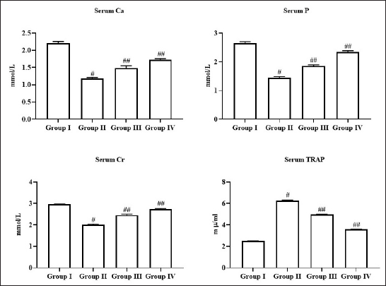
Effect of Cynaropicrin on the Levels of Mineral Contents in the Experimental Rats
Figure 6 reveals the influence of cynaropicrin on the levels of mineral contents such as calcium (Ca), phosphorus (P), creatinine (Cr), and tartrate-resistant acid phosphatase (TRAP) in the serum of control and treated rats. The OVX rats revealed a remarkable depletion in the status of Ca, P, and Cr and elevated status of TRAP in the OVX rats. Meanwhile, the administration of 10 and 20 mg/kg of cynaropicrin effectively augmented the status of Ca, Cr, and P and reduced the TRAP in the OVX rats (Figure 6).
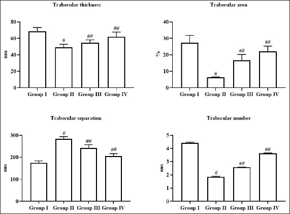
Effect of Cynaropicrin on the Trabecular Microarchitectures in the Experimental Rats
The effects of cynaropicrin on the measurement of trabecular microarchitectures such as trabecular thickness, trabecular area, trabecular separation, and trabecular numbers in the control and experimental rats are represented in Figure 7. The OVX rats displayed a drastic reduction in trabecular thickness, area, and number and an increment in the separation. Interestingly, these alterations were effectively reversed by 10 and 20 mg/kg of cynaropicrin treatment (Figure 7).
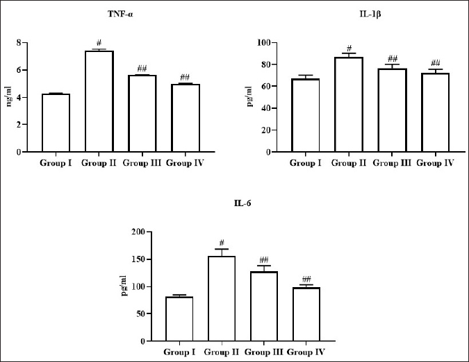
Effect of Cynaropicrin on the Levels of Inflammatory Cytokines in the Experimental Rats
Figure 8 comprises the inflammatory cytokine levels in the experimental rats. The OVX rats revealed a remarkable elevation in the status of TNF-α, IL-6, and IL-1β in the serum. Nonetheless, 10 and 20 mg/kg of cynaropicrin-treated OVX rats demonstrated a remarkable reduction in the status of these cytokines in the serum (Figure 8), which demonstrates its anti-inflammatory activity.
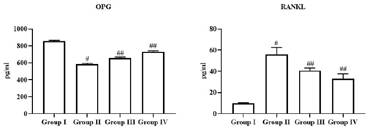
Effect of Cynaropicrin on the Levels of OPG/RANKL in the Experimental Rats
The OPG and RANKL in the control and treated rats were determined and the findings are presented in Figure 9. The OPG level was found to decrease and RANKL level was augmented in the OVX rats. Interestingly, the administration of 10 and 20 mg/kg of cynaropicrin effectively increased the OPG level and decreased the RANKL level in the OVX rats (Figure 9).
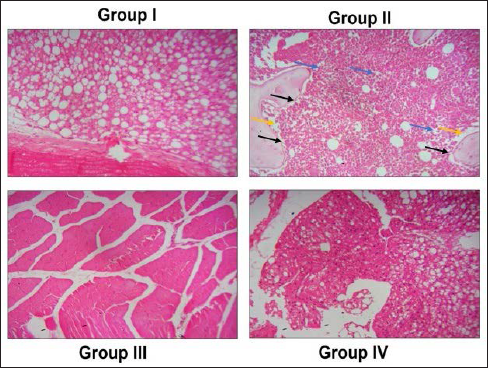
Effect of Cynaropicrin on the Femur Bone Histopathology of the Experimental Rats
The histopathological changes in the femur bone of both control and treated rats were examined and representative images were depicted in Figure 10. The control rats revealed a tissue microarchitecture, and trabecular arrangements. Hoverer, the femur bone of OVX rats revealed a major histological change such as distracted microarchitecture, abnormal thinning of trabeculae, and higher interspace in the trabeculae. Interestingly, these histological changes were effectively ameliorated by 10 and 20 mg/kg of cynaropicrin treatment (Figure 10).
Discussion
Osteoporosis is characterized by the gradual reduction in bone density and mass, and impaired bone trabecular micro-architecture with a higher risk of fracture. 29 It mainly involves dysregulation of balance between the new bone formation by osteoblasts and resorption by osteoclasts. 30 Osteoporotic fractures are connected with high death rates and are responsible for drastic economic burdens. 31 Only a few clinically approved drugs are presently being prescribed to treat osteoporosis, such as bisphosphate and denosumab.32–33 The long-term use of these drugs often comes with a risk of jaw necrosis, which can result in permanent disability. 34 Hence, the need for the exploration and identification of new substitute therapeutic candidates to treat osteoporosis with improved risk-benefit profiles are in great demand. The current study was focused on the evaluation of the anti-osteoporosis properties of cynaropicrin against the OVX rats.
The increase in body weight is an important phenomenon during age-related osteoporosis. 35 Here, we also noticed that the OVX rats demonstrated an appreciable increment in body weight and a reduction in uterine weight. A previous study already reported the depletion of uterine weight in the OVX rats. 36 Our findings ascertained that cynaropicrin remarkably reduced the body weight and improved the uterine weight in the OVX rats (Figure 1), which confirmed the beneficial properties of cynaropicrin.
Inflammation is a major causative factor of osteoporosis, which increases bone resorption. 37 Chronic inflammatory conditions can disturb bone metabolism and speed up the bone loss mechanism. 38 The increased production of inflammatory cytokines inhibits the mineralization of bone nodules, which ultimately leads to bone destruction and bone loss.39–40 The crosstalk between these inflammatory cytokines can synergistically improve bone resorption and osteoclastogenesis41–42 reported that the involvement of TNF-α, IL-1β, and IL-6 in bone resorption, which highlights the connection between bone loss and inflammation. 43 It is already reported that the cytokines were found elevated in osteoporosis patients. In a similar manner, our outcomes also revealed the elevated status of TNF-α, IL-6, and IL-1β in the OVX rats. These findings coincided with the previous report done. 44 Interestingly, the treatment with cynaropicrin appreciably decreased these cytokines in the OVX rats (Figure 8). These findings evidenced the anti-inflammatory potential of cynaropicrin.
TRAP is considered as a critical biomarker of osteoclast phenotype, which is widely utilized to detect the osteoclast precursor cell differentiation. 45 TRAP is generated by the osteoclasts during bone resorption. 46 In comparison with journal bone biomarkers, the TRAP is believed as the most studied bone resorption biomarker with comparatively good specificity and high sensitivity.47–49 In this study, we found that the status of TRAP and BGP was found augmented in the serum of OVX rats; however, this increment was effectively reverted by cynaropicrin (Figures 5 and 6). These findings proved that cynaropicrin effectively inhibited the osteoclast activity by decreasing TRAP levels.
The destruction of trabecular bone micro-architecture ultimately leads to depleted bone strength and higher risks of fracture incidences. 50 Consequently, it is essential to examine the impacts of cynaropicrin on the quality of bone structures and BMD. The BMD and bone density are the significant biomarkers of bone that imitate the integrity and quality of bones.51–52 BMD is a major biomarker to determine the occurrence, prognosis, diagnosis, and treatment followed by the diagnosis of osteoporosis, which affects Ca and P metabolism. 53 Ca is a major nutrient for healthy bone growth. Ca is the crucial player in bone health maintenance and nearly 99% of Ca is found in the bone. 54 It is well known that sufficient consumption of Ca decreases the risks of osteoporosis. The changes in the levels of Ca, P, and Cr are often reported in osteoporosis,55–58 which already been reported that the OVX rats demonstrated the altered levels of Ca, P, and Cr in the serum. Similarly, our findings also demonstrated that the status of Ca, P, and Cr was found depleted in the OVX rats. Meanwhile, the administration of cynaropicrin appreciably escalated the status of Ca, P, and Cr in the OVX rats (Figure 6). Our current results were found similar to the previous report. 59 These findings witnessed that cynaropicrin effectively upholds the bone minerals in the OVX rats. We also found that cynaropicrin effectively increased the trabecular thickness, area, and numbers and decreased the trabecular separation in the OVX rats (Figure 7). These findings revealed the therapeutic potential of cynaropicrin against osteoporosis in rats.
The RANKL/OPG system is a critical player in bone metabolism regulation. 60 The RANKL/OPG signaling is a well-reported pathway, which is essential to regulating the bone formation, resorption, and osteoclast differentiation. 61 It has already been highlighted that the OPG produced by osteoclasts has a suppressive effect on bone development. 62 RANKL is one of the most imperative cytokines produced by osteoblasts, which performs a critical role during the differentiation of osteoclasts. 63 RANKL is expressed in osteoblasts that enhance osteoclast differentiation. 64 Hence, the measurement of RANKL and OPG is important to examine the integrity of bones. Here, we determined the status of OPG and RANKL and outcomes revealed that the OVX rats revealed an increased RANKL and reduced OPG level. A previous report, 65 supported our findings of this study. Interestingly, the treatment with cynaropicrin appreciably depleted the RANKL and improved the OPG status in the OVX rats (Figure 9). These findings witnessed that cynaropicrin has a therapeutic effect against OVX-stimulated bone loss by restoring the OPG/RANKL levels.
Conclusion
The results of this work delivered new prospects into the anti-osteoporosis effects of cynaropicrin. Our results revealed that cynaropicrin increased the femoral weight and length and restored the bone turnover markers in the OVX rats. The mineral profiles and inflammatory markers were also regulated by the cynaropicrin treatment. Cynaropicrin also increased the OPG level and decreased the RANKL level in the OVX rats. These findings revealed a therapeutic potential of cynaropicrin on the OVX rats. Further studies in the future can provide a more evidence on the anti-osteoporosis effects of cynaropicrin and promote it as a promising drug candidate to treat osteoporosis.
Footnotes
Abbreviations
Authors’ Contributions
Hanlu Ye: Experimentations, data organization, and wrote the original draft.
Ke Wang: Proof reading, data validation, and interpretation.
Kai Li: Design and supervision of experiments, data curation, and proofreading of the final draft.
Declaration of Conflicting Interests
The authors declared no potential conflicts of interest with respect to the research, authorship, and/or publication of this article.
Funding
This work was supported by the Department of Orthopedic, The People’s Hospital of Zhong Jiang, Deyang, Sichuan Province 618199, China.
Statement of Informed Consent and Ethical Approval
The animal experiments were conducted based on the approval of institutional animal ethical committee.
Summary
Osteoporosis elevates the risk of bone fragility and fracture, which leads to significant morbidity and poor life quality. The cynaropicrin treatment increased the OPG and reduced the RANKL in the OVX rats. Cynaropicrin increased the femoral weight and length, restored bone turnover markers and mineral profiles, and decreased inflammatory markers.