
Abstract
There has been a considerable increase in natural calamities such as earthquakes and floods occurring across various parts of the world. Apart from causing a high number of fatalities, victim identification poses a significant challenge in mass disaster scenarios. In forensic odontology, biometric information plays a crucial role in identifying deceased individuals. However, soft tissues undergo rapid deterioration following such disasters, making hard tissues a more reliable source for identification.
To evaluate the usefulness of enamel rod end patterns for forensic identification after exposure to conditions simulating flood and earthquake scenarios. Human teeth were subjected to simulated disaster conditions by immersing them in seawater to mimic flood situations and burying them in soil to simulate earthquake conditions. After exposure, enamel rod end patterns were recorded and analyzed using VeriFinger software. Enamel rod end patterns were successfully recorded following exposure to both simulated conditions. The pat- terns demonstrated identifiable characteristics, indicating their resistance to environmental degradation.Tooth enamel, owing to its highly crystalline nature and negligible organic content, serves as a reliable hard tissue for forensic identification. Enamel rod end pattern analysis can be effectively used as a biometric tool for victim iden- tification in mass disaster situations such as floods and earthquakes.
Introduction
The term “forensic” is from the Latin, meaning forum or a place where legal matters are discussed. 1 Forensic medicine is a branch of medicine that deals with the legal aspects of healthcare. According to Pederson, forensic odontology is the branch of odontology which deals with the proper handling and examination of dental evidence and with the proper evaluation and presentation of dental findings in the interest of justice.2, 3
The British Association for Forensic Odontology (BAFO) notes that forensic odontology is the branch of forensic medicine and, in the interest of justice, deals with proper examination, handling, and presentation of dental evidence in a court of law.2, 3
The major area of activity of forensic odontology is the identification of human beings, either dead or alive. This is more so in case of mass disasters, where the corpse is usually badly mutilated. Other areas of application include criminalistics, in cases involving the abuse of children and the elderly.
Tooth enamel is rather specialized and differs from the other calcified tissues in that it is more crystalline and has negligible organic content. Teeth endure postmortem degradation and extreme changes in ambient temperature and pressure better than most human tissues. They form an inert mineralized structure that resists deterioration and hence set up an important method of establishing identity. In a situation involving fire or severe trauma, physical features are often destroyed. Because teeth are heavily calcified, they can resist fire as well as a great majority of traumas. 4
Enamel is a product of ectoderm-derived cells. The process of enamel formation is a complex and organized one, carried out by specialized cells called ameloblasts. These cells lay down enamel rods that are the basic structural unit in an undulating and intertwining path. 5
These enamel rod patterns can be used in forensic sciences for the identification of the diseased individual.
The term “biometrics” refers to an identification technique based on using specific physical characteristics, which transforms a biological, morphological, or behavioral characteristic into a digital value. 6
The unique identification of an individual based on biometric information should have certain desirable prerequisite characteristics: highly unique to each individual, easily transmittable, able to be acquired as noninvasively as possible, and distinguishable by humans without much special training.6, 7
This study intends to determine whether peels of enamel rod end patterns could be used as an aid in forensic odontology for individual identification in the case of mass disasters like earthquakes and floods.
Materials and Methods
The study was conducted using extracted maxillary and mandibular premolars obtained as part of a treatment procedure for orthodontic purposes. The study was approved by the Institutional Ethical Committee (IRB approval number: SRMDC/IRB/2015/MDS/NO.605).
Inclusion Criteria
Maxillary and mandibular premolars were extracted for orthodontic purposes.
Exclusion Criteria
Teeth with decay, attrition, abrasion, erosion, hypoplasia, fractures, or restorations were excluded from the study.
The materials included were a total of 60 extracted teeth, acetone, acid etchant (37% phosphoric acid), Type I Glass Ionomer Cement (GIC) liquid (10% phosphoric acid), cellulose acetate sheets, a light microscope from Labomed, a Nikon digital camera, metal gauge, digital vernier caliper, VeriFinger Standard SDK version 5.0 software, sea water, and a pit with a depth of six feet below the ground level to simulate human burial conditions.
Method Followed to Record the Enamel Rod End Patterns
The 60 teeth selected were scaled and polished. To avoid positional errors, an acetate film was placed over the recording area during serial recordings, and a circle of 5 mm in diameter was drawn on the comparatively flat area (middle thirds) of the buccal surface of the tooth. Following this, the buccolingual measurement was recorded using a metal gauge and confirmed with a digital vernier caliper. The marked area was then etched for 20 minutes. Following this, a thin layer of acetone-coated cellulose film was placed over the marked area for 20 minutes. The film was then removed and observed under the microscope. A photomicrograph of the acetate peel was obtained and subjected to biometric analysis using the VeriFinger Standard SDK version 5.0 software. 6 The software obtains the patterns and sub-patterns of enamel rod endings as a series of lines running in varying directions, assigns a specific identification number and minutiae score, and stores the pattern in the database. These minutiae will be used by the software to compare the similarity or variability of any two patterns. This method will be followed to record the enamel rod patterns after subjecting them to seawater treatment and burial.
Methodology Followed to Simulate Floods and Earthquakes
A set of 30 teeth was buried in soil for 90 days. A pit measuring about six feet in depth was dug out, the teeth were placed at the bottom of the pit, and they were covered with one foot of earth over which a polyethylene bag was placed to help in the easy retrieval of the teeth after 90 days. The pit was closed and a concrete slab was placed over it for identification. After 90 days, the concrete slab was removed, the pit was dug up, and the teeth were retrieved and cleaned. This mimicked the burial of human remains during earthquakes. To recreate a flood-like situation, a set of 30 teeth was immersed in seawater for a month.
Methodology Followed to Obtain Enamel Rod Patterns After Treating with Sea Water and Burial
Blinding was maintained between the groups, and the entire procedure previously used to obtain the enamel rod end patterns was repeated separately for each group. However, instead of 37% phosphoric acid, 10% phosphoric acid was used to condition the teeth before pattern acquisition. The pattern was obtained from the previously recorded site. The enamel rod end patterns obtained before and after were compared for similarity using the minutiae scores as well as the distribution and relationship of these minutiae points obtained using the VeriFinger Standard SDK version 5.0 software. The results were statistically analyzed with a t-test.
Results
The present study was done to determine whether enamel rod end patterns could be used as an aid in forensic odontology for individual identification.
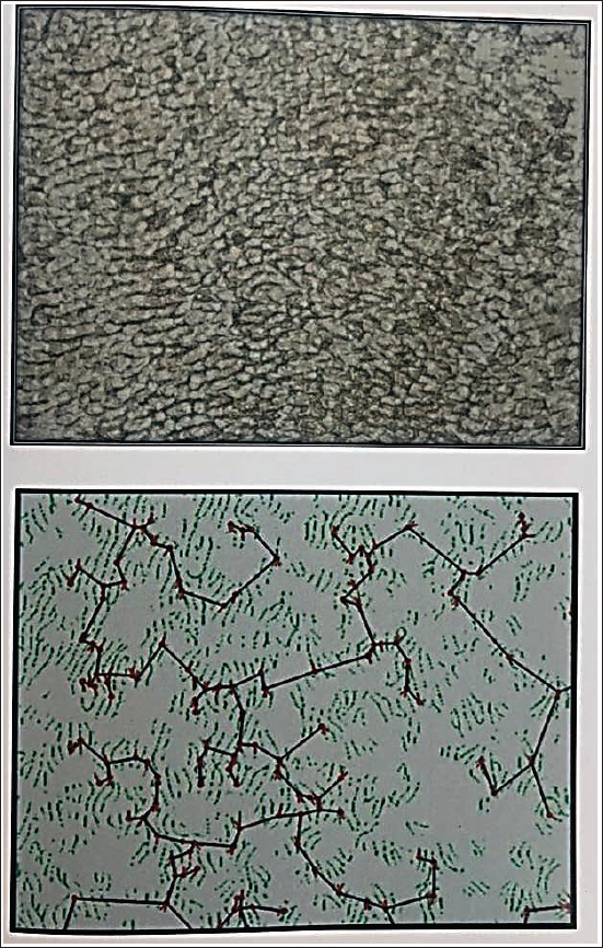
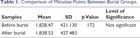
Enamel rod end patterns obtained before and after subjecting these teeth to various alterations were compared using the minutiae points (Figure 1).
Photomicrograph of Acetate Peel and Biometric Generation with Minutiae Points Using the VeriFinger Standard SDK Version 5.0.
Statistical analysis of the data obtained was done using the SPSS software version 17. The descriptive statistics, such as mean and standard deviation (SD) were calculated for individual groups. Comparison between groups was done using a t-test.
Comparison of Minutiae Points Between Burial Groups.
Burial in Soil.
The comparison of minutiae points before and after burial revealed that there is no significant difference (p >.05) (Figure 2 and Table 1).
Comparison of Minutiae Points Between Sea Water Groups.
Sea Water.
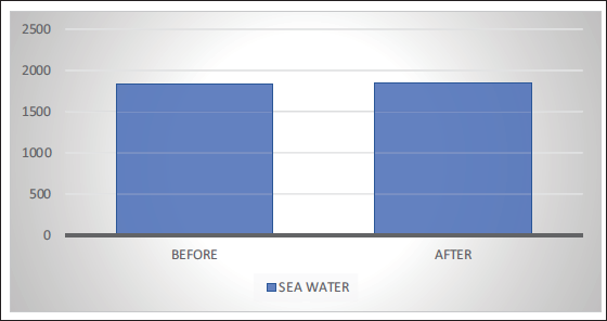
The comparison of minutiae points before and after immersion in seawater revealed that there is no significant difference (p >.05) (Figure 3 and Table 2).
Comparison of Minutiae Points Before and After Groups Burial in Soil and Immersion in Sea Water.
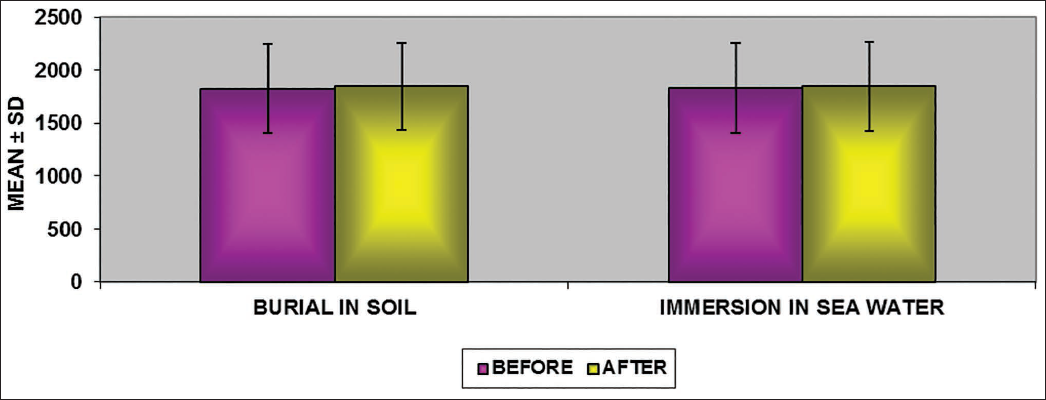
The results reveal that enamel rod end patterns are not significantly altered by environmental changes such as burial in soil and immersion in seawater (Figure 4).
Discussion
Enamel is a product of ectoderm-derived cells called ameloblasts. The basic structural unit of enamel is the enamel rod (enamel prism). Formation of enamel is a highly organized process wherein ameloblasts (cells forming enamel) lay down the enamel rods (enamel prisms) in an undulating and intertwining path. This is reflected on the outer surface of the enamel as patterns of the ends of a series of adjacent enamel rods, resulting in characteristic enamel carvings known as ameloglyphics, commonly referred to as a “tooth print.”
Each enamel rod measures approximately 5–6 µm in diameter and 2.5 mm in length. They follow an undulating course from the dentinoenamel junction to the external tooth surface. Many rods span the entire thickness of the enamel in a straight course, whereas most have a wavy pattern. The acid etching on the surface enamel results in the removal of the surface mineral component in the rod and rod sheath. As the rods and rod sheaths have different mineral densities, the etching results in an uneven dissolution of the surface enamel along with the removal of the smear layer. About 10 µm of surface enamel is removed by acid etching, revealing etching patterns or rod end patterns or tooth prints.
“Tooth prints” are a concept similar to fingerprint analysis. Previous studies have confirmed that tooth prints are unique to an individual. Therefore, the current study was planned to examine the stability of tooth prints in various environmental conditions created artificially (seawater immersion and burial in soil).
Numerous peel techniques, like the plastic peel technique, flexible peel technique, and rigid peel method, which are used to study the surface details of rocks, are used in anthropology and petrology. Scott et al. used metal-shadowed collodion replicas to study tooth surfaces by means of optical microscopy. 8 They observed enamel rod ends, perikymata, scratches, cracks, and various developmental defects and concluded that structural details visualized in these replicas were inaccurate. Fusun et al. used the acetate peel technique to study dental structures, especially enamel, without routine decalcification, sawing, and mounting process. They concluded that the technique is a simple, inexpensive, accurate, and rapid method for studying surface details. 9 So, this technique was selected for our study to record enamel rod endings from tooth surfaces for biometric analysis.
Manjunath et al. analyzed enamel rod end patterns on 30 extracted teeth using the VeriFinger Standard SDK version 5.0 software and concluded that these patterns were unique for each tooth, and they could be used for personal identification. 10 Nidhi et al. used the VeriFinger Standard SDK version 6.0 software to analyze the tooth prints and proved that each tooth has a different pattern. 11 Minutiae points are the identification points marked and stored by the VeriFinger Standard SDK version 6.0 software for the identification, verification, and comparison of patterns. Minutiae points are discontinuities of the lines seen as line endings, dots, very small lines, ponds, bifurcations, and loops.
It was observed that the VeriFinger Standard SDK version 5.0 software was able to identify subsequent records of the same area of the same tooth with the original record stored in the database of the software. Comparison of the minutiae scores using Cronbach’s test also showed that there was no significant difference in the minutiae scores obtained. 6 Therefore, in the present study, we employed the VeriFinger Standard SDK version 5.0 software for the analysis and comparison of enamel rod end patterns for personal identification. The results in the present study showed that the enamel rod end patterns were obtainable for all 60 teeth and were unique for each tooth. This observation is in concordance with the previously reported studies by Manjunath et al. (2008), Nidhi et al. (2009), Manjunath et al. (2012), and Girish et al. (2013).
Our study addressed the effect of adverse environmental conditions on tooth prints by artificially mimicking such conditions using standard laboratory procedures.
In order to simulate human burial conditions in our study, a pit about six feet below the ground level was dug out. After obtaining the initial enamel rod end patterns, a set of 30 teeth was placed at the bottom of the pit and was covered with one foot of earth, over which a polyethylene bag was placed to help in the easy retrieval of the teeth after 90 days. The pit was closed and a concrete slab was placed over it for identification. After 90 days, the concrete slab was removed, the pit was dug up, and the teeth were retrieved, cleaned, and the entire procedure of obtaining the rod end patterns was repeated. We were able to record the enamel rod end patterns from these teeth. The minutiae points and the enamel rod end patterns obtained matched with the data. A buried human body would undergo skeletonization depending upon the duration of burial and the ambient temperature, which can take as little as a month in hot climates and around two months in cold climates.12–14 The teeth are often the only thing left undisturbed, because tooth enamel is the strongest substance in the body. Human teeth are always considered a reliable source in medicolegal identification studies. 15 Our study has shown that teeth, when buried, resist the alterations induced by temperatures, the lack of access to oxygen, and the diversity of various microorganisms. So, enamel rod end patterns can still be obtained from deceased individuals who have been buried for several months.
In order to simulate the chemical action that seawater exerts on human enamel, a set of 30 teeth was placed in a container containing seawater after obtaining the initial enamel rod end patterns. After five days, the teeth were removed from the container, cleaned, and the entire procedure of obtaining the rod end patterns was repeated. We were able to record the enamel rod end patterns from the teeth that were immersed in seawater for five days. The minutiae points and enamel rod end patterns obtained in the study matched the database that had been created from previously recorded minutiae points. This suggests that chemical changes caused due to drowning in seawater do not alter the enamel rod end patterns, even when they body is decomposed beyond recognition, and therefore could be used as a source of individual identification.
Forensic dentistry serves the main objective of victim identification during mass disasters. 16 Tooth prints are unique to each individual, but the patterns change over a period of time (5–6 years) due to physiological or regressive changes. Therefore, the tooth print recording of an individual has to be updated at least once every five years. In the field of forensics, personal identification is an important component and a fundamental aspect in the identification of deceased individuals. 17 Under the adverse conditions of burial and seawater immersion, tooth prints have been found to be useful in the identification of a person. Thus, tooth print recording and the maintenance of a regularly updated tooth print database can be recommended for its use in forensics.
Conclusion
The enamel rod end patterns obtained using the acetate peel technique and automated biometrics were unique for each tooth. Exposing the teeth to seawater to simulate the chemical action of seawater on teeth and burying the teeth in soil to simulate human burial conditions did not alter the enamel rod end patterns. Even though enamel rod end patterns are unique to an individual, they change as age advances due to physiological wear and tear, and so the tooth print should be frequently updated.
Moreover, obtaining an acetate peel from the same area of the tooth in vivo is somewhat difficult. Therefore, future efforts should focus on the development of a fiber-optic laser scanner that can scan the complete tooth surface for enamel rod ends, which could enable the instant identification of an individual.
Footnotes
Declaration of Conflicting Interests
The authors declared no potential conflicts of interest with respect to the research, authorship, and/or publication of this article.
Ethical Approval
Ethical clearance was obtained from the Institutional Ethical Committee.
Funding
The authors received no financial support for the research, authorship, and/or publication of this article.
Informed Consent
Informed consent was obtained from the participating subjects