
Abstract
This pilot study assesses tooth crown metric traits among two geographically related but distinct ethnic groups in South India. The authors aimed to compare the ‘Irula tribal’ and ‘Non-tribal natives’ of Palakkad district using dental metric traits as an adjunct in population identification and to identify sexual dimorphism within them. Mesiodistal (MD) and labio/buccolingual (L/BL) dimensions of selected key teeth, namely, central incisors, canines, first premolars, and first molars were measured using digital calipers in plaster models from 100 participants (50 Irula tribal and 50 Non-tribal natives). The Irula tribal group exhibited significantly larger labiolingual (LL) dimensions in canines for both males and females across both jaws. In contrast, variations in MD dimensions were limited, with only the left maxillary central incisor in Irula females showing statistical significance. Only minimal sexual dimorphism was observed. It is found that L/BL and MD dimensions of maxillary and mandibular teeth can help differentiate the two studied populations and identify gender. Canines of either side of both jaws serve to differentiate the two population groups. In Irula tribals, MD widths of maxillary and mandibular canines aid sex determination. For Non-tribal natives, MD widths of canines, premolars, and first molars are key. Males generally have larger dimensions than females, with tribals showing higher overall measurements.
Introduction
Tooth metrics are a dependable tool in dental anthropology used in both large and restricted-scale studies as population markers. 1 Dental anatomy is the result of a combination of genetic, epigenetic, and environmental factors, with additive genetic variation contributing 59%–62% and environmental factors 8%–29%. 2 Previous research observed such differences among different ethnic groups in mesiodistal (MD) and labio/buccolingual (L/BL) dimensions.3, 4 Odontometry is a simple and cost-effective tool in gender determination when a fractured jaw and incomplete dentition are available. 5
The “Irula tribals” are one of the indigenous groups of Palakkad district in South India, known for their distinct cultural heritage, language, and lifestyle. “Non- tribal natives” are the inhabitants of Palakkad district other than tribals. There are no studies available in the literature on the dental traits of the above populations. Hence, determining the dental metric traits of these two groups is expected to contribute to population and individual identification.
Aims and Objectives
This pilot study aims to identify the differences between “Irula tribals” and “non-tribal natives” and sexual dimorphism (SD) within each group using tooth metric traits.
The objectives are
To assess dental metric traits of selected teeth of Irula tribals and non-tribal natives. To compare the dental metric traits between the “Irula tribal” and “non-tribal native” groups. To identify SD within the “Irula tribal” and “non-tribal native” groups.
Study Design
Cross-sectional descriptive observational study
Materials and Methods
The study was commenced after obtaining approval from the Institutional Ethics Committee. The minimum required sample size was calculated as 50, based on the results of the study by Sravya et al. 3 with 95% confidence level and 80% power.
Fifty Irula tribals and 50 non-tribal natives were taken up for the study after obtaining informed consent. Each group consisted of 25 males and 25 females.
Criteria for division of tribal and non-tribal groups.
The Irula tribal group consisted of individuals who belonged to the Irula tribe for the previous two generations. This was confirmed by the tribal extension officer and tribal promoters of the Irula hamlets. Non tribal natives were those who had been natives of Palakkad district for two generations with no known ancestral history of migration from other places.
The inclusion criteria for participant selection were individuals aged 18–40 years and the presence of intact key teeth (central incisors, canines, first premolars, and first molars) on at least one side of both jaws. Children below 18 were not included, as parental consent will be required. Those above 40 years were not included owing to the probability of increased regressive alterations and missing teeth.
The exclusion criteria were individuals with multiple missing teeth, significant tooth attrition, mobile teeth, decayed teeth, and extruded teeth that could affect measurement accuracy.
Alginate impressions were taken, and stone plaster casts were prepared. The MD and L/BL dimensions of the selected teeth were measured on these casts.
MD measurement was defined as the maximum distance between the mesial and distal proximal surfaces of the tooth crown from the point of contact with adjacent teeth.
L/BL measurement was defined as the greatest distance between the labial/buccal and lingual surfaces of the tooth crown, perpendicular to and bisecting the line that defines the MD dimension.
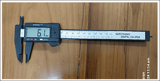
Digital vernier calipers (Figure 1), VERAT Electronic Carbon Fiber Composites Digital Vernier Caliper six-inch TRTA11A (50 mm) were used for precise measurements.2–6 All casts were measured using the same external illumination, which was bright and uniform. The tips of the beaks of the calipers were placed at the contact points. The calipers were held parallel to the incisal/occlusal surface to avoid parallax error, and each measurement was repeated three times to ensure consistency and reduce potential observer error. The mean of the measurements was taken. The measurements were repeated if the difference between them was more than 0.4 mm2. Only a set of 10 teeth was measured at a stretch, and short breaks of five minutes were taken in between to avoid eye fatigue. The total assessment period was three months.
Digital Vernier Calipers.
Data were analyzed using the Statistical Package for the Social Sciences, version 25. Pairwise comparisons of mean MD and L/BL values between the four subgroups (Irula tribal males, Irula tribal females, non-tribal native males, and non-tribal native females) were conducted using the t-test. A p value of <.05 was taken as significant.
The percentage of SD was calculated using Garn’s formula: SD = [(Xm ÷ Xf − 1)] × 100, where Xm represents the mean male canine width and Xf represents the mean female canine width.
Results
The study population consisted of a total of 100 individuals, divided equally in terms of ethnicity and sex.
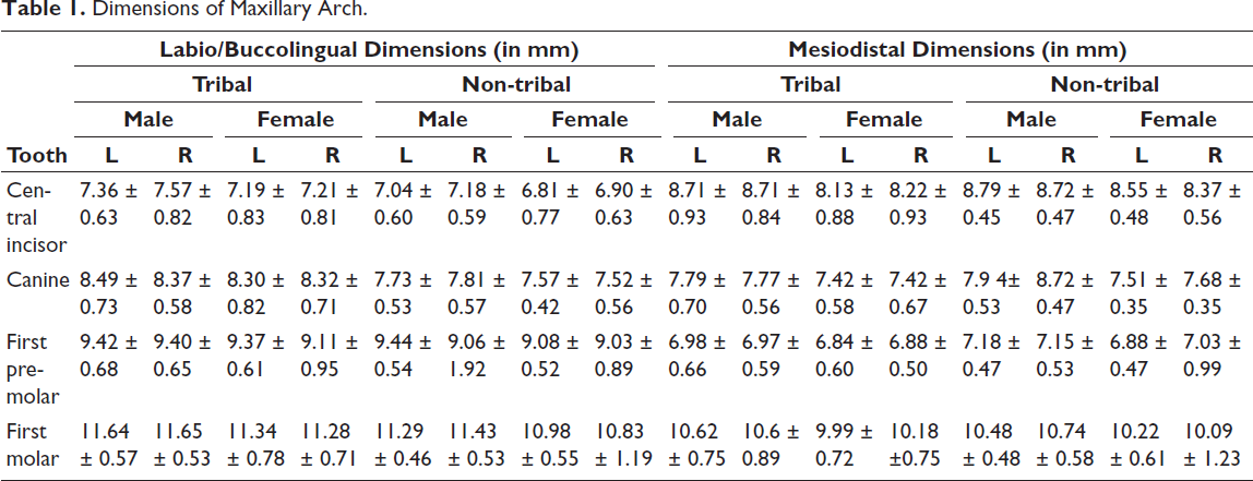
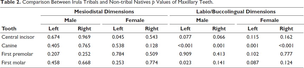
The maxillary canines of both sides in both males and females, and the left maxillary first molars in males of Irula tribals, had significantly larger L/BL widths. The left maxillary central incisors of non-tribal natives showed significantly larger MD widths in females (Tables 1 and 2).
Dimensions of Maxillary Arch.
Comparison Between Irula Tribals and Non-tribal Natives p Values of Maxillary Teeth.
Dimensions of Mandibular Arch.
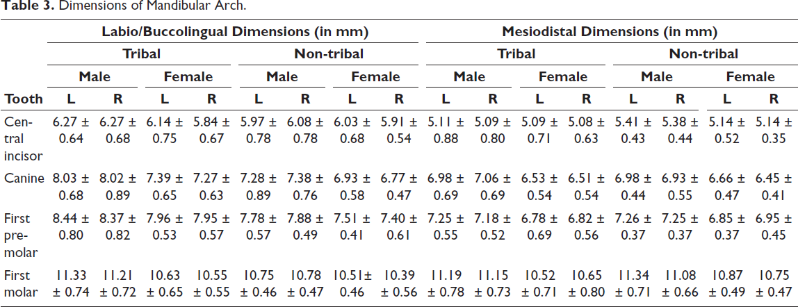
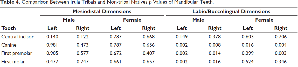
The mandibular canines of both sides of Irula tribal males and females, and the mandibular premolars and molars of both sides of Irula tribal males and the right premolars of Irula tribal females showed significantly larger L/BL widths (Tables 3 and 4).
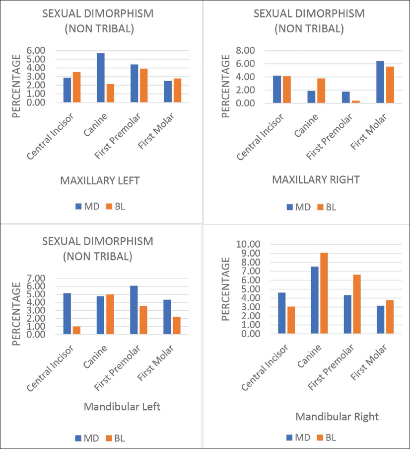
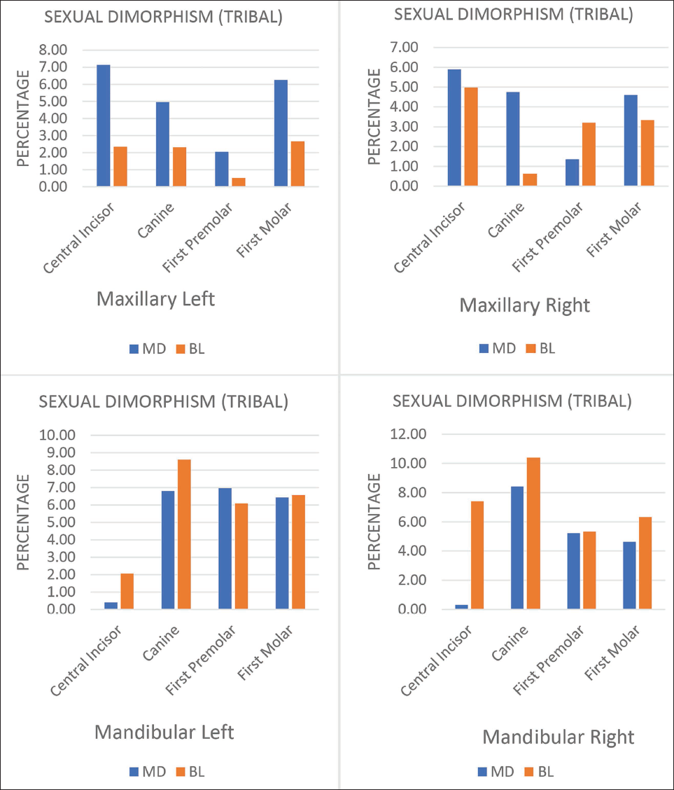
The percentage of SD in both groups is in the range of 0%–10% (Figures 2 and 3).
Percentage of Sexual Dimorphism in Non-tribals in Left and Right Maxillary and Mandibular Teeth.
Percentage of Sexual Dimorphism in Tribals in Left and Right Maxillary and Mandibular Teeth.
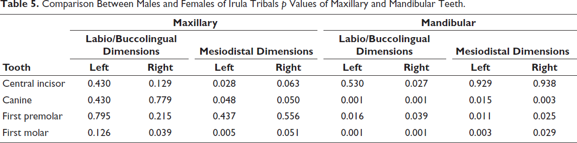
In Irula tribals, the maxillary arch showed significant SD in MD widths of left central incisor, left and right canine and left first molar and BL widths of right first molar while in the mandibular arch the MD and L/BL widths of the canines, first premolars and first molars of both sides and labiolingual (LL) widths of the right central incisor showed significant SD (Table 5).
Comparison Between Irula Tribals and Non-tribal Natives p Values of Mandibular Teeth.
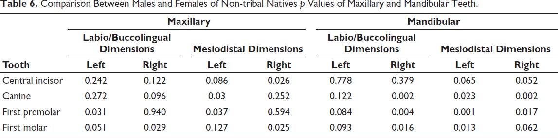
Non tribal natives showed significant SD in the MD widths of the left canine, left first premolar, and the right central incisor and right first molar, and BL widths of left first premolar and left and right first molar in the maxillary arch. The mandibular teeth of this population showed significant SD in the MD widths of the canines and first premolars of both sides, and in the left first molars and in the L/BL lengths of right canine, first premolar, and first molar (Table 6).
Comparison Between Males and Females of Irula Tribals p Values of Maxillary and Mandibular Teeth.
Comparison Between Males and Females of Non-tribal Natives p Values of Maxillary and Mandibular Teeth.
Males had greater widths than females in all measurements in both groups.
Discussion
Maxillary and mandibular canines of the Irula tribal group showed significantly larger LL widths compared to the non-tribal natives in both males and females in both left and right sides. This is a notable finding of this study.
The finding of a significant difference in LL measurements in the Irula group’s canines aligns with research on the stability and inheritance of tooth metrics, as demonstrated in previous studies.6, 7 Minor variations were observed in the MD dimensions of the maxillary central incisors in tribal females.
These could be attributed to genetic isolation, which may have preserved certain dental traits within the Irula population. In contrast, the non-tribal native population contains more sub-communities with greater intermingling among them. Also, anterior teeth are less susceptible to environmental influences. 1
The significant differences in L/BL dimensions, specifically in the mandibular first premolars and first molars, could be the influence of genetic factors coupled with functional adaptation, disease, or dietary habits. 6
Sexual Dimorphism
Determining sex is vital for personal identification and is one of the most utilized applications of forensics. 8 MD and L/BL dimensions are the ones widely used in gender identification. The mandibular canine has been proven to exhibit the maximum SD, and this is reflected in our study. 6
SD could be demonstrated in canines using Garn’s formula in an Arabian sample. 9 Males were found to exhibit significantly higher MD and L/BL dimensions, with canines showing the highest degree of SD in a Latin American sample. 10 In an odontometric study of canines, 64.4% of males and 82.8% of females could be identified with 73.5% accuracy, with lower canines showing more pronounced SD. 11
Bones and teeth are used extensively in forensics, being more resistant to decay than other tissues of the body. 12 The mandible and maxilla are valuable among the facial bones in SD. Measurement of selected mandibular parameters, namely, total mandibular body length, mandibular length, mandibular height, and gonial angle using lateral cephalograms have proved to be decisive in determining gender. 8 Various dimensions of the maxillary sinus, such as height, width, depth, and volume, were measured using computerized tomography scans in a study on Nigerian subjects. The measurements were greater in males, and the most accurate prediction of gender was observed in sinus volume. 13 The MD and BL diameters in the left quadrants were quantified in Arabian, South Asian, and East Asian populations. The BL dimension of the mandibular canine and the MD measurements of the mandibular lateral incisor showed SD. Males exhibited larger teeth. 14 Linear and diagonal odontometric parameters in first molars were found to be increased in males in a sample of 50 males and 50 females. 15 Panda et al. have found SD in metrics of maxillary incisors and canines in Indians, and they attribute this to non-genetic variability. 2
In a retrospective radiographic study of South Indian adults MD width of teeth was significantly greater in males. 5 Though our study population was also South Indian, the variation in our results could be attributed to the low sample size and the ethnic differences.
Males exhibited a significantly larger mandibular intercanine distance in a study involving 50 males and 50 females aged 18–25 years. However, the mean MD width of right mandibular first molars did not show a significant difference between genders 16 which correlates with our study in the case of non-tribal natives.
Our study is in accordance with the one by Tajik et al. on canine teeth using cone beam computed tomographic (CBCT) images in that the MD and BL widths and 10 derived ratios exhibited the lowest accuracy in discriminating gender. The total tooth length and root length were found to be more reliable measurements. 17 Ajmal et al. in a systematic review on odontometric parameters using CBCT as an aid in SD, found the maximum number of reports that had analyzed canines. Most reports favored the use of odontometry in SD, with accuracy ranging from 47.8% to 92.3%. 18
The study population and methodology of our study are similar to the one by Sravya et al. wherein another tribal population of South India has been compared with the urban population of the same district to determine SD. The differences in MD dimensions of canines between genders and population were both statistically significant. 3 In our study, the percentage of SD was below 10%.
Ancestry
The L/BL dimensions were found to be lower than standard values for all anterior maxillary teeth in four ethnic groups of northeastern India. Intra-group variability was noted. 19 Of a mixed population of European, Native American, and African ancestry, European was found to have the strongest influence on dental size. Colombians’ dental dimensions overlapped with those of their parental populations, indicating high dental size diversity within Latin Americans. The Arabian population consistently exhibited larger tooth dimensions compared to South Asian and East Asian populations. 10 A prospective study done on adults has compared tooth metric and skull measurements in an attempt to assess the stature of an individual. A combination of both these measurements has shown a more reliable estimate of stature. 12 In a review on the importance of dental patterns in personal identification, it has been found that the metric and morphologic traits of teeth, along with restorations and wear facets, play a decisive role. 20 Metric variations are highly valuable for identifying relatives in disasters and anthropological studies. 21
Our study supports the role of tooth metrics in determining ancestry, especially in LL dimensions of canines.
Age
The secondary changes occurring in teeth with age have been utilized to a great extent in age estimation. In a South Indian study, the linear and volumetric dimensions of the tooth, as well as the root and root canal, were measured using CBCT. Progressive secondary dentin deposition in the root canals caused a decrease in their dimensions with increasing age. It was found that root canal measurements could predict age with greater accuracy than those of whole teeth. 22 The age of children and adolescents belonging to Indore was calculated using Cameriere’s method, wherein the open apices of permanent mandibular teeth were measured on orthopantomograms. The obtained age exhibited a strong association with the actual age. Minor variations were attributed to population-specific differences, further emphasizing the need to establish normative values for each population. 23 The observed relation between age and lower molar traits is attributed to the interproximal wear, which may cause a reduction of the MD and L/BL dimensions.5, 9 Our study population involved subjects only up to 40 years, reducing the possibility of wear and consequent intra-group variations.
Limitations and Need for Further Studies
The small sample size limits the generalizability of the results. The study focused solely on two ethnic groups within a specific geographical area, and only metric traits were analyzed. Further studies are recommended on a larger scale in Irula tribals and also in other ethnic groups to establish population-specific data.
Conclusion
Statistically significant differences have been observed between the two groups and within the sexes of each group.
The LL dimensions of maxillary and mandibular canines and the BL dimensions of mandibular right premolars may be used as an adjunct means to differentiate the two population groups, irrespective of gender. Males may be assigned to either of these two populations using BL widths of right and left mandibular premolars and first molars, and maxillary left first molar. Females of the two groups may be differentiated using the MD widths of left maxillary central incisors.
Of the maxillary teeth of Irula tribals, the MD widths of maxillary canines of both sides may contribute to discriminating between males and females. MD widths of the maxillary left central incisor and first molar may also be used to aid in determining sex in this group. The MD width of the right central incisor and first molar, and left canine and premolar, and BL widths of left first premolar and right first molar can contribute to the same in the non-tribal native group.
Examination of mandibular teeth shows that the MD and L/BL widths of canines, first premolar, and first molar of both sides, and the LL width of the right central incisor may serve as an aid in sex identification in Irula tribals. In the non-tribal native group, gender can be identified using MD widths of left and right canines, first premolars, and left first molars. The L/BL widths of the right canine, first premolar, and first molar, too, may be used for gender identification.
Footnotes
Acknowledgements
The authors gratefully acknowledge the services of Mr Deepak S, Statistician, and Ms Athira PK, Dental Mechanic.
Declaration of Conflicting Interests
The authors declared no potential conflicts of interest with respect to the research, authorship, and/or publication of this article.
Ethical Approval and Informed Consent
The ethical clearance from the Institutional Ethical Committee and informed consent from the participants have been obtained for assessing tooth crown traits, palatal morphology, mandibular morphology and palatal rugae patterns among Irula tribes and non-tribe natives of Palakkad district as an aid in personal identification.
Funding
The authors received no financial support for the research, authorship, and/or publication of this article.